fig27

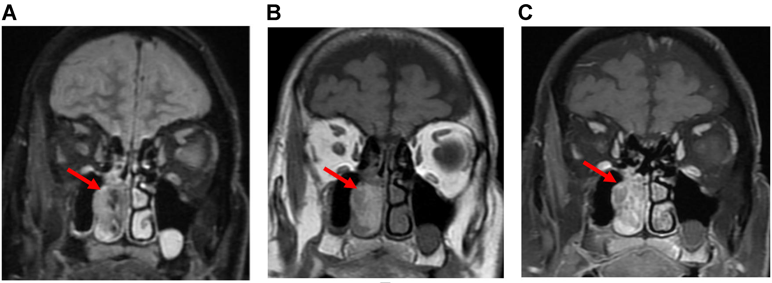
Figure 27. Sinonasal melanoma. Coronal T2 (A), precontrast T1 (B), and post-contrast T1 (C) images showing an ill-defined, T1-hyperintense, enhancing mass expanding the right nasal cavity (arrows).

Figure 27. Sinonasal melanoma. Coronal T2 (A), precontrast T1 (B), and post-contrast T1 (C) images showing an ill-defined, T1-hyperintense, enhancing mass expanding the right nasal cavity (arrows).


All published articles are preserved here permanently:
https://www.portico.org/publishers/oae/