fig2

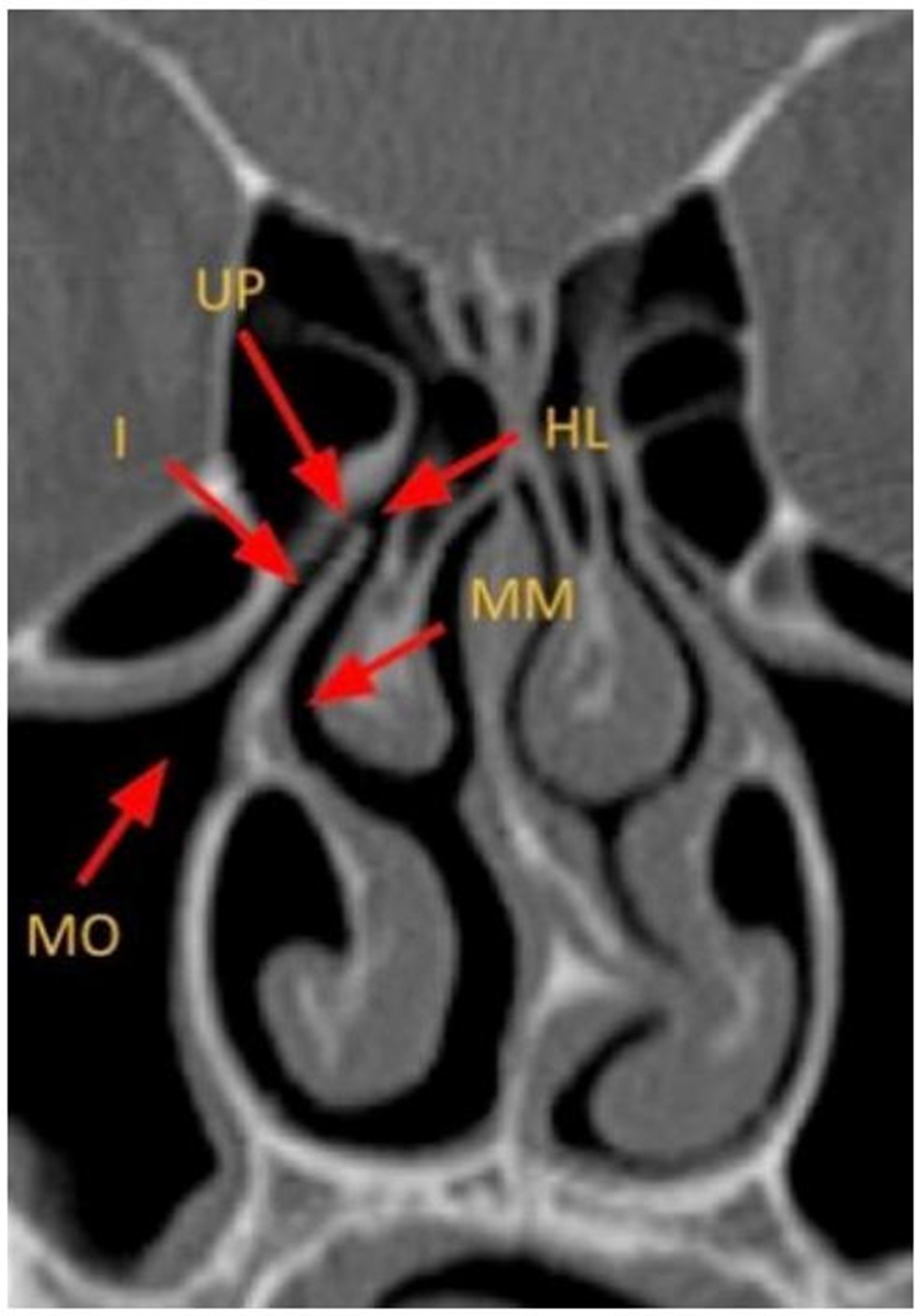
Figure 2. Coronal CT image depicting the key anatomy of the anterior OMU. MO: Maxillary ostium; I: infundibulum; HL: hiatus semilunaris; MM: middle meatus; UP: uncinate process; CT: computed tomography; OMU: ostiomeatal unit.

Figure 2. Coronal CT image depicting the key anatomy of the anterior OMU. MO: Maxillary ostium; I: infundibulum; HL: hiatus semilunaris; MM: middle meatus; UP: uncinate process; CT: computed tomography; OMU: ostiomeatal unit.


All published articles are preserved here permanently:
https://www.portico.org/publishers/oae/