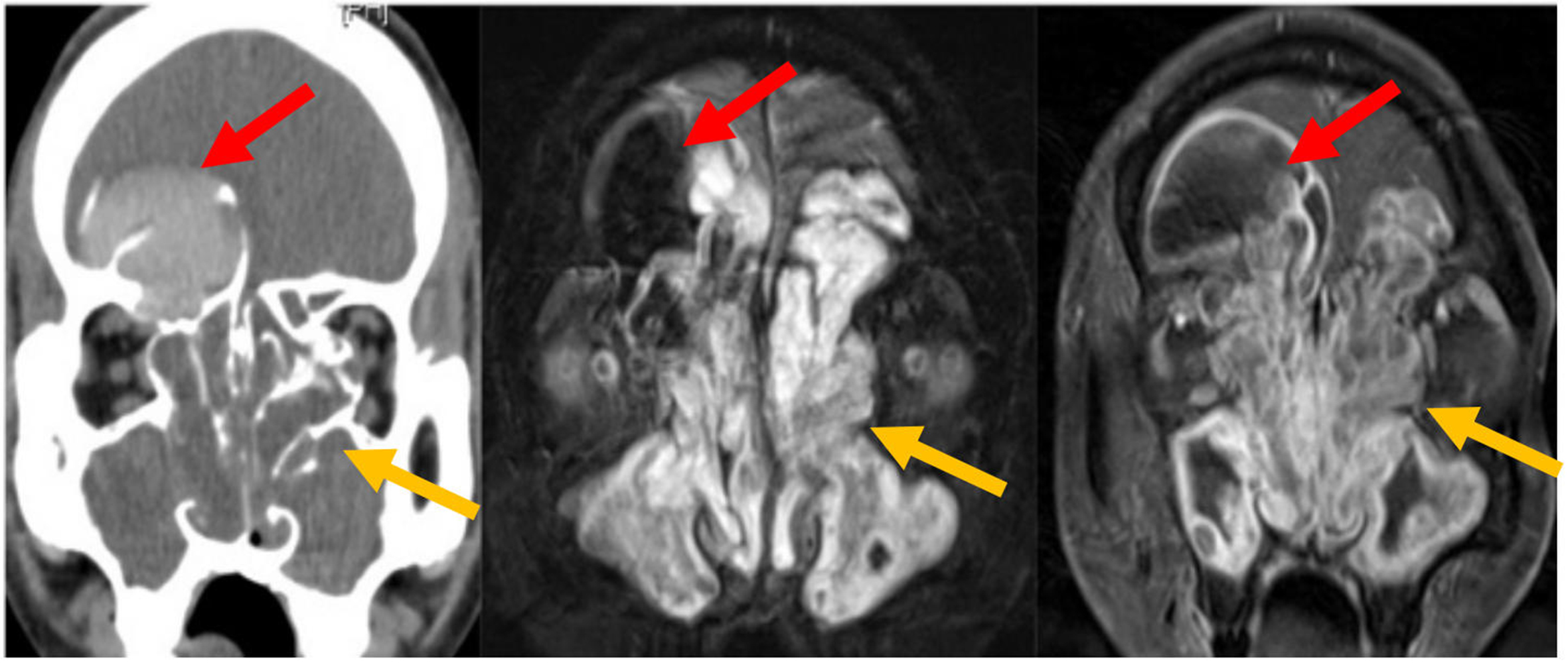
fig12

Figure 12. Coronal CT and MRI images of a right frontal sinus mucocele and sinonasal polyposis. The right frontal sinus is airless and expanded. Hyperdense secretions on CT correspond to the signal void on MRI (red arrows). Bony deficiency surrounds the mucocele. Polyps show low CT attenuation and characteristic T2 hyperintensity with peripheral enhancement on MR (yellow arrows). CT: Computed tomography; MRI: magnetic resonance imaging; MR: magnetic resonance.


