fig1

Imaging evaluation of sinonasal tract neoplasms

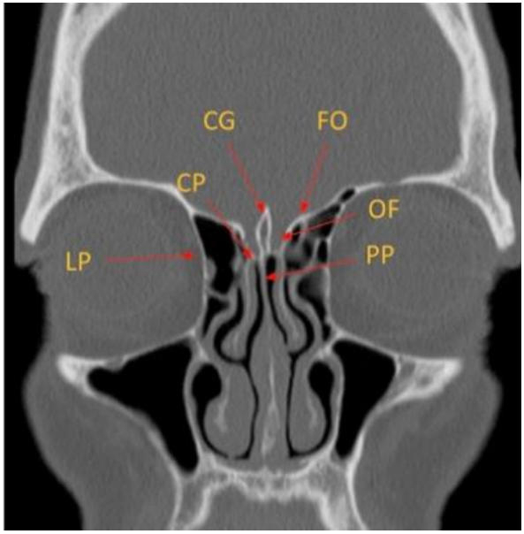
Figure 1. Coronal CT image depicting the key anatomy of the ethmoid bone. CG: Crista galli; FO: fovea ethmoidalis; CP: cribriform plate; OF: olfactory fossa; LP: lamina papyracea; PP: perpendicular plate of the ethmoid bone; CT: computed tomography.

Mini-invasive Surgery
ISSN 2574-1225 (Online)

Portico

All published articles are preserved here permanently:

https://www.portico.org/publishers/oae/

Portico

All published articles are preserved here permanently:

https://www.portico.org/publishers/oae/