


Next-generation vascular organoids: multi-lineage and immune integration for disease modeling and regenerative medicine
 0
0 Abstract
Traditional human blood vessel organoids, built primarily around endothelial monocultures, fail to replicate the multicellular architecture and dynamic immunological functions of native microvasculature. This review synthesizes an emerging paradigm shift toward third-generation, multi-lineage, and immune-competent vascular organoids. Multicellular integration, combining perivascular mural lineages (pericytes and vascular smooth muscle cells) with functional immune populations (macrophages, microglia, and lymphocytes), is delineated. Its role in reinstating baseline barrier tightness, contractility, and tissue-level immunosurveillance is highlighted. Critical bioengineering workflows are examined, with emphasis on fluidic shear stress in microfluidic platforms, the spatial precision afforded by three-dimensional (3D) bioprinting, and multiplex CRISPR gene editing. These technologies resolve lineage-specific media conflicts and enable off-the-shelf, hypoimmunogenic vascular constructs. Furthermore, the capacity of these systems to recapitulate complex pathophysiology is evaluated. Such pathophysiology includes complement-driven immunothrombosis in SARS-CoV-2 infection, neurovascular degeneration in Alzheimer’s disease, genetic small-vessel disorders, and tumor-immune barriers that govern chimeric antigen receptor T (CAR-T) cell infiltration. Finally, persistent translational hurdles are outlined, including metabolic bottlenecks, diffusion limits, scale‑up challenges, and the lack of large‑animal efficacy and safety data. Strategies that may help transition these models from research tools toward clinically relevant platforms are also discussed.
Keywords
INTRODUCTION
The human vasculature is a dynamic homeostatic hub that extends beyond blood conduction. In addition to delivering oxygen and nutrients, and clearing metabolic waste, blood vessels also direct tissue morphogenesis, organ-specific functional specialization, and regenerative capacity[1]. Structural stability in native blood vessels relies on a specialized architecture in which a continuous endothelial cell (EC) monolayer is enveloped by perivascular mural cells - microvascular pericytes and vascular smooth muscle cells (VSMCs) in larger vessels - and anchored within a dense basement membrane. This multicellular assembly depends on reciprocal crosstalk among ECs, mural lineages, and tissue-resident immune cells to maintain barrier function and homeostasis[1]. Crucially, the vasculature exhibits striking organ-specific heterogeneity, adapting to distinct physiological demands[2].
Disruption of this coordination precipitates severe pathologies. Endothelial dysfunction, aberrant vessel remodeling, and compromised barrier integrity underpin metabolic syndromes, stroke, neurodegeneration, viral infections, and tumor metastasis[3]. Despite decades of biomedical research, modeling human vascular pathophysiology in vitro remains challenging. Traditional two-dimensional (2D) monolayer cultures lack essential cell-matrix interactions and shear stress, which rapidly triggers endothelial dedifferentiation and functional decline[4]. Animal models, although physiologically intact, frequently fail to predict human responses because of interspecies differences in receptors, inflammatory cascades, and drug metabolism[5].
The emergence of human-induced pluripotent stem cell (hiPSC) technology, coupled with advanced bioengineering, has redefined this landscape. Human blood vessel organoids (BVOs) now provide a physiologically relevant, three-dimensional (3D) platform that recapitulates aspects of human vascular development and pathology. First-generation BVOs established self-assembling endothelial networks[6]. Second-generation models incorporated mural cells, thereby improving basement membrane deposition, lumen formation, and mechanical stability[7,8].
A critical limitation persists: the omission of functional immune populations. In vivo, immune cells, particularly macrophages and T cells, are integral to the vascular niche and drive physiological remodeling, inflammation resolution, and tissue repair[9,10]. Omitting immune lineages severely limits current vascular organoids, restricting the modeling of immune-mediated vascular pathologies, infectious barrier breaches, and tumor-immune-vascular microenvironment.
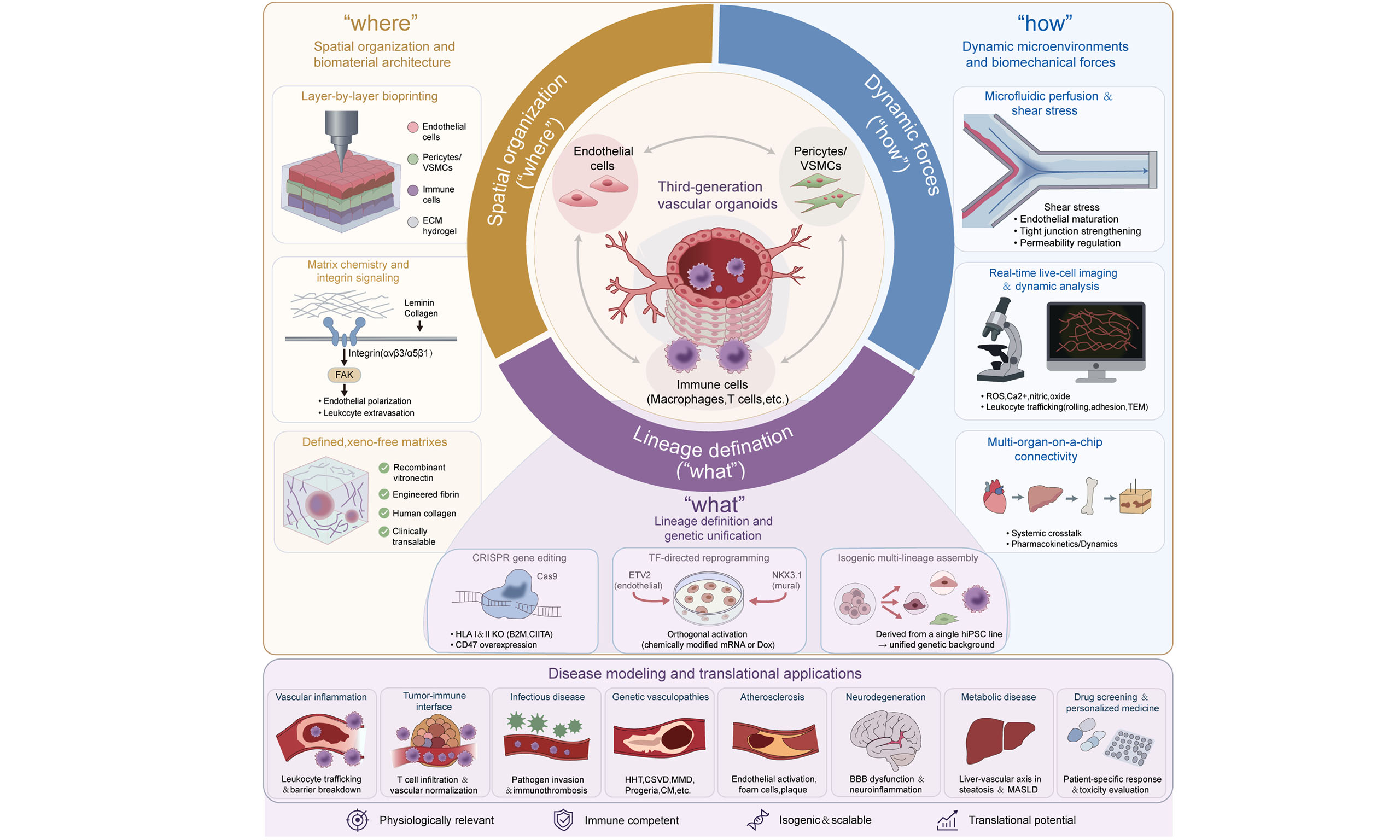
Next-generation bioengineering is driving a paradigm shift toward third-generation, multi-lineage, immune-competent vascular organoids [Table 1]. In this review, we synthesize recent advances in establishing these complex assemblies. We explore crosstalk among endothelial, mural, and immune lineages, evaluate engineering enablers, from microfluidic organ-on-a-chip to 3D bioprinting, and highlight applications in disease modeling and regenerative therapeutics. Finally, we outline key biofabrication bottlenecks and challenges in scalability, current Good Manufacturing Practice (cGMP) compliance, engraftment, and long-term safety.
Proposed generational framework of vascular organoid complexity
| Generation | Cellular composition | Functional hallmarks | Representative advances | Period |
| First | Self-assembled endothelial networks | Baseline lumen formation; static 2D/3D culture | Early BVO protocols[6] | ~2014-2018 |
| Second | Endothelium + mural cells (pericytes/VSMCs); basement membrane deposition | Structural stabilization; tight-junction reinforcement; limited barrier function | Disease-modeling CC[7]; mural-integration studies[8] | 2019-2023 |
| Third | Endothelium + mural cells + functional immune populations; dynamic immune interactions | Shear-dependent barrier integrity; leukocyte trafficking; immune-regulated remodeling; perfusion-enabled stability | Immune-integrated organ-on-chip and tri-culture platforms[12,13] | 2024-present |
Several reviews have already summarized BVO differentiation and its applications[6,11-13]. However, none has systematically examined functional immune integration as a design principle. This review fills that gap by framing third-generation vascular organoids through three interdependent dimensions:
• Compositional complexity: simultaneous presence of endothelial, mural, and functional immune lineages alone is insufficient. The key is the dynamic heterotypic crosstalk among all three compartments, which generates emergent properties (e.g., immune-regulated barrier permeability, pericyte-mediated control of leukocyte trafficking) that binary co-cultures cannot reproduce;
• Functional capabilities: Beyond cell types, third-generation vascular organoids acquire tissue-level functions, including shear-dependent barrier integrity, contractile responsiveness, and dynamic immunosurveillance, that are absent in earlier generations. These requires both the cellular triad and appropriate biomechanical cues (e.g., fluid shear stress, cyclic stretch), bridging the gap between structural complexity and physiological relevance;
• Engineering enablers: Third-generation systems are architected by advanced bioengineering platforms, microfluidic perfusion, 3D bioprinting with spatial precision, and clustered regularly interspaced short palindromic repeats (CRISPR)-based isogenic lineage generation, which coordinate spatial organization, deliver physiological forces, and unify genetic backgrounds. These engineering solutions are what make the multi-lineage integration functionally meaningful.
To define the scope, we searched PubMed and Web of Science for English-language studies since 2000, using combinations of “vascular organoid”, “blood vessel organoid”, “organ-on-a-chip”, “pericyte co-culture”, and “immune integration”, prioritizing primary studies from 2019 onward.
BUILDING MULTI-LINEAGE AND IMMUNE-COMPETENT VASCULAR ORGANOIDS
Recapitulating the perivascular niche: mural cells and basement membrane assembly
Functional vascular maturation requires precise spatiotemporal coordination and heterotypic signaling between ECs and mural lineages[14]. In late-stage angiogenesis, pericytes stabilize nascent endothelial sprouts[15], strengthen tight junctions through paracrine factors and direct contact[16], and deposit basement membrane components[17]. Heterocellular gap junctions such as Connexin 43 (Cx43) mediate these interactions and stabilization events[8]. Mural cell recruitment thus directly determines microvascular integrity, compliance, and selective permeability.
To recreate this niche in vitro, stem cell bioengineering employs two main strategies.
• Spontaneous co-differentiation vs. directed sequential induction: This approach mimics embryogenesis by guiding mesodermal progenitors through wingless-related integration site (WNT) and transforming growth factor-beta (TGF-β) signaling modulations to generate distinct endothelial and mural fates[18]. Early protocols by Patsch et al.[19] and Orlova et al.[20] demonstrated the concurrent expansion of hiPSC-derived ECs with VSMCs or pericytes, respectively. Beyond soluble factor cocktails, the biophysical and spatial cues of the surrounding microenvironment actively instruct endothelial fate determination. Zhang et al. demonstrated that a 3D differentiation protocol not only enhances the efficiency of hiPSC-EC generation but also preserves a stable endothelial phenotype, highlighting that spatiotemporal 3D context - rather than biochemical signals alone - is a critical determinant of EC specification[21]. More recently, the identification of shared mesenchymoangioblast (MAB) progenitors has accelerated pericyte differentiation[22,23]. PDGFRβ+ CD271+CD73- progenitors derived from MABs further differentiate into pericytes {cluster of differentiation 274 [CD274]+ capillary or delta-like homolog 1 [DLK1]+ arteriolar}, VSMCs and stromal cells. Self-assembling vessel organoids differentiated via WNT activation and BMP4 stimulation exhibit endothelial lumens, covered by a continuous basement membrane and interact closely with pericytes[6,24].
• Orthogonal transcription factor (TF) reprogramming: To bypass conflicting morphogenetic signals, recent studies have turned to direct TF-driven fate engineering[25]. Gong et al. demonstrated that orthogonal activation of TF ETV2 (endothelial) and NKX3.1 (mural) using Dox-inducible or modified mRNA (modRNA) systems induces balanced co-differentiation within five days[26]. Delivering these TFs via chemically modified mRNA produces footprint-free, physiologically relevant organoids, offering a scalable platform for disease modeling[27]. However, forced TF overexpression may bypass normal developmental checkpoints[28,29], raising concerns about incomplete epigenetic remodeling and off-target effects[29]. For ETV2/NKX3.1 systems, independent control of endothelial and mural compartments remains challenging due to the shared trigger, and absent perfusion limits maturation[26].
Parallel to lineage specification, accommodating these multicellular networks requires animal-origin-free extracellular matrices (ECMs) to replace batch-variable Matrigel preparations[30]. Recombinant vitronectin, engineered fibrin and human-derived collagen hydrogels match Matrigel in driving sprouting and capillary morphogenesis[30]. Paired with ultra-low attachment U-bottom or “sitting drop” configurations, these platforms enable standardized, high-throughput biofabrication of biomimetic vascular architectures[31] [Table 2].
Summary of multi-lineage and immune integration strategies
| Integration type | Approach | Cell types | Key technologies | Advantages | Limitations | References |
| Multi-lineage: pericytes/VSMCs | Spontaneous co-differentiation and directed sequential lineage induction | ECs + pericytes/VSMCs | Sequential modulation of WNT and TGF-β signaling | Mimics in vivo developmental spatiotemporal sequence | Time-consuming, asynchronous maturation, high batch-to-batch heterogeneity | [18-20,22,23] |
| Multi-lineage: organotypic vascularization | Co-development of mesoderm and endoderm | ECs + organ-specific parenchymal cells (lung/gut epithelia) | Simultaneous differentiation of mesodermal and endodermal lineages within same spheroid | Captures organ-specific endothelial specialization and tissue-resident immune niches | Complexity of multi-lineage co-differentiation; limited to specific organ systems | [24] |
| Multi-lineage: pericytes/VSMCs | Orthogonal transcription factor activation | ECs + pericytes/VSMCs | ETV2 (endothelial) + NKX3.1 (mural), chemically modified mRNA or Dox-inducible systems | Rapid generation within 5 days, footprint-free, controllable ratio | Long-term stability and in vivo functionality require further validation; forced TF overexpression may introduce non-physiological phenotypes or bypass normal developmental checkpoints | [25-29] |
| Multi-lineage: pericytes/VSMCs | Modular assembly with animal-origin-free matrix culture | Pre-differentiated ECs + pre-differentiated pericytes/VSMCs | Recombinant vitronectin, fibrin-based hydrogels, human-derived collagen, “sitting drop” method | Enhanced batch-to-batch consistency, compatible with high-throughput automation, xeno-free, clinically translatable | Matrix composition optimization requires systematic investigation | [30,31] |
| Immune: myeloid (monocytes/macrophages) | hiPSC-EC co-culture with monocytes/macrophages | ECs + monocytes/macrophages | TNF-α-induced ICAM-1/VCAM-1 upregulation; MMP-9 and VEGF secretion | Recapitulates leukocyte adhesion and migration under inflammatory conditions | Precise control of macrophage polarization (M1/M2) remains challenging | [13,35] |
| Immune: T cells/NK cells | Vascular chip co-culture | ECs + Pericytes + T cells/NK cells | Microfluidic perfusion, TNF-α-mediated endothelial activation, TEM quantification | Enables quantitative assessment of T-cell rolling, adhesion, and TEM | Device complexity, limited throughput, requirement for specialized equipment | [36,37] |
| Immune: organ-specific (neurovascular unit) | Multi-cell co-culture with glial components | ECs + pericytes + astrocytes + microglia | Co-culture-induced BBB phenotype (tight junctions, restricted solute permeability) | Recapitulates tissue-specific vascular-immune interactions and CNS immunosurveillance | Complex cell sourcing, optimization of multi-cell ratios and maturation kinetics required | [38-40] |
| Dual integration (multi-lineage + immune) | Full-spectrum tri-culture platforms | ECs + pericytes/VSMCs + macrophages/T cells | Multi-cell hydrogel encapsulation + microfluidic perfusion | Offer increased physiological complexity, enables triadic (EC-mural-immune) crosstalk studies | Media incompatibility remains a critical bottleneck, scaling and standardization challenges | [44-46] |
Integrating the immune compartment: leukocyte trafficking and vascular-immune crosstalk
Beyond the physical barrier, the vascular endothelium functions as a dynamic immunological interface coordinating homeostasis, repair, and inflammatory cascades[32,33]. Under inflammatory conditions or altered shear stress, ECs upregulate adhesion molecules such as intercellular adhesion molecule-1 (ICAM-1) and vascular cell adhesion molecule-1 (VCAM-1), capturing circulating leukocytes for rolling, adhesion, and transendothelial migration (TEM)[13].
Single-cell transcriptomics revealed that aged splenic endothelial subpopulations acquire immune-like intermediate states with antigen-presentation signatures[34]. This vascular-immune coupling is most evident at the blood-brain barrier (BBB), where ECs collaborate with pericytes and astrocytes to regulate central nervous system (CNS) immunosurveillance[33].
Modern vascular organoid models incorporate immune compartments by co-differentiating or embedding myeloid and lymphoid lineages into 3D constructs.
• Myeloid integration (monocytes/macrophages): hiPSC-ECs closely mimic primary human umbilical vein endothelial cells (HUVECs) in inflammatory responsiveness. In microfluidic systems exposed to tumor necrosis factor-alpha (TNF-α), hiPSC-ECs support increased monocyte adhesion, capturing leukocyte extravasation[13]. Once recruited via endothelial chemokines, monocytes and macrophages drive ECM remodeling, endothelial sprouting, and lumen anastomosis by secreting Matrix Metalloproteinase-9 (MMP-9) and vascular endothelial growth factor (VEGF)[9,35].
• Lymphoid integration [T cells/ natural killer (NK) cells]: Modeling adaptive immune cell trafficking through 3D vascularized matrices is essential for evaluating cancer immunotherapies and chronic inflammation[36]. Perfused microfluidic platforms with arterial-venous ECs wrapped by pericytes allow real-time tracking of T-cell rolling and TEM after cytokine stimulation[37]. These systems provide quantitative models to test the homing, extravasation efficiency, and tumor-penetration kinetics of engineered cell therapies, including chimeric antigen receptor (CAR) T cells.
• Organ-specific immune microenvironments: Replicating specialized pathology requires tailored multicellular design. Modeling the neurovascular unit, for instance, requires the integration of microglia, the brain’s resident myeloid population. Co-culturing hiPSC-derived brain microvascular ECs with astrocytes induces mature BBB phenotypes with tight junctions and restrictive permeability[38,39]. Adding microglia enables precise dissection of heterocellular communication and cellular dynamics under neuroinflammatory conditions[40].
Synergistic homeostasis: the triadic crosstalk driving vascular maturation
Mural and immune integration are functionally interdependent. Mural cells regulate immune trafficking by controlling vessel diameter, tone, and basal permeability, governing leukocyte TEM kinetics[16]. Conversely, integrated macrophages secrete pro-angiogenic factors (e.g., VEGF) to direct tip-cell sprouting while recruiting and anchoring pericytes.
Tri-culture systems (EC + mural + immune) have been reported to enhance network complexity, basement membrane assembly, and long-term stability[41-43]. Defining and exploiting this triadic crosstalk is key to next-generation organoids, though translating these advances into therapeutic applications will require overcoming substantial barriers in scale, safety, and functional validation[44-46].
MULTI-LINEAGE COMPLEXITY IN DISEASE MODELING
The translational utility of immune-integrated vascular organoids centers on their capacity to model disease mechanisms that simpler platforms cannot capture. Unlike single lineage endothelial cultures, these multi-lineage systems reconstruct dynamic, multicellular pathological events under highly controlled conditions [Table 3].
Representative applications of multi-lineage vascular organoids in disease modeling
| Disease area | Integrated cell types | Key pathological processes | Major mechanisms | References |
| Vascular inflammation | ECs + pericytes + monocytes (BBB-on-a-chip) | Leukocyte rolling, firm adhesion, TEM; biphasic barrier opening; MMP-mediated basement membrane degradation | Cytokine-driven barrier disruption and leukocyte diapedesis | [48-51] |
| Tumor-immune interface | ECs + pericytes + CAR-T/NK cells (vascularized tumor-on-a-chip) | CAR-T cell homing, rolling, firm adhesion, and transendothelial infiltration into tumor parenchyma; immunosuppressive vascular barrier | Pericyte-governed permeability and tumor-vascular crosstalk | [52-56] |
| SARS-CoV-2 infection | hiPSC-derived ECs + pericytes/VSMCs | Microvascular endothelitis; immunothrombosis | Complement-mediated microvascular occlusion and thrombotic microangiopathy | [57] |
| Bacterial meningitis | ECs + astrocytes + neurons (BBB-on-a-chip) | Pathogen crossing of endothelial barrier; junctional protein impairment; monocyte extravasation across compromised barrier | Endothelial barrier disruption and leukocyte TEM | [58-60] |
| SARS-CoV2 intestinal viral infection | Vascularized colon organoids-on-chip (intestinal epithelium + vascular endothelium) | Increased viral signals within vascular lumens upon STEAP3 depletion | Host-factor-mediated viral dissemination from intestinal epithelium into circulation | [61] |
| Placental viral infection | Human placental organoids MPS + vascular endothelium | Trophoblast-vascular interface; viral infection modeling | Pathogen breach of the trophoblast-vascular barrier leading to vertical transmission | [62] |
| HHT | ECs + VSMCs | Capillary structure thickening; decreased EC-SMC interaction; reduced cell viability | ENG haploinsufficiency-driven Notch signaling disruption | [63] |
| CSVD | ECs + VSMCs + pericytes | Lower growth density; earlier blood vessel sprouting; longer and thinner vascular filaments; smaller final vascular organoids | NOTCH3-driven transcriptional repression of vascular maturation genes | [64] |
| Atherosclerosis | iPSC-derived BVOs (ECs + VSMCs) + shear stress + LDL + pro-inflammatory cytokines + monocyte co-culture | Endothelial dysfunction; inflammatory responses; foam cell formation; fibrous plaque; plaque calcification | M2 polarization and lipid-lowering intervention | [65] |
| Capillary malformation (CM) | ECs + VSMCs (patient iPSC-derived) | Increased EC and SMC density; elongated vascular branches | Endothelial-to-mesenchymal transition features; aberrant NR2F2 expression; adherens junction disruption | [66] |
| Moyamoya disease (MMD) | ECs + neural crest-derived VSMCs (RNF213 mutant/KO patient iPSCs) | Reduced vascular branching; impaired branch elongation; CD31+ endothelial network and Col IV+ basement membraned disruption | RNF213-related ECM dysregulation and vascular branching defects | [67,68] |
| Hutchinson-Gilford progeria syndrome (HGPS) | ECs + VSMCs (LMNA mutant hESC) | Accelerated vascular aging phenotype | SRF pathway repression and mechanosignaling disruption | [69] |
| Diabetic vasculopathy | hESC-derived BVO (ECs + pericytes/VSMCs) | Basement membrane thickening; excessive ECM deposition; microvascular rarefaction | Hyperglycemia-driven pericyte-endothelial dysfunction and remodeling | [7,70-72] |
| Alzheimer’s disease | Vascularized neuroimmune organoids (neurons + microglia + astrocytes + blood vessels) | Aβ plaque-like aggregates; tau tangle-like aggregates; neuroinflammation; elevated microglial synaptic pruning; synapse/neuronal loss; impaired neural network activity | AD pathology induction and amyloid-directed vascular inflammation | [73,74] |
| MASLD | Hepatobiliary organoids + vascular organoids (liver-vascular co-culture) | Hepatic steatosis; foam cell formation in vascular organoids | Liver-vascular metabolic crosstalk and foam cell formation | [75,76] |
Recapitulating vascular inflammation and cytokine dynamics
Leukocyte rolling, adhesion, TEM, and secondary leakage are hallmarks of inflammatory conditions[47]. Next-generation organoid systems move beyond static end-point measurements and capture these shear-dependent cellular dynamics in real time.
In advanced BBB models exposed to ischemia/reperfusion, the introduction of circulating monocytes triggers a classic biphasic barrier breakdown via upregulation of pro-inflammatory cytokines, including TNF-α, interleukin-6 (IL-6), and interleukin-1 beta (IL-1β), driving MMP release and basement membrane degradation[48,49]. Notably, monocyte migration across the damaged barrier has been documented in inflammatory BBB models, where chemokine gradients and endothelial adhesion molecules orchestrate leukocyte diapedesis[50]. Real-time imaging shows monocytes migrating across destabilized junctions toward the abluminal space. These emergent properties materialize only when endothelium, perivascular stroma, and immune cells interact simultaneously[51].
Deconstructing the tumor-immune-vascular interface
In tumor microenvironments, aberrant, leaky vasculature sustains immunosuppression, impairing adoptive immunotherapy delivery. Coupling vascular organoids with microfluidic chips provides a controllable system to deconstruct this interface[52].
When hiPSC-ECs are cultured in 3D matrices exposed to tumor-conditioned medium, they assemble into perfusable networks that adopt tumor-like transcriptional and structural signatures. Here, perivascular mural cells (pericytes and VSMCs) directly determine vessel permeability and tight junction integrity, thereby controlling how effectively immune cells penetrate the matrix. These platforms allow quantitative tracking of the homing, rolling, adhesion, and transendothelial extravasation efficiency of engineered cellular therapeutics, including chimeric antigen receptor T (CAR-T) cells or NK cells[53-56]. By capturing the interplay between tumor-derived angiogenic factors [e.g., vascular endothelial growth factor-A (VEGF-A), Angiopoietin-2] and the vessel wall, these models help clarify why immunotherapies fail in solid tumors and serve as screening tools for vascular normalization drugs.
Modeling infectious disease pathology and barrier invasion
Vascular networks serve as both primary target tissues and physical barriers for diverse pathogens, which makes multi-lineage organoids valuable for infectious disease modeling.
In human vascular organoids infected with SARS-CoV-2, complement factor D (CFD) has been identified as a critical driver of microvascular endothelitis and immunothrombosis, providing a potential link between viral infection, complement cascade activation, and vessel occlusion[57]. Similarly, dynamic BBB-on-a-chip models have been employed to replicate the stepwise passage of diverse blood-borne pathogens across restrictive human endothelia. In bacterial meningitis models, pathogens disrupt tight junction proteins and activate local endothelial cells[58]. These platforms also capture fungal neurotropism[59] and viral encephalitis pathology[60].
Vascularized organoid platforms extend infection modeling to tissue interfaces beyond endothelium. In vascularized colon organoids-on-a-chip models, STEAP3 depletion increased viral signals within the vascular lumen, demonstrating host-factor facilitation of viral dissemination[61]. Likewise, vascularized human placental organoids microphysiological systems (MPS) maintain trophoblast viability, proliferation, and differentiation while enabling the study of vertical transmission at the trophoblast-vascular interface[62].
Modeling hereditary and vascular disorders
Multi-lineage vascular organoids are also advancing the study of genetic vasculopathies and metabolic disorders.
• Hereditary hemorrhagic telangiectasia (HHT): Organoids generated from endoglin (ENG)-haplo-insufficient hiPSCs display microvascular wall thickening, disrupted endothelial-smooth muscle interactions, and reduced cell survival. Single-cell transcriptomics in these models identified dysregulated Notch signaling as a core driver of the phenotype[63].
• Cerebral small vessel disease (CSVD): Vascular organoids carrying the NOTCH3 p.R141C mutation, modeling the most common monogenic cerebral small vessel disease, exhibit reduced capillary density, premature sprouting, and stunted overall organoid growth. Transcriptomic profiling indicates widespread downregulation of genes associated with cell adhesion, ECM assembly, and vessel maturation[64].
• Atherosclerosis: In vitro models of atherosclerosis combine iPSC-derived vessel organoids with fluid shear stress, low-density lipoprotein (LDL) exposure, pro-inflammatory cytokines, and circulating monocytes. This setup captures key pathological events that require both mural and immune components, including endothelial activation, monocyte-to-foam cell transition, fibrous plaque formation, and calcification[65].
• Other vascular disorders: Capillary malformation organoids show increased EC/smooth muscle cell (SMC) density, elongated branches, and endothelial-to-mesenchymal transition features[66]. Moyamoya disease organoids (RNF213-mutant) exhibit reduced branching with ECM dysregulation[67,68]. Lamin A/C (LMNA)-mutant organoids model progeroid vasculopathy via serum response factor (SRF) pathway repression[69]. Finally, diabetic vasculopathy organoids reproduce basement membrane thickening, excessive ECM deposition, and microvascular rarefaction under hyperglycemia[7,70-72].
Modeling neurodegenerative and metabolic disease
Vascularized neuroimmune organoids containing neurons, microglia, astrocytes, and blood vessels develop Amyloid-beta (Aβ) plaque-like and tau tangle-like aggregates, neuroinflammation, synaptic pruning, and network dysfunction within four weeks of exposure to brain extracts from patients with sporadic Alzheimer’s disease. Notably, lecanemab reduced amyloid burden but increased vascular inflammation, with direct drug-safety implications [73,74]. Liver-vascular co-culture shows that effluent from steatotic hepatic organoids induces foam cell formation, revealing a liver-vascular axis in metabolic dysfunction-associated steatotic liver disease (MASLD) and providing a system to test interventions such as sirolimus[75,76].
Together, these models illustrate the generality of the multi-lineage concept: wherever a pathogen must cross a tissue barrier to reach the bloodstream, an organoid that reconstructs both the barrier and its vascular exit route becomes a uniquely informative platform. Combining multi-lineage organoids with single-cell multi-omics and CRISPR gene editing establishes a precise, human-relevant framework to map how cell-cell interactions drive disease phenotypes and to identify target-directed therapeutics.
ENGINEERING PLATFORMS ENABLING MULTI-LINEAGE INTEGRATION
Orchestrating multi-lineage cellular assemblies alongside functional immune networks requires bioengineering platforms that extend beyond static culture dishes. Modern approaches combine biomaterial science, microfluidics, 3D bioprinting, and targeted gene editing into a unified biofabrication pipeline[77]. This workflow addresses three architectural challenges: spatial arrangement (“where”), biomechanical force regimes (“how”), and cellular lineage origin (“what”) [Figure 1].
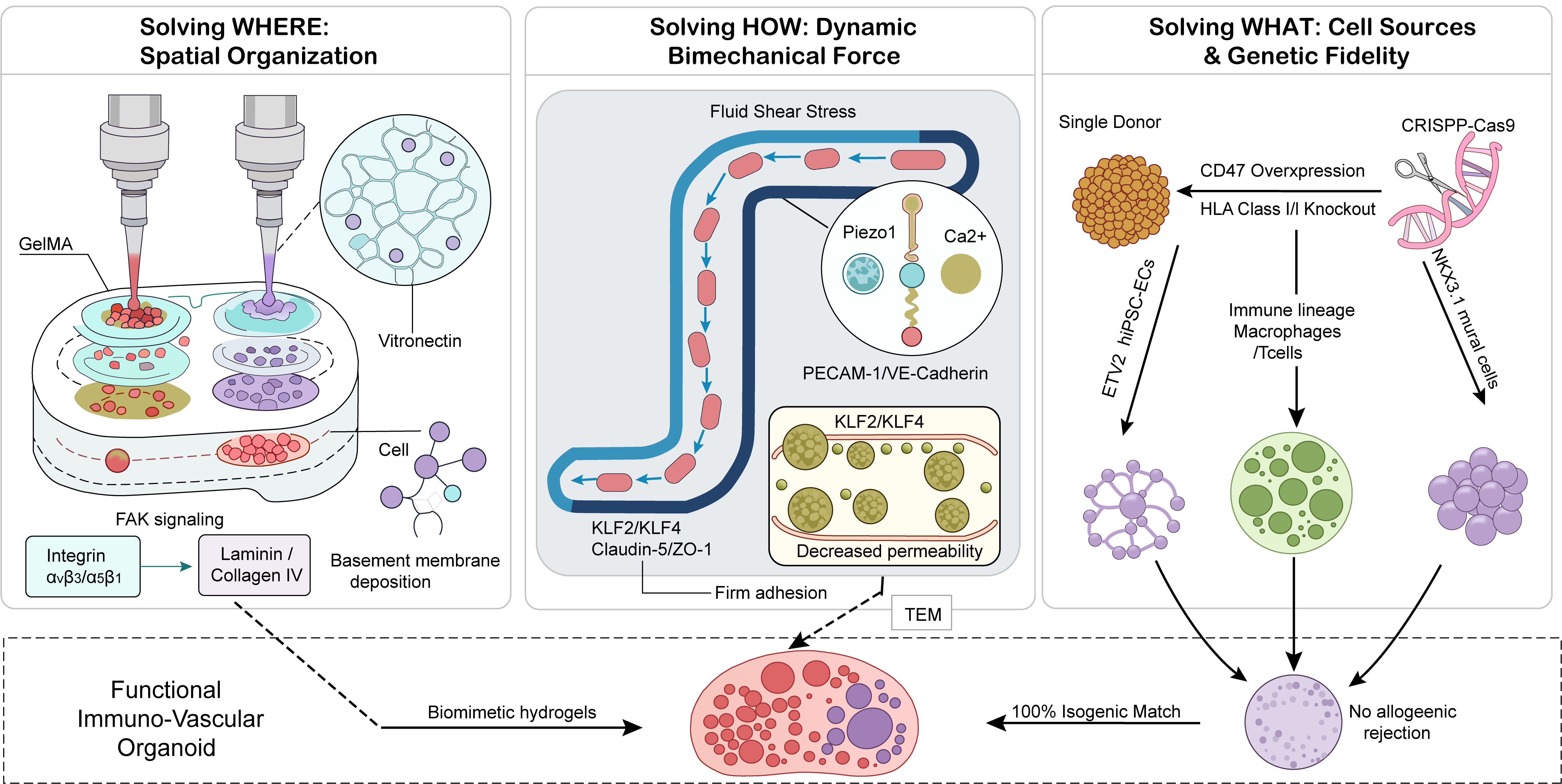
Figure 1. Bioengineering platforms driving multi-lineage and immune integration in vascular organoids. The biofabrication pipeline addresses three architectural challenges: (left) spatial organization, 3D bioprinting and engineered matrix chemistry position mural and immune cells around the abluminal endothelial surface; (center) force regimes, microfluidic perfusion applies physiological shear stress and soluble gradients; (right) lineage definition, CRISPR editing and transcription-factor reprogramming unify the genetic identity of all compartments derived from a single donor line. TEM: Transendothelial migration; FAK: focal adhesion kinase; GelMA: gelatin methacryloyl; HLA: human leukocyte antigen; CRISPR-Cas9: clustered regularly interspaced short palindromic repeats/CRISPR-associated protein 9; PECAM-1: platelet endothelial cell adhesion molecule-1; VE: vascular endothelial; KLF2/4: Krüppel-like factors 2 and 4.
Solving “where”: spatial organization and biomaterial architecture
Recreating native microvasculature requires micro-scale spatial control: mural cells must wrap the abluminal surface of endothelial capillaries, whereas immune cells require functional routes from luminal fluid into perivascular tissues.
• Layer-by-layer bioprinting: Modern 3D bioprinting addresses this spatial challenge by depositing diverse cell types embedded in customized bioinks with micrometer-level precision. For example, in skin-on-a-chip models, multi-layered extrusion printing embeds perfusable microvascular networks within dermal equivalents, which are subsequently capped with stratified epidermal layers to build functional, organotypic barrier models[78].
• Matrix chemistry and integrin signaling: These spatial approaches are complemented by biocompatible hydrogels, such as gelatin methacryloyl (GelMA) and alginate derivatives, engineered with tunable mechanical stiffness and viscoelasticity to mirror native tissue compliance and guide cell morphodynamics[79,80]. Functionalizing these matrices with basement membrane proteins, such as laminin and type IV collagen, engages cellular integrins (e.g., αvβ3/α5β1), triggering focal adhesion kinase (FAK) signaling to direct endothelial polarization and leukocyte extravasation[81].
• Translational ECM formulations: Non-swelling, degradable hydrogels matching the viscoelastic properties of brain tissue provide physical space for organoid expansion without imposing compressive mechanical stress. Replacing batch-variable Matrigel with defined recombinant human vitronectin and engineered fibrin matrices provides standardized, animal-origin-free scaffolds compatible with clinical-grade, high-throughput biofabrication[30,82].
Solving “how”: dynamic microenvironments and biomechanical force regimes
Vascular network assembly depends on physical forces. The incorporation of cellular complexity into 3D matrices must therefore be paired with microfluidic technologies that provide physiological fluid dynamics.
• Mechanotransduction and junctional tightening: Perfusable microchannels generate physiological pressure gradients and fluid shear stress, both of which are essential for endothelial maturation and barrier permeability regulation[83]. Shear stress stimulates endothelial mechanosensors, driving downstream activation of the TFs, which regulate tight junction proteins. When pericytes and perivascular fibroblasts are introduced at the abluminal surface under flow, these systems induce the self-assembly of stable, non-leaky networks with robust basement membrane deposition[84,85].
• Perfusion dynamics and live-cell diagnostics: Microfluidic architectures with perfusable channels generate uniform cytokine gradients and steady shear stress profiles across the organoid network. These perfusion systems enable real-time imaging of reactive oxygen species (ROS) levels[86], leukocyte recruitment, rolling, adhesion, and TEM under controlled hemodynamic regimes[87].
• On-chip multi-omics and multi-organ linkage: Beyond mechanical stimulation, microfluidic platforms with integrated automated processing modules can isolate high-viability single-cell suspensions directly from dense 3D matrices without conventional enzymatic digestion. When coupled with on-chip single-cell RNA transcriptomics, these platforms track dynamic phenotypic drift and spatial heterogeneity in integrated immune cells in real time[88-90]. At a systemic level, multi-organ-on-a-chip setups connect distinct tissue modules (e.g., cardiac, hepatic, osseous, and cutaneous compartments) via endothelialized fluidic channels, maintaining organ-specific endothelial phenotypes while modeling human pharmacokinetic profiles and systemic immune trafficking[91-94].
Solving “what”: defining cell sources and unifying genetic identity via fate engineering
A persistent challenge in engineering third-generation vascular networks is sourcing scalable, donor-matched, and immune-compatible endothelial, mural, and immune cell lineages[4]. Precision gene editing and TF driven reprogramming are increasingly addressing this limitation.
• CRISPR editing and single-cell droplet selection: CRISPR-Cas9 genome editing allows precise targeted modification of hiPSCs before directed differentiation into discrete vascular or hematopoietic branches[95-97]. Encapsulating these edited hiPSCs within microfluidic droplet platforms enables single-cell clonal selection and rapid line validation[98].
• TF-directed reprogramming: The transition from empirical cytokine cocktails to TF-guided fate engineering is informed by single-cell multi-omics, which maps the molecular checkpoints governing vascular lineage decisions. Large-scale perturbation screens and single-cell multi-omic analyses have identified TFs governing early mesodermal-to-endothelial fate commitment[99]. Furthermore, TF-directed reprogramming has enabled the generation of organ-specific endothelial subtypes, including brain-specific barrier phenotypes with functional efflux transporter activity[99].
• Isogenic multicellular assembly: By temporally modulating the VEGF-Notch signaling axis, hiPSC-ECs can be directed toward specific arterial (e.g., EphrinB2+) or organ-specific barrier states[100]. Recent advances have demonstrated the feasibility of generating endothelial, mural, and immune compartments from a single hiPSC donor line, providing an isogenic platform for disease modeling and therapeutic screening[101]. This shared genetic background reduces the risk of confounding allogeneic immune responses, providing an isogenic system for disease modeling and therapeutic screening.
Single-cell and spatial multi-omics for quality assessment and mechanistic dissection
As vascular organoids increase in complexity, bulk analysis methods become insufficient. Emerging single-cell and spatial multi-omics are essential for evaluating organoid quality, maturity, and physiological relevance.
Evaluating cellular identity
Single-cell multi-omics enables unbiased characterization of cell type composition and lineage trajectories. Nikolova et al. used this approach to reconstruct human brain vascular organoid (hBVO) development, identifying TFs such as MDS1 and EVI1 complex locus (MECOM) that play critical roles in endothelial and mural specification[99].
Assessing maturation and heterogeneity
Murine BVOs single-cell RNA sequencing revealed heterogeneous and dynamic trajectory of endothelial, mesenchymal, and macrophage clusters, with ECs adopting arterial, venous, capillary, and tip/stalk cell identities - recapitulating in vivo diversity[102].
Deciphering cell-cell communication
Spatial transcriptomics platforms such as Slide-seq allow unbiased mapping of ligand-receptor pair distributions within intact organoid architectures. In vascularized liver organoids, spatial transcriptomic profiling identified insulin-like growth factor 2 (IGF2)-insulin-like growth factor 1 receptor (IGF1R)-protein kinase B (AKT)/mitogen-activated protein kinase (MAPK) signaling as a key axis governing spatial organization and functional maturation[103].
Resolving spatial organization
Spatial multi-omics preserves 3D architecture, enabling researchers to map molecular dynamics, uncover novel spatial patterns, and analyze cell-cell interactions within an intact architectural context, enabling analysis of EC polarization, mural positioning, and immune cell compartmentalization - criteria essential for physiological relevance[104].
Quality control applications
These technologies serve as molecular “fingerprinting” tools to benchmark batches, identify aberrant cell states, and guide protocol optimization. As spatial technologies continue to advance in resolution, throughput, and accessibility, they are poised to become standard components of the vascular organoid biofabrication pipeline - transforming organoid engineering from empirical optimization into data-driven, quality-controlled biomanufacturing.
CHALLENGES, FUTURE DIRECTIONS, AND TRANSLATIONAL BOTTLENECKS
Despite clear conceptual and technical progress in engineering third-generation vascular organoids, key biological, engineering, and immunological barriers must be overcome before these multicellular systems can be routinely deployed for industrial screening or clinical applications[105]. Beyond the biochemical and engineering obstacles discussed below, the integration of functional immune populations introduces a distinct set of biological challenges that merit separate consideration.
Navigating biochemical antagonism, media incompatibility, and metabolic paradoxes
A central biological obstacle in constructing complex tissue models arises from the distinct, and often conflicting microenvironmental, biochemical, and metabolic requirements of different cell lineages.
• Divergent lineage dependencies: Under normal homeostasis, ECs, mural cells, and immune populations operate in tight synchrony. In co-culture, however, their survival and maturation requirements diverge[106]. Quiescent ECs require continuous laminar fluid shear stress and baseline VEGF signaling to maintain barrier integrity. Conversely, perivascular mural cells rely on specific ECM cues and TGF-β signaling to sustain a contractile, non-fibrotic phenotype[107]. These divergent dependencies can be partially reconciled through targeted modulation of shared intracellular signaling nodes[108]. Adding tissue-resident macrophages introduces another layer of complexity, because their functional plasticity and survival depend on localized cytokines that can directly oppose the anti-inflammatory, quiescent state required for a baseline endothelial barrier[109].
• Diffusion limits and nutrient gradients: When these cell types are assembled at physiological densities, nutrient and oxygen diffusion limits emerge, causing necrotic core formation and network regression[110,111]. While microfluidic channels help partition lineages and establish nutrient or oxygen gradients, dynamically balancing these signals, such as promoting angiogenic sprouting without triggering hyper-inflammatory immune responses, remains difficult. This is further complicated by donor-specific transcriptomic variation in hiPSC-derived cells[18].
• Integrated multi-omic solutions: Resolving these limitations requires single-cell multi-omics (transcriptomics, metabolomics, and fluxomics) to map the metabolic consumption and signaling networks of each cell subpopulation in real time. These datasets can inform the design of switchable, computer-controlled perfusion media and precision delivery systems[106].
Scaling, automation, and the transition to predictive biomanufacturing
To move multi-lineage vascular organoids from small-scale laboratory prototypes to standardized platforms compliant with cGMP guidelines, the field must overcome limitations in scalability and batch-to-batch variability.
• Artificial intelligence (AI)-driven robotic assembly: Manual and semi-automated fabrication methods suffer from inconsistent embryoid body size, variable cellular aggregation, and uneven ECM crosslinking. Automated, AI-guided paradigms, such as “pick-place-perfuse” (Bio-P3), enable robotic systems to assemble pre-formed microtissue modules (e.g., ring- or honeycomb-shaped perivascular aggregates) with high spatial accuracy while maintaining continuous luminal perfusion to preserve cell viability[112].
• Systemic multi-organ linkage: When coupled with microfluidic chips, these platforms may help establish biomimetic branching, hierarchy, and shear-induced maturation under physiological flow conditions. Interconnecting distinct organoid modules (e.g., heart, liver, bone, and skin) via vascularized channels helps preserve organ-specific endothelial identities over extended durations (exceeding four weeks) while capturing systemic pharmacokinetic and pharmacodynamic profiles[113].
• Closed-loop feedback systems: Combining solid-state sensors with deep-learning models enables continuous tracking of dissolved oxygen, pH, and metabolite levels (e.g., lactate, glucose). Autonomous, closed-loop adjustments convert vascular organoid engineering from empirical optimization to predictive biomanufacturing[114].
Immunological compatibility and immune cell integration challenges
For clinical applications, such as using vascularized constructs as therapeutic grafts for ischemic disease, allogeneic rejection remains a critical hurdle[115]. Additionally, immune cell integration introduces distinct biological challenges.
• Universal hypoimmunogenic master lines: Patient-specific hiPSCs offer an autologous source, but are time- and cost- prohibitive for acute interventions such as myocardial infarction or stroke[116]. Universal off-the-shelf allogeneic cell lines are being developed via CRISPR-Cas9 genome editing[117]. Disruption of human leukocyte antigen (HLA) class I and II complexes {via β-2-microglobulin [B2M] and class II transactivator [CIITA] knockouts} combined with CD47 overexpression (“don’t-eat-me” signaling) helps constructs evade both adaptive T cell responses and innate NK-cell surveillance[118].
• Biomaterial encapsulation and immunomodulation: Semi-permeable, immune-protective biomaterials allow nutrient exchange while shielding constructs from immunoglobulins and cytotoxic T cells[119]. Additionally, embedding tolerogenic subsets, such as regulatory T cells or M2-polarized macrophages, can promote local immune tolerance and facilitate integration with the host microvasculature after transplantation[120].
• Immune cell maturation and maintenance: induced pluripotent stem cell (iPSC)-derived macrophages often exhibit fetal-like phenotypes with immature cytokine profiles[121]. Maintaining long-term function remains difficult since niche signals, such as colony-stimulating factor 1 (CSF-1), interleukin-34 (IL-34), and TGF-β, are often absent in conventional organoid culture medium, limiting chronic disease modeling[122].
• Context-dependent effects: The impact of immune cells is highly context-dependent. In a 3D tri-culture model, pro-inflammatory (M1) microglia inhibited neuronal differentiation and vascular development, while anti-inflammatory (M2) microglia supported neurovascular maturation via the stromal cell-derived factor-1/C-X-C chemokine receptor type 4 (SDF-1/CXCR4) signaling axis[40]. While appropriately polarized immune cells can support vascular maturation, uncontrolled or pro-inflammatory immune populations may actively disrupt organoid development and homeostasis.
• Tissue-specific immune phenotypes: Organ-specific macrophages (microglia, Kupffer cells, alveolar macrophages) require tissue-specific cues to instruct appropriate immune cell differentiation, making recapitulation of tissue-specific inflammatory or infectious pathologies challenging[123,124].
• Immune disruption of vascular homeostasis. While quiescent macrophages support vascular homeostasis and remodeling, their activation, whether by pathogens, or damage-associated signals, can rapidly destabilize the vascular network[125]. SARS-CoV-2 infection activates inflammatory macrophages and upregulates interferon signaling, driving cytokine release and vascular damage[126]. Balancing baseline stability with on-demand inflammatory responses remains a critical challenge.
Table 4 summarizes these challenges and potential solutions.
Practical challenges in immune cell integration and potential solutions
| Challenge | Underlying Mechanism | Potential Solutions | References |
| Immune cell maturation | iPSC-derived macrophages exhibit fetal-like phenotype; incomplete surface marker expression and cytokine profiles | Optimize differentiation protocols with tissue-specific niche signals (CSF-1, IL-34, TGF-β); co-culture with target tissue cells to drive maturation | [121] |
| Maintenance of immune function | Conventional organoid medium lack survival factors for long-term immune cell persistence; static culture limits nutrient/gas exchange | Develop immune-supportive medium; integrate microfluidic perfusion for continuous nutrient delivery | [122] |
| Phenotype-dependent effects | M1/pro-inflammatory immune cells inhibit vascular development and disrupt homeostasis; M2 cells support maturation | Precise control of polarization state via cytokine modulation; use of inducible systems for on-demand activation | [40] |
| Tissue-specific immune phenotypes | Organ-specific macrophage identities (microglia, Kupffer cells) require local environmental cues not present in generic cultures | Co-differentiation with organ-specific parenchymal cells; use of tissue-derived ECM scaffolds | [123,124] |
| Batch-to-batch variability | Heterogeneity in immune cell differentiation and polarization across differentiations | Implement quality control checkpoints; use AI-driven image analysis for phenotype validation | [125] |
| Immune disruption of vascular homeostasis | Activated immune cells secrete inflammatory cytokines (TNF-α, IL-6, IFN) that compromise endothelial barrier integrity and promote thrombosis | Incorporate negative feedback mechanisms; use hypoimmunogenic or regulatable immune cell lines | [126] |
AI-driven quality control and phenotypic screening
Deep learning models and computer vision pipelines are transforming quality control and phenotypic screening in vascular bioengineering[127].
• Label-free image analysis: Because multi-lineage organoids exhibit high structural complexity, manual quantification of network connectivity, capillary diameter, and spatial organization is inefficient[128]. Convolutional neural networks, such as the Vessel Connectivity Network (VC-Net), enable label-free, automated classification of normal versus disrupted vascular networks directly from brightfield or phase-contrast images[129].
• High-throughput sandbox for precision medicine: Applying these AI vision systems during manufacturing provides real-time quality control, flagging defective batches and guiding protocol adjustments. Combining patient-derived multi-lineage organoids with microfluidic chips under physiological flow creates a robust platform for precision medicine. These integrated platforms allow researchers to study how hemodynamics influence patient-specific disease phenotypes and drug responses, connecting genetic variation to functional human pathophysiology.
Beyond the current blueprint: engraftment, missing lineages, and aging
Several frontiers remain largely unexplored. First, in vivo functionality: although hypoimmunogenic master lines address immune rejection, the decisive translational test is whether transplanted vascular constructs anastomose with the host circulation and sustain perfusion; engraftment, inosculation, and long-term patency should become standard reporting criteria. An alternative or complementary approach to circumvent the engraftment and rejection challenges is the use of cell-free therapeutics. Wei et al. recently demonstrated that nanovesicles derived from hiPSC-ECs effectively promote angiogenesis and restore perfusion in ischemic limb disease models, suggesting that the paracrine/vesicular payload of vascular organoids - rather than the cells themselves - may offer a safer, more scalable route to clinical translation[130]. Second, the current multilineage blueprint omits perivascular nerves (regulating tone beyond the neurovascular unit) and lymphatic endothelium (coordinating fluid homeostasis and immune trafficking); integration of these lineages may define the next level of physiological completeness. Third, aging: organoids derived from progeria patients[69] and aged endothelial populations[34] indicate that vascular organoids can serve as tunable models of vascular aging, opening a route to test senolytic and rejuvenation strategies.
CONCLUSION
The evolution of vascular organoid bioengineering reflects a shift from structurally reducible models toward functionally integrated human microvasculature, a transition defined not merely by the addition of cell types, but by the convergence of compositional complexity, functional maturation under physiologically relevant biomechanics, and engineering-enabled architectural control. Early endothelial-centric systems provided valuable insight into lumen formation, but they lacked the cellular diversity, mechanical resilience, and immunological capacity of native tissues. Third-generation vascular organoids address these limitations by establishing a dual-integration framework: incorporating mural lineages (pericytes and VSMCs) to reinforce barrier function and vascular tone, alongside functional myeloid and lymphoid populations to capture dynamic immunosurveillance, angiogenic remodeling, and inflammatory cascades.
Realizing the translational potential of these multi-lineage systems requires bridging the gap between stem cell biology and advanced bioengineering. Convergence across microfluidics, 3D bioprinting, biomaterial design, and targeted gene editing provides the technical foundation for controlling the spatial arrangement of cells, simulating physiological shear stress, and overcoming lineage-specific media conflicts. Furthermore, combining multiplex CRISPR editing to produce off-the-shelf hypoimmunogenic cell lines with deep-learning-driven quality control elevates these platforms from empirical setups to standardized, predictive biomanufacturing systems.
As these multilineage and immune-integrated models continue to mature, they already offer a versatile platform for studying complex human pathophysiology, from acute infectious barrier breaches and neurovascular degeneration to tumor angiogenesis and metabolic vasculopathies. When paired with single-cell multi-omics and patient-specific hiPSCs, these organoids hold promise for drug discovery, mechanobiological research, and the development of personalized therapeutic strategies.
However, clinical translation, particularly as implantable grafts, faces formidable hurdles that should not be underestimated. These include the lack of standardized cGMP‑compliant protocols; insufficient large‑animal efficacy data; unknown immunogenicity even with hypoimmunogenic modifications; thrombosis and leakage risk; high cost and time for patient‑specific organoid generation; and no benchmark criteria for “sufficient” functional maturity.
Moreover, while TF-driven reprogramming offers rapid lineage specification, it is important to acknowledge its inherent limitations. Unlike physiological development, where lineage commitment unfolds through sequential, dose-dependent signaling with built-in quality control checkpoints, forced TF overexpression imposes an abrupt and non-physiological instruction[28,29]. This bypass of normal developmental intermediates raises concerns about incomplete epigenetic remodeling, residual donor-cell gene expression, and suboptimal functional maturation[29]. Moreover, TF overexpression may cause nonspecific transcriptional interference (“squelching”) and off-target gene suppression, with potential safety implications including chromosomal abnormalities and oncogenic transformation. For ETV2/NKX3.1-based orthogonal activation systems specifically, independent control of endothelial and mural compartments remains challenging due to the shared Dox-inducible trigger, and the absence of perfusion in current suspension cultures limits endothelial maturation[26]. These considerations underscore that TF-driven approaches, while powerful, require rigorous functional benchmarking and safety validation before clinical translation.
Until these challenges are systematically addressed, the primary translational value of multi-lineage vascular organoids will remain in disease modeling, target discovery, and preclinical drug screening. The path toward regenerative therapeutics will require sustained interdisciplinary effort to bridge the gap between in vitro complexity and in vivo functionality.
DECLARATIONS
Acknowledgment
The Graphical Abstract was drawn by Adobe Illustrator 2026 v30.4. We thank OAE Publishing Inc. for language editing.
Authors’ contributions
Conceptualization, methodology, writing-review & editing, resources, supervision: Li J, Li G, Kantawong F
Investigation, writing-original draft: Chen Q, Jin J
Funding acquisition: Li G, Li J
Availability of data and materials
Not applicable.
AI and AI-assisted tools statement
During the preparation of this manuscript, Google (version Gemini, 3.5 Flash, released 2026-05-19) was used solely for language polishing and editorial refinement. The tool did not influence the study design, data collection, analysis, interpretation, or the scientific content of the work. All authors take full responsibility for the accuracy, integrity, and final content of the manuscript.
Financial support and sponsorship
This work was funded by the Natural Science Foundation of Sichuan Province (2026NSFSCZY0039) and the Southwest Medical University Clinical Medicine Project (2024LCYXZX51).
Conflicts of interest
All authors declared that there are no conflicts of interest.
Ethical approval and consent to participate
Not applicable.
Consent for publication
Not applicable.
Copyright
© The Author(s) 2026.
REFERENCES
1. Red-Horse K, Crawford Y, Shojaei F, Ferrara N. Endothelium-microenvironment interactions in the developing embryo and in the adult. Dev Cell. 2007;12:181-94.
2. Minami T, Muramatsu M, Kume T. Organ/tissue-specific vascular endothelial cell heterogeneity in health and disease. Biol Pharm Bull. 2019;42:1609-19.
3. Augustin HG, Koh GY. A systems view of the vascular endothelium in health and disease. Cell. 2024;187:4833-58.
4. Lancaster MA, Knoblich JA. Organogenesis in a dish: modeling development and disease using organoid technologies. Science. 2014;345:1247125.
5. Seok J, Warren HS, Cuenca AG, et al. Genomic responses in mouse models poorly mimic human inflammatory diseases. Proc Natl Acad Sci U S A. 2013;110:3507-12.
6. Naderi-Meshkin H, Cornelius VA, Eleftheriadou M, Potel KN, Setyaningsih WAW, Margariti A. Vascular organoids: unveiling advantages, applications, challenges, and disease modelling strategies. Stem Cell Res Ther. 2023;14:292.
7. Wimmer RA, Leopoldi A, Aichinger M, et al. Human blood vessel organoids as a model of diabetic vasculopathy. Nature. 2019;565:505-10.
8. Payne LB, Tewari BP, Dunkenberger L, et al. Pericyte progenitor coupling to the emerging endothelium during vasculogenesis via connexin 43. Arterioscler Thromb Vasc Biol. 2022;42:e96-e114.
9. Cahill TJ, Sun X, Ravaud C, et al. Tissue-resident macrophages regulate lymphatic vessel growth and patterning in the developing heart. Development. 2021;148:dev194563.
10. Bizou M, Itier R, Majdoubi M, et al. Cardiac macrophage subsets differentially regulate lymphatic network remodeling during pressure overload. Sci Rep. 2021;11:16801.
11. Mu X, Gerhard-Herman MD, Zhang YS. Building blood vessel chips with enhanced physiological relevance. Adv Mater Technol. 2023;8:2201778.
12. Salewskij K, Penninger JM. Blood vessel organoids for development and disease. Circ Res. 2023;132:498-510.
13. Hirth E, Cao W, Peltonen M, et al. Self-assembled and perfusable microvasculature-on-chip for modeling leukocyte trafficking. Lab Chip. 2024;24:292-304.
14. Bergers G, Song S. The role of pericytes in blood-vessel formation and maintenance. Neuro Oncol. 2005;7:452-64.
15. Armulik A, Abramsson A, Betsholtz C. Endothelial/pericyte interactions. Circ Res. 2005;97:512-23.
16. Winkler EA, Bell RD, Zlokovic BV. Central nervous system pericytes in health and disease. Nat Neurosci. 2011;14:1398-405.
17. Stratman AN, Malotte KM, Mahan RD, Davis MJ, Davis GE. Pericyte recruitment during vasculogenic tube assembly stimulates endothelial basement membrane matrix formation. Blood. 2009;114:5091-101.
18. Jiao YC, Wang YX, Liu WZ, et al. Advances in the differentiation of pluripotent stem cells into vascular cells. World J Stem Cells. 2024;16:137-50.
19. Patsch C, Challet-Meylan L, Thoma EC, et al. Generation of vascular endothelial and smooth muscle cells from human pluripotent stem cells. Nat Cell Biol. 2015;17:994-1003.
20. Orlova VV, van den Hil FE, Petrus-Reurer S, Drabsch Y, Ten Dijke P, Mummery CL. Generation, expansion and functional analysis of endothelial cells and pericytes derived from human pluripotent stem cells. Nat Protoc. 2014;9:1514-31.
21. Zhang S, Dutton JR, Su L, Zhang J, Ye L. The influence of a spatiotemporal 3D environment on endothelial cell differentiation of human induced pluripotent stem cells. Biomaterials. 2014;35:3786-93.
22. Aisenbrey EA, Torr E, Johnson H, Soref C, Daly W, Murphy WL. A protocol for rapid pericyte differentiation of human induced pluripotent stem cells. STAR Protoc. 2021;2:100261.
23. Kumar A, D’Souza SS, Moskvin OV, et al. Specification and diversification of pericytes and smooth muscle cells from mesenchymoangioblasts. Cell Rep. 2017;19:1902-16.
24. Miao Y, Pek NM, Tan C, et al. Co-development of mesoderm and endoderm enables organotypic vascularization in lung and gut organoids. Cell. 2025;188:4295-4313.e27.
25. Ng AHM, Khoshakhlagh P, Rojo Arias JE, et al. A comprehensive library of human transcription factors for cell fate engineering. Nat Biotechnol. 2021;39:510-9.
26. Gong L, Zhang Y, Zhu Y, et al. Rapid generation of functional vascular organoids via simultaneous transcription factor activation of endothelial and mural lineages. Cell Stem Cell. 2025;32:1200-1217.e6.
27. Zhao Y, Sun M, Zhang K, Melero-Martin JM, Wang X, Wang K. Generation of synthetic vascular organoids via orthogonal programming of human pluripotent stem cells. Cell Regen. 2026;15:23.
28. Zaret KS. Cell fate conversion: a chromatin remodeling checkpoint revealed. Cell Res. 2017;27:598-9.
29. Li F, Hu J. Direct lineage conversion with pluripotency factors: a risky detour through transient pluripotency? Genes Dis. 2015;2:286-7.
30. Giles R, Meijer EM, Maas RGC, van Dijk CGM, Verhaar MC, Cheng C. Animal-free alternatives for matrigel in human iPSC-derived blood vessel organoid culture. Sci Rep. 2025;15:36042.
31. Xu C, He S, Zhu Y, et al. Vascular organoid generation from human-induced pluripotent stem cells. J Vis Exp. 2024;214:10.3791/67125.
32. Adams WJ, Zhang Y, Cloutier J, et al. Functional vascular endothelium derived from human induced pluripotent stem cells. Stem Cell Reports. 2013;1:105-13.
34. Huang Y, Liu Z, Li M, et al. Deciphering the impact of aging on splenic endothelial cell heterogeneity and immunosenescence through single-cell RNA sequencing analysis. Immun Ageing. 2024;21:48.
35. Wang L, Xiang M, Liu Y, et al. Human induced pluripotent stem cells derived endothelial cells mimicking vascular inflammatory response under flow. Biomicrofluidics. 2016;10:014106.
36. Alcaide P. Mechanisms regulating T cell-endothelial cell interactions. Cold Spring Harb Perspect Med. 2022;12:a041170.
37. de Haan L, Suijker J, van Roey R, et al. A microfluidic 3d endothelium-on-a-chip model to study transendothelial migration of T cells in health and disease. Int J Mol Sci. 2021;22:8234.
38. Sonninen TM, Peltonen S, Kälvälä S, et al. From inserts to chips: microfluidic culture and 3D astrocyte co-culture drive functional and transcriptomic changes in hiPSC-derived endothelial cells. Fluids Barriers CNS. 2025;22:58.
39. Qi D, Wu S, Lin H, et al. Establishment of a human iPSC- and nanofiber-based microphysiological blood-brain barrier system. ACS Appl Mater Interfaces. 2018;10:21825-35.
40. Han Y, Guo L, Wang M, et al. Bioengineering an improved three-dimensional vascularized co-culture model for studying neuron-microglia interactions. Bioact Mater. 2025;54:813-28.
41. Kong SY, Kim DH. Human iPSC-derived vascularized organoids: strategy-based insights from 2D endothelial cells to 3D blood vessel organoids. Biofabrication. 2026.
42. Gordiichuk P, Bai J, Bolonduro OA, et al. Development of a human 3D immune-competent neurovascular model enabling time-resolved monitoring of neuroinflammatory dynamics and neuroimmune interactions. Adv Sci. 2026;13:e75461.
43. Singh R, Mori N, Kida YS. Methodological innovations in perfusable vascular networks: Advancing high-density tissue models. Regen Ther. 2026;32:101109.
45. Sweet DR, Lam C, Jain MK. Evolutionary protection of krüppel-like factors 2 and 4 in the development of the mature hemovascular system. Front Cardiovasc Med. 2021;8:645719.
46. Pasqualini FS, Emmert MY, Parker KK, Hoerstrup SP. Organ chips: quality assurance systems in regenerative medicine. Clin Pharmacol Ther. 2017;101:31-4.
47. Ley K, Laudanna C, Cybulsky MI, Nourshargh S. Getting to the site of inflammation: the leukocyte adhesion cascade updated. Nat Rev Immunol. 2007;7:678-89.
48. Cucullo L, Marchi N, Hossain M, Janigro D. A dynamic in vitro BBB model for the study of immune cell trafficking into the central nervous system. J Cereb Blood Flow Metab. 2011;31:767-77.
49. Eltaibany AA, McGovern K, Nzou G, Porada D, Seeds MC, Atala A. Modeling neuroimmunological interactions at the blood-brain barrier using in vitro 3D human organoids: inflammation and ischemia-reperfusion injury. Cells. 2026;15:1173.
50. Gu L, Mao X, Tian C, et al. Engineering blood-brain barrier microphysiological systems to model Alzheimer’s disease monocyte penetration and infiltration. Biomater Sci. 2025;13:3650-61.
51. Birtele M, Lancaster M, Quadrato G. Modelling human brain development and disease with organoids. Nat Rev Mol Cell Biol. 2025;26:389-412.
52. Sontheimer-Phelps A, Hassell BA, Ingber DE. Modelling cancer in microfluidic human organs-on-chips. Nat Rev Cancer. 2019;19:65-81.
53. Liu H, Noguera-Ortega E, Dong X, et al. A tumor-on-a-chip for in vitro study of CAR-T cell immunotherapy in solid tumors. Nat Biotechnol. 2026;44:959-75.
54. Li K, Liu C, Sui X, et al. An organoid co-culture model for probing systemic anti-tumor immunity in lung cancer. Cell Stem Cell. 2025;32:1218-1234.e7.
55. Baisiwala S, Fazzari E, Li MX, et al. A human tumor-immune organoid model of glioblastoma. Cell Rep. 2026;45:116790.
56. Cai L, Xiao Y, Xie F, Yang Z. Tumor organoid-immune cell co-culture systems for precision oncology. Front Cell Dev Biol. 2026;14:1865487.
57. Kawakami E, Saiki N, Yoneyama Y, et al. Complement factor D targeting protects endotheliopathy in organoid and monkey models of COVID-19. Cell Stem Cell. 2023;30:1315-1330.e10.
58. Rauti R, Navok S, Biran D, et al. Insight on bacterial newborn meningitis using a neurovascular-unit-on-a-chip. Microbiol Spectr. 2023;11:e0123323.
59. Bennett AB, Lanser DM, Vu K, Sahoo AR, Buck M, Gelli A. Macropinocytosis mediates neurotropism of cryptococcus neoformans in a human organoid model of the blood-brain barrier. Commun Biol. 2026.
60. Boghdeh-Olson NA, Barrera MD, Britt CM, et al. Application of a blood-brain barrier organ-on-a-chip model for assessment of countermeasure efficiency against eastern equine encephalitis virus. Viruses. 2026;18:548.
61. Chen YW, Chiang HJ, Liu KT, et al. Integration of biomimetic organoid-on-chip and 2D models advances the mechanistic understanding of STEAP3-mediated regulation in intestinal viral infection. Sci Rep. 2025;15:30582.
62. Wang Y, Guo Y, Wang P, et al. An engineered human placental organoid microphysiological system in a vascular niche to model viral infection. Commun Biol. 2025;8:669.
63. Koh H, Kang W, Mao YY, et al. Employment of diverse in vitro systems for analyzing multiple aspects of disease, hereditary hemorrhagic telangiectasia (HHT). Cell Biosci. 2024;14:65.
64. Wang Z, Yin J, Chao W, Zhang X. Inducing mononuclear cells of patients with CADASIL to construct a CSVD disease model. Eur J Med Res. 2025;30:227.
65. Kong D, Ryu JC, Shin N, et al. In vitro modeling of atherosclerosis using iPSC-derived blood vessel organoids. Adv Healthc Mater. 2025;14:e2400919.
66. Nguyen V, Harper A, Azuero M, et al. Vascular organoids derived from capillary malformation-induced pluripotent stem cells exhibit disease-relevant phenotypes. Stem Cell Rev Rep. 2026;22:668-75.
67. He S, Zhang J, Wang X, et al. Organoid modeling and single-cell profiling reveal smooth muscle cell migration in moyamoya disease. Commun Biol. 2026;9:198.
68. Wu M, Li S, Liu W, Guo Z, Guo Z, Zhang D. RNF213 deficiency in human iPSC-derived vascular organoids captures key feature of moyamoya disease vasculopathy. Microvasc Res. 2026;167:104979.
69. Sun X, Che S, Wang H, et al. Vascular organoid model of hutchinson-gilford progeria syndrome uncovers repression of the SRF pathway in premature aging. Dev Cell. 2026;61:505-517.e7.
70. Naderi-Meshkin H, Wahyu Setyaningsih WA, Yacoub A, et al. Unveiling impaired vascular function and cellular heterogeneity in diabetic donor-derived vascular organoids. Stem Cells. 2024;42:791-808.
71. Su L, Kong X, Loo SJ, et al. Diabetic endothelial cells differentiated from patient iPSCs show dysregulated glycine homeostasis and senescence associated phenotypes. Front Cell Dev Biol. 2021;9:667252.
72. Su L, Kong X, Loo S, et al. Thymosin beta-4 improves endothelial function and reparative potency of diabetic endothelial cells differentiated from patient induced pluripotent stem cells. Stem Cell Res Ther. 2022;13:13.
73. Ji Y, Chen X, Wang Z, et al. Alzheimer’s disease patient brain extracts induce multiple pathologies in novel vascularized neuroimmune organoids for disease modeling and drug discovery. Mol Psychiatry. 2025;30:4558-75.
74. Uzoechi SC, Johnson-Campbell C, Badeaux CJ, Kwak SS, Kim DY, Yun Y. Recapitulating Alzheimer’s disease pathophysiology with a microfluidic neurospheroid-grafted endothelial barrier model. Mol Brain. 2026;19:25.
75. Aleman J, Vernetti L, Schurdak ME, et al. Recapitulation of clinical and molecular hallmarks of lipid-induced hepatic insulin resistance in a zonated, vascularized human liver acinus microphysiological system during metabolic dysfunction-associated steatotic liver disease (MASLD) progression. BMC Biotechnol. 2026;26:70.
76. Kota VG, Yi JLJ, Zhang Z, Yooyuen A, Yu H, Leo HL. Organoid models reveal mechanistic connections and sirolimus efficacy in liver-vascular steatosis and foam cell formation. Atherosclerosis. 2026;417:120762.
77. Ingber DE. Human organs-on-chips for disease modelling, drug development and personalized medicine. Nat Rev Genet. 2022;23:467-91.
78. Juraski AC, Sharma S, Sparanese S, et al. 3D bioprinting for organ and organoid models and disease modeling. Expert Opin Drug Discov. 2023;18:1043-59.
79. Yue K, Trujillo-de Santiago G, Alvarez MM, Tamayol A, Annabi N, Khademhosseini A. Synthesis, properties, and biomedical applications of gelatin methacryloyl (GelMA) hydrogels. Biomaterials. 2015;73:254-71.
80. Chaudhuri O, Gu L, Klumpers D, et al. Hydrogels with tunable stress relaxation regulate stem cell fate and activity. Nat Mater. 2016;15:326-34.
81. Kosyakova N, Kao DD, Figetakis M, et al. Differential functional roles of fibroblasts and pericytes in the formation of tissue-engineered microvascular networks in vitro. NPJ Regen Med. 2020;5:1.
82. Zheng F, Chen Y, Xue G, et al. Differentiation of arterioles and capillaries in human blood vessel organoids with decellularized splenic matrix. Bioact Mater. 2026;64:731-43.
83. Mazari-Arrighi E, Boyreau A, Chaillot L, et al. A versatile microfluidic extrusion-based hydrogel platform for self-organization and long-term maintenance of engineered 3D lymphatic endothelium. Adv Healthc Mater. 2026;15:e71307.
84. Jung O, Tung YT, Sim E, et al. Development of human-derived, three-dimensional respiratory epithelial tissue constructs with perfusable microvasculature on a high-throughput microfluidics screening platform. Biofabrication. 2022;14:025012.
85. Luu RJ, Hoefler BC, Gard AL, et al. Fibroblast activation in response to TGFβ1 is modulated by co-culture with endothelial cells in a vascular organ-on-chip platform. Front Mol Biosci. 2023;10:1160851.
86. Hu SY, Xu XY, Xue CD, Qin KR. A microfluidic system for modeling endothelial dysfunction under combined physiological pulsatile shear stress and oscillatory hyperglycemia. J Vis Exp. 2026;231.
87. Zhu L, Zhao W, Shen M, et al. Human BBB-brain organoid on a millifluidic plate for modeling brain parenchymal pathology-induced barrier dysfunction. J Adv Res. 2026:S2090-1232(26)00506.
88. Lombardo JA, Aliaghaei M, Nguyen QH, Kessenbrock K, Haun JB. Microfluidic platform accelerates tissue processing into single cells for molecular analysis and primary culture models. Nat Commun. 2021;12:2858.
89. Wills QF, Mellado-Gomez E, Nolan R, et al. The nature and nurture of cell heterogeneity: accounting for macrophage gene-environment interactions with single-cell RNA-Seq. BMC Genomics. 2017;18:53.
90. Jones CFE, Di Cio S, Connelly JT, Gautrot JE. Design of an integrated microvascularized human skin-on-a-chip tissue equivalent model. Front Bioeng Biotechnol. 2022;10:915702.
91. Novak R, Ingram M, Marquez S, et al. Robotic fluidic coupling and interrogation of multiple vascularized organ chips. Nat Biomed Eng. 2020;4:407-20.
92. Quintard C, Tubbs E, Jonsson G, et al. A microfluidic platform integrating functional vascularized organoids-on-chip. Nat Commun. 2024;15:1452.
93. Jun Y, Nguyen-Ngoc KV, Sai S, et al. Engineered vasculature induces functional maturation of pluripotent stem cell-derived islet organoids. Dev Cell. 2025;60:2455-2469.e7.
94. Marder M, Remmert C, Perschel JA, et al. Stem cell-derived vessels-on-chip for cardiovascular disease modeling. Cell Rep. 2024;43:114008.
95. Acun A, Zorlutuna P. CRISPR/Cas9 edited induced pluripotent stem cell-based vascular tissues to model aging and disease-dependent impairment. Tissue Eng Part A. 2019;25:759-72.
96. Skowronek D, Pilz RA, Saenko VV, et al. High-throughput differentiation of human blood vessel organoids reveals overlapping and distinct functions of the cerebral cavernous malformation proteins. Angiogenesis. 2025;28:32.
97. Guo H, Yu X, Liu Y, et al. SGLT2 inhibitor ameliorates endothelial dysfunction associated with the common ALDH2 alcohol flushing variant. Sci Transl Med. 2023;15:eabp9952.
98. Sarmah H, Iannello G, Wantroba R, et al. Protocol for clonal isolation of gene-edited hiPSCs using droplet and microfluidic sorting. STAR Protoc. 2026;7:104518.
99. Nikolova MT, He Z, Seimiya M, et al. Fate and state transitions during human blood vessel organoid development. Cell. 2025;188:3329-3348.e31.
100. Korbmacher F, Long RKM, Fleckenstein H, et al. ETS-guided iPSC-endothelial models recapitulate malaria pathogenesis. EMBO Mol Med. 2026;18:3389-418.
101. Shen AN, Matazel KS, Gill WD, Ewart L, Daughters RS, Rosas-Hernandez H. Modeling neurovascular dysfunction in Alzheimer’s disease using an isogenic brain-chip model. Fluids Barriers CNS. 2026;23:1.
102. Guelfi S, Gopalan KS, Maene P, et al. Murine vascular organoids are responsive and adaptable 3D systems with cellular heterogeneity and dynamic plasticity. Sci Adv. 2025;11:eady4738.
103. Kim DH, Lee Y, Kim MJ, Lee AC, Kwon S, Kang KS. Leveraging IGF signaling to improve the spatial organization and regenerative potential of iPSC-derived vascularized liver organoids. Sci Adv. 2026;12:eaea3814.
104. Zhang X, Jeya Vandana J, Greenspun B, Chen S. Dissecting human organ development using spatial technologies. Dev Cell. 2026;61:1363-91.
105. Marx U, Akabane T, Andersson TB, et al. Biology-inspired microphysiological systems to advance patient benefit and animal welfare in drug development. ALTEX. 2020;37:365-94.
106. Helle E, Ampuja M, Dainis A, et al. HiPS-endothelial cells acquire cardiac endothelial phenotype in co-culture with hiPS-cardiomyocytes. Front Cell Dev Biol. 2021;9:715093.
107. Huang NF, Chaudhuri O, Cahan P, et al. Multi-scale cellular engineering: from molecules to organ-on-a-chip. APL Bioeng. 2020;4:010906.
108. Su L, Kong X, Lim S, et al. The prostaglandin H2 analog U-46619 improves the differentiation efficiency of human induced pluripotent stem cells into endothelial cells by activating both p38MAPK and ERK1/2 signaling pathways. Stem Cell Res Ther. 2018;9:313.
109. Gordon S, Martinez FO. Alternative activation of macrophages: mechanism and functions. Immunity. 2010;32:593-604.
110. Miller JS. The billion cell construct: will three-dimensional printing get us there? PLoS Biol. 2014;12:e1001882.
111. Smith Q, Gerecht S. Going with the flow: microfluidic platforms in vascular tissue engineering. Curr Opin Chem Eng. 2014;3:42-50.
112. Blakely AM, Manning KL, Tripathi A, Morgan JR. Bio-pick, place, and perfuse: a new instrument for three-dimensional tissue engineering. Tissue Eng Part C Methods. 2015;21:737-46.
113. Ronaldson-Bouchard K, Teles D, Yeager K, et al. A multi-organ chip with matured tissue niches linked by vascular flow. Nat Biomed Eng. 2022;6:351-71.
114. Zhang YS, Aleman J, Shin SR, et al. Multisensor-integrated organs-on-chips platform for automated and continual in situ monitoring of organoid behaviors. Proc Natl Acad Sci U S A. 2017;114:E2293-302.
115. Petrus-Reurer S, Romano M, Howlett S, Jones JL, Lombardi G, Saeb-Parsy K. Immunological considerations and challenges for regenerative cellular therapies. Commun Biol. 2021;4:798.
116. Trounson A, DeWitt ND. Pluripotent stem cells progressing to the clinic. Nat Rev Mol Cell Biol. 2016;17:194-200.
117. Deuse T, Hu X, Gravina A, et al. Hypoimmunogenic derivatives of induced pluripotent stem cells evade immune rejection in fully immunocompetent allogeneic recipients. Nat Biotechnol. 2019;37:252-8.
118. Hu X, White K, Olroyd AG, et al. Hypoimmune induced pluripotent stem cells survive long term in fully immunocompetent, allogeneic rhesus macaques. Nat Biotechnol. 2024;42:413-23.
119. Vegas AJ, Veiseh O, Gürtler M, et al. Long-term glycemic control using polymer-encapsulated human stem cell-derived beta cells in immune-competent mice. Nat Med. 2016;22:306-11.
120. Raheem A, Khan I, Ahmad I, et al. The emerging role of tissue regulatory T cells in tissue repair and regeneration. Front Immunol. 2025;16:1640113.
121. Buchrieser J, James W, Moore MD. Human induced pluripotent stem cell-derived macrophages share ontogeny with MYB-independent tissue-resident macrophages. Stem Cell Reports. 2017;8:334-45.
122. Lohasz C, Häfeli T, Hasanova D, et al. A microfluidic platform for the co-culturing of microtissues with continuously recirculating suspension cells. Microsyst Nanoeng. 2025;11:184.
123. Battumur E, Clegg JR, Acar H. Exploring macrophage polarization: biological insights, key laboratory techniques and research perspectives. Front Immunol. 2026;17:1837287.
124. Lee CZW, Tasnim F, Huang X, et al. Direct contact between iPSC-derived macrophages and hepatocytes drives reciprocal acquisition of Kupffer cell identity and hepatocyte maturation. Elife. 2026;14:RP108938.
125. Klepikova A, Nenasheva T, Sheveleva O, et al. iPSC-derived macrophages: the differentiation protocol affects cell immune characteristics and differentiation trajectories. Int J Mol Sci. 2022;23:16087.
126. Chau CW, To A, Au-Yeung RKH, et al. SARS-CoV-2 infection activates inflammatory macrophages in vascular immune organoids. Sci Rep. 2024;14:8781.
127. Badai J, Bu Q, Zhang L. Review of artificial intelligence applications and algorithms for brain organoid research. Interdiscip Sci. 2020;12:383-94.
128. Borten MA, Bajikar SS, Sasaki N, Clevers H, Janes KA. Automated brightfield morphometry of 3D organoid populations by OrganoSeg. Sci Rep. 2018;8:5319.
129. Yun E, Kim J, Jeong D. Lightweight deep learning model for classification of normal and abnormal vasculature in organoid Images. Sensors. 2025;26:112.
Cite This Article
 Open Access
How to Cite
Download Citation
Export Citation File:
Type of Import
Tips on Downloading Citation
Citation Manager File Format
Type of Import
Direct Import: When the Direct Import option is selected (the default state), a dialogue box will give you the option to Save or Open the downloaded citation data. Choosing Open will either launch your citation manager or give you a choice of applications with which to use the metadata. The Save option saves the file locally for later use.
Indirect Import: When the Indirect Import option is selected, the metadata is displayed and may be copied and pasted as needed.
About This Article
Copyright

Data & Comments
Data
0









Comments
Comments must be written in English. Spam, offensive content, impersonation, and private information will not be permitted. If any comment is reported and identified as inappropriate content by OAE staff, the comment will be removed without notice. If you have any queries or need any help, please contact us at [email protected].