

fig3
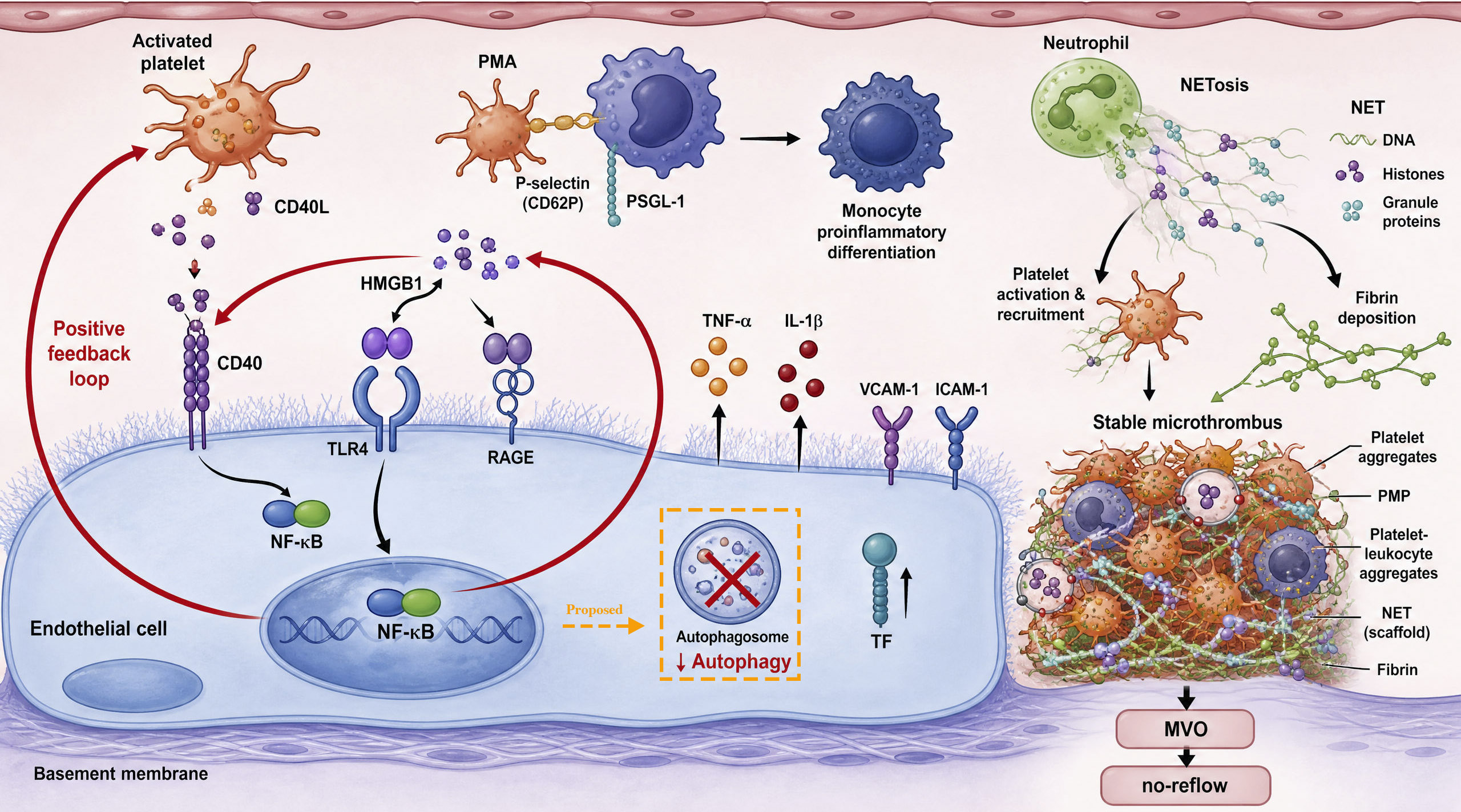
Figure 3. Platelet-mediated inflammation, HMGB1/TLR4/NF-κB axis, and microthrombosis. The central positive feedback loop between inflammation and platelet activation is driven by the HMGB1/TLR4/NF-κB pathway in the endothelial cell, which upregulates pro-inflammatory cytokines (TNF-α, IL-1β) and adhesion molecules (VCAM-1, ICAM-1) while suppressing protective autophagy. Within the vascular lumen, activated platelets release CD40L and IL-1β that further amplify this axis via endothelial CD40, form proinflammatory PMAs through P-selectin/PSGL-1 interaction, and shed PMPs into blood circulation. Neutrophils release NETs (DNA with bead-like histones) that capture platelets and promote fibrin deposition. All components converge within the lumen to form a stable microthrombus containing platelet aggregates, PMPs, platelet-leukocyte aggregates, NET scaffolds, and fibrin strands, ultimately causing MVO and no-reflow. The orange dashed box/arrow and the “Proposed” label indicate that the illustrated HMGB1/TLR4/NF-κB-mediated autophagy suppression is based on the evidence from whole-myocardium studies (predominantly cardiomyocytes), and definitive endothelial-specific verification is yet to be collected. Solid arrows denote established endothelial signaling events. HMGB1: High mobility group box 1; TLR4: toll-like receptor 4; RAGE: receptor for advanced glycation end products; NF-κB: nuclear factor kappa-B; TNF-α: tumor necrosis factor-α; IL-1β: interleukin-1β; VCAM-1: vascular cell adhesion molecule-1; ICAM-1: intercellular adhesion molecule-1; TF: tissue factor; CD40L: CD40 ligand; CD40: CD40 receptor; PSGL-1: P-selectin glycoprotein ligand-1; PMPs: platelet-derived microparticles; PMAs: platelet-monocyte aggregates; NET: neutrophil extracellular trap; MVO: microvascular obstruction.