


Pathological remodeling driven by the cardiac autoantigen-dendritic cell-CD4+ T-cell axis under sustained mechanical stress load
 0
0 Abstract
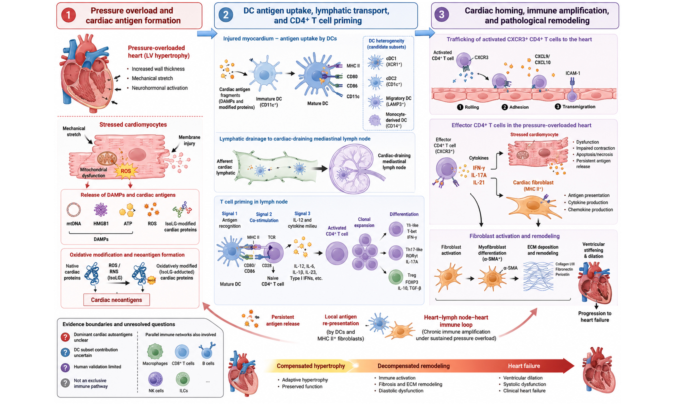
Pressure overload-induced cardiac remodeling is a shared pathological basis in the progression of hypertension, aortic stenosis, aortic coarctation, and related diseases, and represents an important antecedent stage in the development of heart failure. This process is not driven by a single injurious event, but evolves gradually from early compensatory hypertrophy to late decompensated remodeling. In the early phase, the heart can maintain short-term hemodynamic stability through cardiomyocyte hypertrophy, ventricular wall thickening, and a certain degree of contractile compensation. However, when abnormal loading persists, mechanical stretch, perfusion imbalance, and metabolic stress progressively accumulate, leading to a decline in the intrinsic adaptive capacity of cardiomyocytes. This is followed by ventricular structural rearrangement, reduced compliance, aggravated interstitial fibrosis, and impaired systolic and diastolic function. Thus, pressure overload-induced cardiac remodeling is not merely a mechanical adaptation, but a dynamic pathological process that progresses from limited compensation to sustained injury and functional imbalance. Recent studies have shown that the progression of this disease process cannot be explained solely by hemodynamic burden, as immune responses are also involved. The chronically overloaded myocardium is not a passive recipient of stress. Rather, under sustained stress, it continuously releases danger signals, reshapes the local inflammatory milieu, and increases the likelihood that cardiac components will be recognized by the immune system. Therefore, immune involvement is not simply a secondary phenomenon following pressure overload, but an important factor that drives the transition from compensation to decompensation. Compared with acute and transient inflammatory responses, a more critical issue is whether local injury signals can be converted into a sustained adaptive immune response, because this step determines whether remodeling evolves from a relatively confined tissue reaction into a persistent chronic pathological process.
Keywords
INTRODUCTION
Pressure overload-induced cardiac remodeling is a common pathological basis in the progression of hypertension, aortic stenosis, aortic coarctation, and related diseases, and represents an important antecedent event in the development of heart failure[1]. This process is not driven directly by a single injury, but progresses from compensatory hypertrophy to decompensated remodeling. In the early stage, the heart can maintain hemodynamic stability through cardiomyocyte hypertrophy, ventricular wall thickening, and contractile compensation[2]; however, when abnormal loading persists, mechanical stretch, perfusion imbalance, and metabolic stress progressively accumulate, leading to a decline in cardiomyocyte adaptive capacity, followed by ventricular structural rearrangement, reduced compliance, interstitial fibrosis, and impaired systolic and diastolic function[3]. Thus, pressure overload-induced cardiac remodeling is not merely a mechanical adaptation, but a dynamic process in which limited compensation gradually gives way to sustained injury. Recent studies suggest that immune responses are not secondary bystanders in this disease process, but important contributors to the transition from compensation to decompensation. The persistently overloaded myocardium can release danger signals, reshape the local inflammatory milieu, and increase the likelihood that cardiac components are recognized by the immune system[4]. Compared with transient inflammation, the more critical issue is whether local injury signals can be converted into sustained adaptive immunity, as this determines whether remodeling evolves from a confined tissue reaction into a chronic pathological process[5].
Against this background, the immune process formed by dendritic cells (DCs) and cluster of differentiation 4-positive (CD4+) T cells has particular research significance, although it is not the only immune pathway operating in the pressure-overloaded heart[6]. Macrophages, monocytes, cluster of differentiation 8-positive (CD8+) T cells, B cells, natural killer (NK) cells, natural killer T (NKT) cells, and innate lymphoid cells (ILCs) may all participate in remodeling through cytokine production, antibody responses, cytotoxicity, or matrix regulation[7]. Some studies also suggest that certain myeloid cells may not be required to sustain remodeling once pressure overload-induced heart failure has been established[8], and that cardiomyocyte antigen-specific CD8+ T cells can be activated without necessarily accelerating heart failure progression[9]. Therefore, this review does not present the DC-CD4+ T cell axis as an exclusive explanation, but instead considers it a working model that connects cardiac antigens, major histocompatibility complex class II (MHC II)-dependent recognition, costimulation, and C-X-C chemokine receptor type 3 (CXCR3)-mediated homing. The strength of this model lies in its relatively coherent evidence chain, which helps explain how local injury is converted into chronic adaptive immunity; its limitation is that causal validation still derives mainly from murine transverse aortic constriction (TAC) models, whereas evidence from human samples remains largely correlative. This review will also discuss other immune pathways, opposing or inconsistent findings, and uncertainties in key mechanisms, and will evaluate the different risks associated with broad immunosuppression, antigen-specific tolerance, and homing blockade in the intervention section, thereby avoiding a direct equation of a single axis observed in animal models with the clinical disease course.
TISSUE BASIS FOR PRESSURE OVERLOAD-INDUCED ACTIVATION OF THE DC-CD4+ T CELL AXIS
Mechanical stretch-associated danger signals and formation of a pro-inflammatory microenvironment
When pressure overload persists, the myocardium is first exposed to cellular stress caused by the combined effects of mechanical stretch and perfusion imbalance, rather than to a purely structural burden[1]. Under repeated stretch and limited energy metabolism, cardiomyocytes gradually develop mitochondrial dysfunction, redox imbalance, and impaired membrane integrity, leading to the release of damage-associated molecular patterns such as mitochondrial DNA (mtDNA), high mobility group box 1, and adenosine triphosphate. These signals drive sterile inflammation from focal injury toward a tissue-level response[10]. Pressure overload-induced oxidative stress can amplify pre-existing inflammatory signals and promote the modification of cardiac proteins by lipid peroxidation products, thereby increasing their likelihood of being recognized by the immune system. In this way, simple stress-related injury is converted into an immunologically recognizable signal capable of triggering antigen presentation[11]. In parallel, cytosolic accumulation of mtDNA can activate the cGAS-STING pathway, induce interferon-like responses and inflammatory cytokine transcription, and sustain the local pro-inflammatory environment[12].
On this basis, the local microenvironment of the overloaded heart is no longer characterized merely by increased inflammation, but gradually acquires the conditions required for immune cell recruitment, antigen processing, and intercellular communication[13]. On the one hand, enhanced adhesion molecule expression and chemokine signaling can promote the entry of peripheral immune cells into the myocardium and prolong their local retention[14]. On the other hand, resident macrophages and infiltrating monocytes may participate in damage clearance, inflammatory amplification, and the release of profibrotic signals, whereas fibroblasts shape the local niche through chemokine production and matrix remodeling[15]. Under the influence of signals such as interferon-gamma (IFN-γ), a subset of fibroblasts may acquire features related to MHC II-associated antigen processing. CD8+ T cells, B cells, NK cells, and ILCs may also participate in disease progression through cytotoxicity, antibody responses, or cytokine networks; however, their causal roles, temporal windows, and cellular origins remain inconsistent across studies. Accordingly, the increase in cluster of differentiation 11c-positive (CD11c+), MHC II-positive (MHC II+) DCs should be understood as one of the conditions that enables adaptive immune activation, rather than as the source of all inflammation or the sole pathogenic entry point.
DC heterogeneity, recruitment, maturation, and cardiac antigen loading
As pressure overload continues to progress, DCs gradually assume a bridging role between local myocardial injury and subsequent activation of adaptive immunity; however, DCs in this context do not represent a functionally homogeneous cell population[16]. Previous studies have often used CD11c+ and MHC II+ as broad markers, showing that these cells begin to expand early in the disease course and exhibit a biphasic pattern of accumulation within the heart. Corresponding cell populations also increase in the spleen and peripheral blood, suggesting that DC recruitment occurs not only at the local lesion site but also involves mobilization of peripheral immune compartments[6]. As pressure overload persists, the local microenvironment promotes the upregulation of antigen presentation-related molecules, including MHC II, in DCs, thereby establishing a more mature antigen-presenting phenotype[17]. Depletion of bone marrow-derived CD11c+ DCs markedly attenuates TAC-induced myocardial inflammation, hypertrophy, and fibrosis, suggesting that DCs as a whole exert a disease-promoting role[6]. Nevertheless, these findings remain largely confined to the level of total DC populations and do not yet distinguish the respective contributions of type 1 conventional dendritic cells (cDC1), type 2 conventional dendritic cells (cDC2), monocyte-derived DCs, and migratory DCs.
Furthermore, current studies support the ability of DCs to take up, process, and present cardiac-derived antigens, although different DC subsets may perform distinct tasks. cDC1 cells are generally more efficient in the uptake of necrotic cell-associated antigens and cross-presentation, and may participate in T helper 1 (Th1)-like responses[18]; cDC2 cells are more closely associated with MHC II-dependent antigen presentation and CD4+ T cell polarization[19]; monocyte-derived DCs often expand in inflammatory settings and may amplify local responses through robust cytokine output[20]; and migratory DCs are more likely to mediate antigen transport from the myocardium to draining lymph nodes[19]. It should be noted that previous studies have commonly defined total DCs by CD11c and MHC II expression[6], making it difficult to exclude the influence of monocytes, macrophages, and differences in maturation status, and also insufficient to demonstrate that all antigen-presenting cells originate from the same lineage. Both homogenates from pressure-overloaded cardiac tissue and isolevuglandin (IsoLG)-modified proteins can enhance the ability of DCs to induce T cell receptor (TCR)-dependent CD4+ T cell activation. However, these findings only indicate that antigen burden and oxidative modifications are immunogenic, and do not yet define the dominant antigen repertoire, the key presenting subsets, or their stage-specific contributions.
Candidate cardiac autoantigens and their directed transport to lymph nodes
Myocardial stress induced by mechanical loading does not end with local injury, but may generate autoantigenic information that can be recognized and transported by the peripheral immune system[21]. Among the existing evidence, α-myosin is one of the earliest recognized candidate cardiac autoantigens[22]; Under steady-state conditions, it can be presented by conventional DCs in the cardiac-draining mediastinal lymph nodes, suggesting the presence of basal antigen exchange between the heart and mediastinal lymph nodes[23]. After pressure overload, cardiomyocyte injury and changes in protein structure may increase the release of antigenic substrates[24]. Cardiomyocyte-restricted antigen models further show that, as long as a recognizable myocardial antigen is locally present, TAC can drive antigen-specific CD4+ T cell activation and accelerate deterioration of cardiac function[25]. However, this model relies on an artificially defined antigen system and therefore cannot demonstrate that the true dominant antigens in pressure overload have been identified. Clues regarding cardiac neoantigens or humoral immunity also suggest that B cell and antibody responses may coexist in parallel[26].
The feasibility of this spatial transition depends critically on the continuous uptake and transport of interstitial fluid, macromolecular components, and antigenic burden by the cardiac lymphatic drainage system. Cardiac lymphatic vessels can direct macromolecules in the interstitial fluid to draining lymph nodes, and vascular endothelial growth factor C- and vascular endothelial growth factor D-associated lymphatic responses during pressure overload are particularly important for maintaining lymphatic vessel density and limiting the retention of inflammatory components[27]. As early as 2 days after TAC, T cell activation first appears in the cardiac-draining mediastinal lymph nodes, whereas no synchronous increase is observed in other non-draining lymphoid organs. This suggests that antigens and danger signals derived from the pressure-overloaded myocardium preferentially enter local draining lymphoid organs[28]. Therefore, the sequence from autoantigen formation, local uptake and processing, to directed transport toward the mediastinal lymph nodes is currently better understood as a set of interconnected candidate steps [Figure 1], rather than as a single pathway that has been fully established. Future studies combining antigen fate tracing, DC subset-specific labeling, and time-resolved analysis are still needed to determine whether specific antigens generated by the pressure-overloaded heart enter the draining lymph nodes through the same antigen-presenting cell population and directly initiate antigen-specific CD4+ T cell responses.
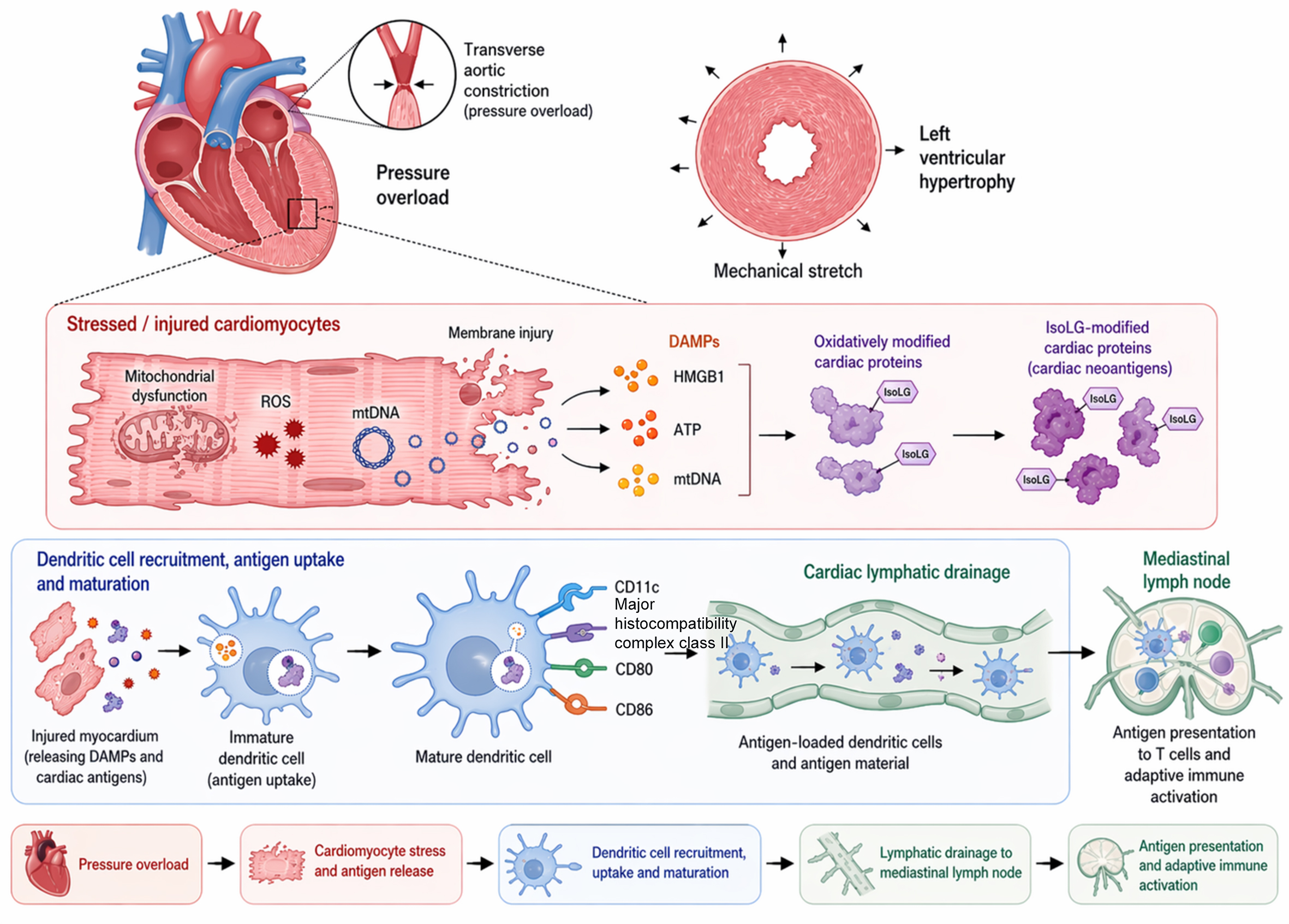
Figure 1. Model of cardiac antigen formation and activation of the DC-CD4+ T cell axis under pressure overload. Pressure overload induces the release of cardiac injury signals, the formation of candidate antigens, and DC-mediated antigen transport. This figure summarizes the candidate processes involving myocardial stress, DAMP release, generation of oxidatively modified antigens, DC uptake and maturation, and cardiac lymphatic drainage to the mediastinal lymph nodes. DC: Dendritic cell; ROS: reactive oxygen species; mtDNA: mitochondrial DNA; DAMPs: damage-associated molecular patterns; HMGB1: high mobility group box 1; ATP: adenosine triphosphate; CD11c: cluster of differentiation 11c; MHC II: major histocompatibility complex class II; CD80: cluster of differentiation 80; CD86: cluster of differentiation 86.
DC-DEPENDENT CD4+ T CELL PRIMING AND FUNCTIONAL DIFFERENTIATION UNDER PRESSURE OVERLOAD
Antigen recognition and costimulation set the activation threshold for CD4+ T cells
In pressure overload-induced cardiac remodeling, the initial activation of CD4+ T cells is not a passive consequence of intensified local inflammation, but an important starting point that drives disease progression from compensatory hypertrophy toward decompensation[29]. This step primarily depends on antigen-specific recognition, rather than being driven solely by increased inflammatory cytokines or tissue injury signals. In TAC models, mice lacking adaptive immunity, as well as mice deficient in MHC II, show markedly attenuated subsequent deterioration of cardiac function, suggesting that helper T cells and their capacity for antigen recognition play a critical role in disease progression[11]. When CD4+ T cells are unable to recognize antigens associated with the pressure-overloaded heart, typical pathogenic responses are difficult to establish even under persistent mechanical loading[30]. Conversely, when a recognizable antigen is present within cardiomyocytes, pressure overload is sufficient to markedly enhance CD4+ T cell activation and accelerate the transition from cardiac hypertrophy to heart failure. These findings indicate that whether the pressure-overloaded heart can generate and continuously provide immunologically relevant autoantigenic information is an important prerequisite for determining whether CD4+ T cells enter a pathogenic program. However, only a limited number of cardiac antigens can currently be clearly included as candidates, and α-myosin, oxidatively modified proteins, and experimental model antigens cannot fully represent the true antigen repertoire in pressure overload[25]. Mechanistically, the key transition at this stage may occur during the uptake, processing, and presentation of cardiac-derived antigens by DCs. DCs process antigens released from injured tissue into peptides and present them to naïve CD4+ T cells as MHC II complexes, allowing the TCR to receive the first antigen-specific recognition signal and thereby converting local mechanical stress and tissue injury into an initiating event for adaptive immunity[11].
However, antigen recognition between MHC II and the TCR alone is still insufficient for naïve CD4+ T cells to achieve stable activation and enter sustained expansion[31]. For naïve T cells, costimulatory signaling is required to cross the full activation threshold, with the central event being the interaction between cluster of differentiation 80 (CD80) and cluster of differentiation 86 (CD86) on DCs and cluster of differentiation 28 (CD28) on T cells[32]. Related studies have shown that T cell activation can already be detected in the cardiac-draining mediastinal lymph nodes at a very early stage after pressure overload. When CD80/CD86-mediated costimulation is blocked, the proportion of activated T cells decreases markedly, followed by parallel reductions in inflammatory cell infiltration, cardiomyocyte injury, fibrosis, and deterioration of cardiac function[28]. This indicates that costimulation is not an accessory factor after response initiation, but a key step by which DCs convert antigen recognition into initial T cell priming. However, this process should not be interpreted as being carried out by a single DC population. Migratory DCs may be responsible for transporting cardiac antigens to draining lymph nodes; cDC2 cells are more likely to mediate MHC II-dependent CD4+ T cell priming; cDC1 cells may have potential roles in cross-presentation, necrotic cell antigen processing, and Th1-like responses; and monocyte-derived DCs may contribute more to inflammatory amplification and maintenance of costimulatory signals[23]. Pressure overload-induced heart failure may be accompanied by autoimmune-like responses against cardiac autoantigens and clonal expansion of activated CD4+ T cell populations. Taken together, current evidence supports the view that initial CD4+ T cell activation is a layered process jointly determined by antigen burden, DC subset composition, MHC II-dependent recognition, and costimulation[4]. Therefore, the entire priming process cannot be explained solely by changes in total CD11c+ or MHC II+ cell populations. Further clarification is needed to determine which DC subsets carry which cardiac antigens and where they establish effective contact with naïve T cells. In addition, although the functional significance of MHC II-dependent recognition and costimulatory blockade is relatively clear, direct evidence linking specific cardiac antigens, specific DC subsets, and specific T cell clones remains lacking. DCs are therefore better described as candidate regulatory nodes composed of multiple functional subsets, rather than as a fully established single upstream controlling cell population.
The inflammatory microenvironment shapes CD4+ T cell effector differentiation and pathogenic phenotypes
During cardiac remodeling driven by sustained pressure overload, the role of DCs in CD4+ T cells is not limited to initial activation; more importantly, DCs determine the functional direction in which these cells subsequently undergo stable differentiation[6]. At this stage, the key issue that truly influences disease progression is no longer simply whether T cells are activated, but which type of DC presents the antigenic information continuously released from the pressure-overloaded heart, and under which cytokine context this information guides T cells toward a particular effector program[33]. In general, the more persistent the antigen burden and the more stable the presentation, the more likely naïve CD4+ T cells are to shift from transient activation to a relatively stable differentiation program[5]. However, different DC subsets do not generate identical outputs. cDC2 cells are more commonly associated with MHC II-dependent CD4+ T cell polarization[34], cDC1 cells may participate in necrotic cell antigen processing and Th1-like responses through features such as XCR1 and CLEC9A[35], and monocyte-derived DCs may produce interleukin-6 (IL-6), tumor necrosis factor-alpha (TNF-α), and interleukin-12 (IL-12) family-related signals in inflammatory settings, thereby amplifying T cell effector responses[36]. Studies have shown that IL-12α deficiency markedly attenuates cardiac inflammation, hypertrophy, fibrosis, and functional deterioration after pressure overload[37]. Therefore, a more reasonable interpretation is that the antigen-presenting capacity and cytokine output of different DC subsets in the pressure-overloaded heart jointly shape the trajectory by which CD4+ T cells progress from early activation toward Th1-like, T helper 17 (Th17)-like, or regulatory T-cell (Treg) phenotypes.
This DC-driven fate divergence ultimately determines the pathological output of CD4+ T cells within the tissue. Studies have shown that, in the context of non-ischemic heart failure, the cell population most closely associated with cardiac fibrosis and dysfunction is not T cell infiltration in a general sense, but IFN-γ-producing Th1 effector cells[38]. More importantly, these cells do not amplify injury only at a distance through the secretion of inflammatory mediators. They can also directly establish adhesive contact with cardiac fibroblasts, induce the expression of α-smooth muscle actin, and promote their transition toward myofibroblasts with profibrotic features[39,40]. In vivo, adoptive transfer of Th1 cells, but not naïve T cells or activated T cells lacking IFN-γ, is sufficient to reinduce elevated left ventricular end-diastolic pressure, impaired systolic function, and pathological remodeling in T cell-deficient mice[41]. These findings indicate that DC-directed functional polarization is not merely an abstract immunological classification, but directly determines whether CD4+ T cells acquire the executive capacity to promote fibrosis and decompensation[16]. In other words, among activated CD4+ T cells, only those that further develop a Th1-like differentiation bias under specific cytokine and costimulatory signals provided by DCs are more likely to become pathogenic populations that drive structural remodeling.
In addition, the balance between costimulatory and coinhibitory signals not only determines the magnitude of the response, but also alters the direction of differentiation. Under pressure overload, cytotoxic T-lymphocyte-associated protein 4 (CTLA-4) blockade can promote Th17 cell activation and enhance interleukin-17A-related inflammatory responses, while aggravating cardiac remodeling and dysfunction[42]; In contrast, selective expansion of Tregs can alleviate inflammation and organ remodeling in chronic pressure overload-associated heart failure[43]. These two lines of evidence indicate that the effects of CD4+ T cells are not fixed toward a pro-inflammatory outcome, but depend on the balance among antigenic stimulation, the costimulatory threshold, and regulatory restraint. However, it should also be noted that CTLA-4 blockade represents an artificial release of an inhibitory checkpoint and is not equivalent to a major driving event in the natural disease course. Similarly, the protective effects of Treg expansion may also arise from broad anti-inflammatory and tissue-reparative effects, rather than being entirely dependent on cardiac antigen specificity. Therefore, Th1, Th17, and Treg responses should be viewed as competing differentiation branches rather than as a linear progression. Future studies need to assess antigen specificity, cytokine profiles, clonal expansion, and tissue localization simultaneously within the same model to determine which type of CD4+ T cell truly governs the decompensation process, and to avoid extrapolating a single phenotype to the entire disease mechanism. This approach preserves the central role of CD4+ T cells while also accounting for evidence showing that effector directions are not uniform.
Cardiac homing, local reactivation, and maintenance of effector CD4+ T cell responses
After completing initial activation and acquiring effector features in the draining lymph nodes, CD4+ T cells do not remain within peripheral immune compartments for long, but instead enter a phase of directed recirculation. After leaving lymphoid organs, they re-enter the circulation, by which time the expression profiles of chemokine receptors and adhesion-related molecules on their surface have already been shaped during the preceding priming and functional polarization processes[28]. Thereafter, the key determinant of whether these cells can re-enter the heart is not the passive diffusion of inflammatory mediators in the blood, but whether the pressure-overloaded myocardium continues to provide recognizable homing signals[44]. Studies have shown that myeloid cells and fibroblasts in the injured heart can upregulate chemokines such as C-X-C motif chemokine ligand 9 (CXCL9) and C-X-C motif chemokine ligand 10 (CXCL10), thereby preferentially attracting CXCR3-expressing effector CD4+ T cells to accumulate locally[14]. At the same time, increased expression of adhesion molecules such as intercellular adhesion molecule 1 (ICAM-1) on activated endothelial cells further provides the structural conditions required for circulating T cells to slow down, adhere, and undergo transendothelial migration[45]. Thus, chemokines determine the direction of return, adhesion molecules determine the efficiency of entry, and the inflammatory activation state of the local vascular wall determines whether this migration can actually be completed. As a result, CD4+ T cells that have already acquired effector characteristics in the draining lymph nodes can be redirected to cardiac lesions, allowing the preceding peripheral immune priming to be further converted into local immune infiltration and translating antigen recognition, functional differentiation, and cardiac return into tissue-level cellular accumulation [Figure 2].
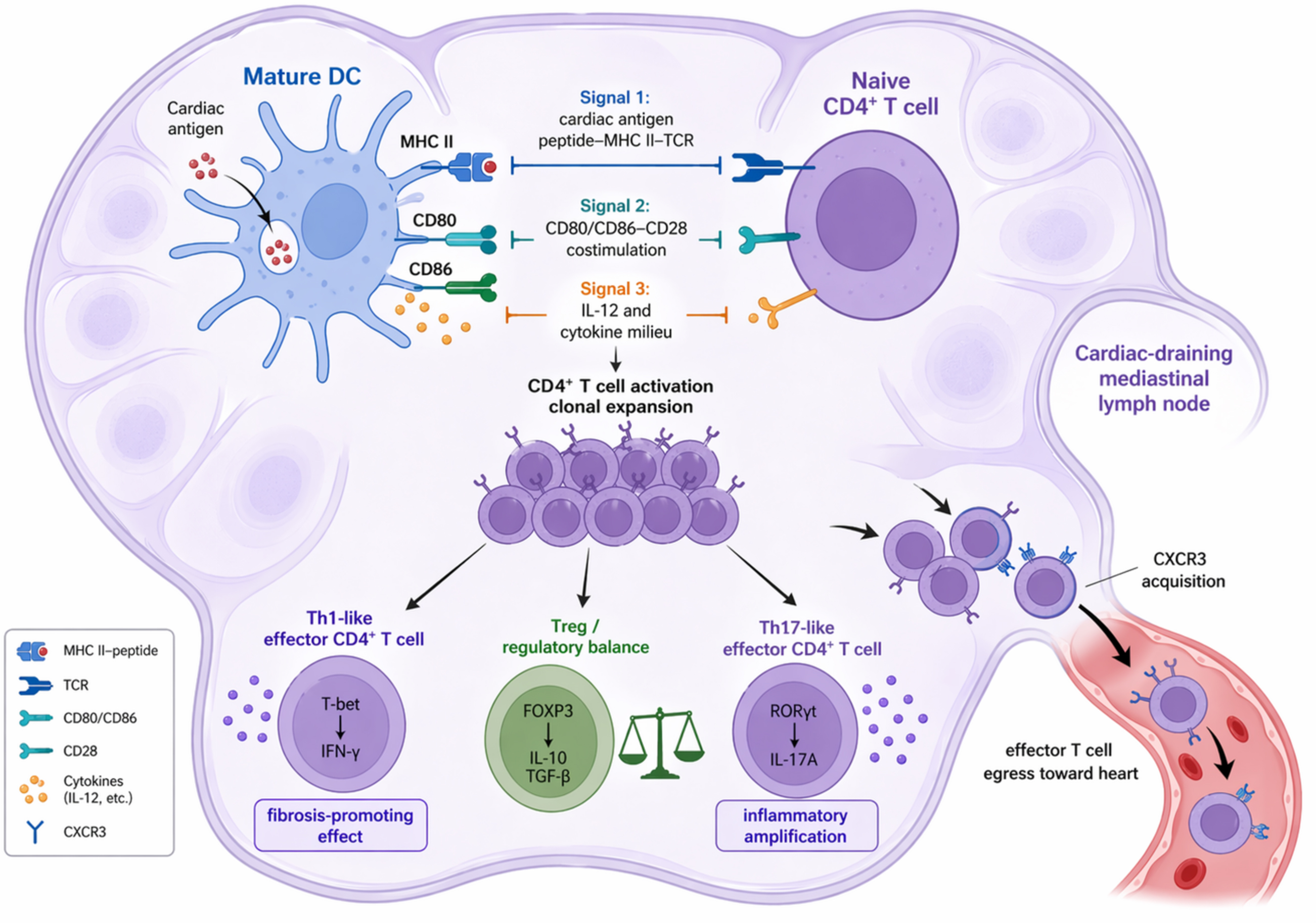
Figure 2. Candidate processes by which different DC subsets mediate CD4+ T cell priming and functional polarization under pressure overload. This figure summarizes the process by which cardiac antigens are processed by migratory DCs, cDC1, cDC2, and monocyte-derived DCs, and subsequently induce CD4+ T cell activation, clonal expansion, and differentiation toward Th1-like, Th17-like, or Treg phenotypes in the draining lymph nodes through MHC II-TCR recognition, CD80/CD86-CD28 costimulation, and cytokine signaling. TCR: T cell receptor; CD28: cluster of differentiation 28; IL-12: interleukin-12; Th1: T helper 1 cell; IFN-γ: interferon-gamma; FOXP3: forkhead box protein 3; IL-10: interleukin-10; TGF-β: transforming growth factor-beta; Th17: T helper 17 cell; RORγt: RAR-related orphan receptor gamma t; IL-17A: interleukin-17A; CXCR3: C-X-C chemokine receptor type 3; DC: dendritic cell; cDC1: type 1 conventional dendritic cell; cDC2: type 2 conventional dendritic cell; MHC II: major histocompatibility complex class II; CD80: cluster of differentiation 80; CD86: cluster of differentiation 86; Treg: regulatory T cell.
However, cardiac homing represents only spatial return and is not the endpoint of this response. After entering the myocardium, CD4+ T cells often cannot maintain long-term activation without local restimulation. In contrast, if antigen presentation and inflammatory responses persist within the lesion, their effector program can be reinforced and sustained[46]. As the disease progresses, TCR-related antigen recognition signals within the heart are further enhanced, suggesting that returning T cells may continue to receive antigenic stimulation within the tissue[39]. Notably, this reactivation process is not restricted to recruited professional antigen-presenting cells. Local stromal cells, especially fibroblasts, may also acquire MHC II-dependent antigen processing and presentation capacity under the influence of inflammatory cytokines such as IFN-γ, thereby working together with DCs to maintain restimulation sites and form a relatively stable local stimulatory platform[47]. However, direct evidence remains lacking as to whether these cells possess complete costimulatory capacity and whether they can independently sustain pathogenic T cell responses in the absence of professional antigen-presenting cells such as DCs and macrophages. Therefore, a more cautious interpretation is that fibroblasts may act as part of the local restimulation platform, prolonging antigen exposure and T cell retention, rather than independently replacing professional antigen-presenting cells. At the same time, persistent antigenic fragments, cellular injury signals, and an incompletely resolved chemokine environment within local tissue can extend the residence time of effector T cells in the lesion, allowing them to undergo repeated restimulation through antigen re-presentation[6,46]. In this setting, the heart is no longer merely the terminal target organ in which effector T cells act, but may also serve as a local immune site that supports their reactivation, retention, and functional maintenance[48]. Thus, cardiac antigen release, lymphatic drainage, T cell activation in draining lymph nodes, and the return of effector CD4+ T cells together constitute an explanatory spatial model. However, this model is mainly assembled from evidence derived from different studies and should not yet be equated with a closed loop that has been continuously demonstrated in pressure overload models. Future studies are still needed to further distinguish the temporal sequence and degree of dependence between professional antigen-presenting cells and fibroblasts in local restimulation.
MECHANISMS BY WHICH CD4+ T CELLS AND IMMUNE NETWORKS DRIVE CARDIAC DECOMPENSATION
Local cardiac antigen re-presentation: DC-T cell interactions and fibroblast involvement
As effector CD4+ T cells re-enter the pressure-overloaded heart, DC-mediated antigen presentation may no longer serve only as an upstream step in the initiation of the early immune response. Instead, it may interact with local T-cell effector programs, thereby providing a tissue basis for sustained immune amplification[49]. Studies have shown that bone marrow-derived CD11c+ DCs exert pathogenic effects in left ventricular inflammation, hypertrophy, and fibrosis induced by hemodynamic overload, suggesting that DCs may continue to contribute to the maintenance of local inflammation and structural injury during the later stages of remodeling[6]. Further distinction is needed among locally retained cDC1, cDC2, inflammation-induced monocyte-derived DCs, and returning migratory DCs, as these subsets may have different roles in antigen representation, cytokine production, T-cell restimulation, and inflammatory amplification[23]. At present, there is still no evidence showing that the same population of DCs repeatedly presents the same antigen within the heart and continuously drives clonal expansion of the same T-cell population. Therefore, the relationship between DCs and CD4+ T cells is better described as a subset-dependent local interaction rather than as an established closed positive-feedback loop.
After effector CD4+ T cells return to the lesion site, their role may extend beyond amplifying inflammatory output and may also reshape the local landscape of antigen presentation[50]. Studies have shown that antigen presentation in the pressure-overloaded heart is not restricted to professional antigen-presenting cells. Under the influence of IFN-γ, cardiac fibroblasts can take up and process antigens and present them to CD4+ T cells through MHC II[51]; Conditional deletion of the MHC II pathway in fibroblasts markedly attenuates remodeling and cardiac dysfunction[46]. These findings support the involvement of fibroblasts in local restimulation, but they do not establish fibroblasts as fully competent professional antigen-presenting cells. Key limitations remain, including the uncertain expression of costimulatory molecules, antigen-processing efficiency, and correspondence with specific T-cell clones. In addition, MHC II deletion may also alter the inflammatory phenotype of fibroblasts. Therefore, DCs, macrophages, and fibroblasts together constitute a local antigen-presenting network that sustains T-cell retention and restimulation at different stages, rather than chronic T-cell responses being maintained independently by a single non-immune cell population.
Superimposed immune pressure and cardiomyocyte stress decompensation
Under pressure overload, cardiac dysfunction can develop independently of the extent of hypertrophic growth when cardiomyocyte stress-adaptive and prosurvival signaling is impaired[52]. The previously established immune axis involving DCs and CD4+ T cells may alter the adaptive outcome of the pressure-overloaded myocardium at this stage. However, the immune pressure imposed on cardiomyocytes does not arise solely from CD4+ T cells[53]. Macrophages and monocytes can amplify local injury through TNF-α, interleukin-1 beta, IL-6, and profibrotic mediators[15]; CD8+ T cells may aggravate myocardial stress through cytotoxic molecules and inflammatory cytokines[54]; B cells can influence adaptive immunity through antibodies, antigen presentation, and cytokine production[55]; and NK cells, NKT cells may reshape the early cytokine milieu and immune balance[56]. Therefore, CD4+ T cells are better viewed as an antigen-specific amplification node within a multicellular inflammatory network, rather than as the sole effector source. Compensatory hypertrophy is, in essence, a limited protective remodeling response, and its maintenance depends on intact energy supply, contractile reserve, and injury repair capacity[57]. Once the CD4+ T-cell response persists, cardiomyocytes are no longer exposed to mechanical stretch alone, but to a long-term superposition of mechanical load, innate immunity, and adaptive immunity[14]. During this process, local proinflammatory mediators, oxidative stress, and sustained antigenic stimulation continue to accumulate, converting cellular stress that could originally be buffered in the short term into a chronic state of exhaustion that is difficult to reverse. The adaptive margin of cardiomyocytes is thereby progressively narrowed[58]. Studies have shown that, under pressure overload, once a CD4+ T-cell response against cardiac antigens is established, the transition from hypertrophy to functional failure is markedly accelerated[25]. The significance of this process lies in the ability of CD4+ T cells to link antigen recognition, cytokine output, and fibroblast activation, thereby converting an inflammatory background into sustained adaptive immune pressure. This long-standing immune injury weakens the ability of cardiomyocytes to tolerate pressure overload, causing cells that might otherwise maintain function through hypertrophic remodeling to gradually develop impaired contraction, restricted diastolic relaxation, and reduced survival capacity.
At the same time, once this immune axis is established, cardiomyocytes themselves may in turn become an important source driving disease progression. Pressure-overloaded and injured cardiomyocytes can continuously release new damage signals and antigenic substrates, making it difficult for the local immune response to terminate spontaneously[59]. Thus, cardiomyocytes are not merely passive targets affected by inflammation; after injury, they continue to provide new stimuli to the immune system, further sustaining DC-mediated activation and local reinitiation of CD4+ T-cell responses[60]. As a result, the local response fails to enter the resolution phase smoothly, and a self-reinforcing vicious cycle gradually develops between persistent inflammation and cardiomyocyte injury[61]. Each new round of cardiomyocyte stress imbalance leads to further antigen exposure and danger signaling, while each round of immune amplification further compromises cardiomyocyte survival and compensatory potential[62]. Therefore, the effect of the DC-CD4+ T-cell immune axis on cardiac function is essentially mediated by its sustained erosion of cardiomyocyte stress tolerance. This process causes cardiomyocytes to lose the functional reserve required to maintain compensation earlier under chronic load, and converts local inflammation from a relatively reversible response into persistent tissue stress[63]. As the number of surviving cardiomyocytes gradually decreases, the remaining cells are forced to bear a greater workload, further increasing ventricular wall stress and ultimately driving the transition from compensatory hypertrophy to decompensated remodeling[64]. Therefore, the link between this immune axis and cardiac dysfunction is not merely indirect; rather, by continuously weakening the adaptive capacity of cardiomyocytes, this axis directly participates in the central process of heart failure progression.
Fibroblast immune activation, extracellular matrix imbalance, and fibrosis progression
As the disease process shifts from immune activation to structural remodeling, DC-mediated antigen presentation and costimulation can sustain CD4+ T-cell responses, causing cardiac remodeling to further manifest as fibroblast activation, extracellular matrix imbalance, and fibrosis progression[65]. At this stage, fibroblasts show a clear phase-dependent change in function[48]. In the early phase, these cells may participate in chemokine release and maintenance of the inflammatory microenvironment, thereby facilitating local immune-cell accumulation. As effector CD4+ T cells continue to enter and remain within the heart, fibroblasts gradually become the principal executors of structural remodeling. Their proliferation, activation, and myofibroblast-like transition are progressively enhanced, accompanied by abnormal accumulation of collagen and other extracellular matrix components. Under inflammatory conditions, a subset of fibroblasts may also acquire MHC II-dependent antigen-presenting features; however, this property should be regarded as an extension of their immunomodulatory function rather than a complete replacement of professional antigen-presenting cells[46]. CD4+ T cells and their antigen-recognition capacity play a critical role in the transition from compensatory hypertrophy to decompensated heart failure[29], suggesting that subsequent fibrosis is not determined solely by mechanical stimulation, but is progressively amplified under sustained involvement of adaptive immunity. Consistent with this view, ICAM-1-mediated persistent leukocyte recruitment can markedly affect left ventricular inflammatory infiltration, the extent of fibrosis, and changes in cardiac function, indicating that repeated entry of immune cells into the lesion site continuously provides pro-activation signals to fibroblasts and promotes long-term imbalance in matrix turnover[63]. Studies have shown that interleukin-21-related signaling can disturb the balance between tissue inhibitor of metalloproteinases 4 and matrix metalloproteinase 9, accelerate disordered matrix degradation and redeposition, and ultimately promote abnormal interstitial collagen accumulation and reduced ventricular compliance[66]. Therefore, at this stage, fibroblasts act both as executors of structural remodeling and as immunomodulatory participants within the inflammatory milieu, although their antigen-presenting role should still be interpreted in the context of the coordinated actions of DCs and other professional antigen-presenting cells.
As this process continues, fibroblasts themselves may in turn amplify inflammation and structural injury, creating a mutually reinforcing loop between immune responses and tissue remodeling. Studies have shown that pressure overload can activate specific fibroblast subsets and recruit inflammatory Ly6C-high monocytes into the heart through fibroblast-intrinsic nuclear factor kappa B signaling. This indicates that fibroblasts are not merely passive targets of immune stimulation, but important participants in maintaining a chronic inflammatory milieu[67]. In this context, persistently activated fibroblasts promote collagen deposition and matrix rearrangement on the one hand, while on the other hand releasing chemokines and inflammatory mediators that prolong local immune-cell retention and enhance intercellular signaling within the lesion. Together, these processes reinforce the coupling between interstitial remodeling and immune amplification[68]. Therefore, pressure overload-associated fibrosis is not simply an increase in collagen content. Rather, it reflects the transition of fibroblasts from a short-term reparative response to long-term pathological activation under sustained immune drive, and also provides an important tissue basis for the failure of local inflammation to resolve. This process gradually converts an initially localized repair program into persistent structural output, thereby aggravating ventricular wall stiffness, reduced compliance, and fixation of adverse remodeling. In summary, the ability of the DC-CD4+ T-cell axis to drive the heart from immune imbalance toward structural imbalance lies in its continuous provision of pro-activation and pro-matrix-remodeling signals to fibroblasts. Activated fibroblasts, in turn, further consolidate the local pathological environment through inflammatory-cell recruitment, extracellular matrix rearrangement, and increased tissue stiffness, ultimately promoting the progression of myocardial fibrosis and ventricular remodeling [Figure 3].
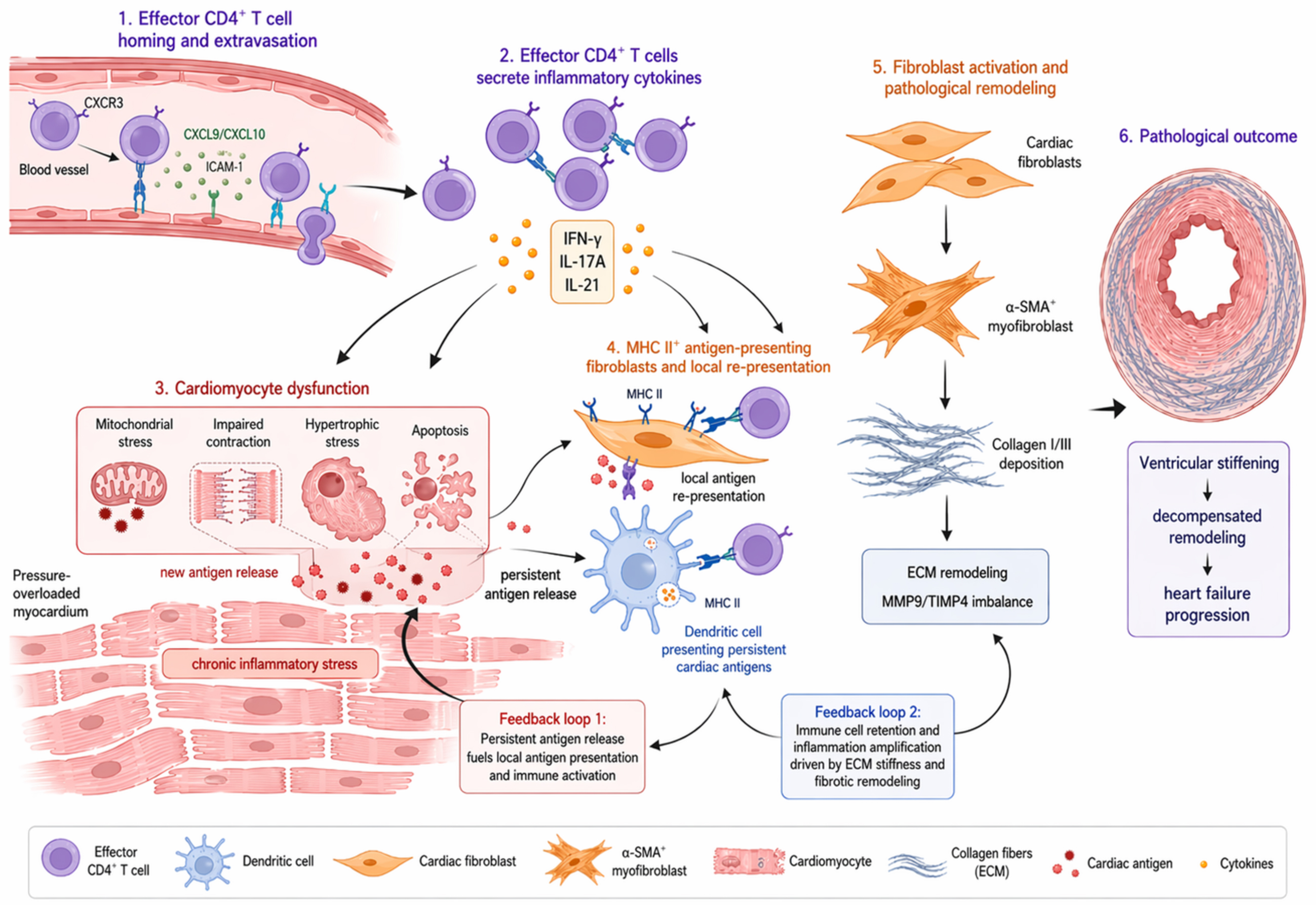
Figure 3. Model of effector CD4+ T cells, fibroblast antigen presentation, and structural remodeling in the pressure-overloaded heart. Local interactions among effector CD4+ T cells, cardiomyocyte injury, and fibroblast-mediated matrix remodeling in the pressure-overloaded heart. This figure summarizes the process by which, after homing to the heart, effector CD4+ T cells interact locally with cardiomyocytes, DCs, and fibroblasts in the presence of antigenic and inflammatory signals, accompanied by cardiomyocyte stress, fibroblast activation, collagen deposition, and ventricular remodeling. DC: Dendritic cell; CXCL9: C-X-C motif chemokine ligand 9; CXCL10: C-X-C motif chemokine ligand 10; ICAM-1: intercellular adhesion molecule 1; IL-21: interleukin-21; α-SMA: alpha-smooth muscle actin; ECM: extracellular matrix.
TRANSLATIONAL VALUE, RISKS, AND RESEARCH LIMITATIONS OF IMMUNOMODULATORY INTERVENTIONS
Stratified targeting of costimulation, cytokines, homing, and immune tolerance
Potential intervention strategies targeting DC-CD4+ T-cell coupling should not simply aim to suppress inflammation, but rather to attenuate this immune axis stepwise at key points of response initiation, effector differentiation, and cardiac homing. More upstream strategies include reducing the formation of cardiac autoantigens and limiting the increase in their immunogenicity, for example by decreasing the burden of oxidatively modified neoantigens, or by inducing tolerogenic DCs, using antigen-delivery nanoparticles, or applying oral or mucosal antigen exposure. These approaches may bias cardiac antigens toward the induction of Treg responses, anergy, or clonal deletion, rather than the expansion of proinflammatory T cells[4]. Compared with broad immunosuppression, antigen-specific immune tolerance is theoretically more tissue-selective and better suited to the long-term management required for chronic pressure overload. However, this strategy depends on prior clarification of the dominant cardiac antigens, the cellular sources of antigen presentation, and the effective delivery window. Because the dominant antigenic repertoire in pressure overload remains undefined, such approaches are currently more appropriate as early exploratory directions and should not yet be regarded as mature translational strategies.
At a level closer to the effector phase, costimulatory blockade, IL-12-related interventions, and CXCR3 inhibition constitute more specific immunomodulatory approaches. Blockade of the CD28-CD80/CD86 axis can reduce initial T-cell priming, subsequent expansion, and the efficiency of cardiac infiltration, and pressure-overload models have shown that this strategy can delay the deterioration of cardiac function[65]. IL-12α deficiency or pharmacological inhibition of IL-12β can attenuate Th1-like polarization and inflammatory amplification, and may therefore reduce profibrotic T-cell effects[36]; CXCR3 inhibition mainly targets the cardiac homing of activated effector T cells and can block CXCL9/CXCL10-driven local accumulation[14]. In addition, tolerogenic DCs can bias cardiac antigen presentation toward immune tolerance by downregulating MHC class II and costimulatory molecules, enhancing IL-10/indoleamine 2,3-dioxygenase-mediated immunoregulation, or inducing Tregs[69]. Antigen-specific tolerance targeting cardiac myosin or troponin has shown feasibility in models of autoimmune myocarditis and ischemic myocardial injury, but its efficacy in pressure overload-induced cardiac remodeling remains to be established. These approaches correspond, respectively, to peripheral priming, functional polarization, tissue infiltration, and restoration of immune homeostasis. Their shared limitation is the lack of cardiac specificity. Long-term use may impair anti-infective and antitumor immunity, as well as tissue repair responses, thereby increasing the risk of systemic immunosuppression. Therefore, future translational studies should emphasize short-course, stage-specific, low-dose, or locally delivered interventions, rather than prolonged suppression of T-cell function.
Human relevance, levels of evidence, safety, and unresolved questions
At present, evidence regarding the DC-CD4+ T-cell axis is still derived mainly from animal models of pressure overload, and most studies have validated individual steps of the pathway separately. Existing studies suggest that cardiac antigen generation, DC activation, CD4+ T-cell priming, fibroblast antigen presentation, and pathogenic T-cell responses may all contribute to cardiac remodeling. However, the complete sequence has not yet been continuously demonstrated within a single experimental system. Human-related evidence currently comes mainly from findings of immune-cell infiltration, CXCR3-associated homing signals, fibroblast MHC II expression, and autoimmune-like responses in samples from pressure overload or non-ischemic heart failure, but overall it remains largely correlative. Table 1 summarizes the major models, findings, and boundaries of evidence. Inconsistent findings should also be considered in parallel; for example, certain myeloid cells may not be essential at specific stages, antigen-specific CD8+ T cells can be activated but may not necessarily promote heart failure progression, and B-cell or antibody responses may occur alongside the CD4+ T-cell axis. Overall, although costimulatory blockade, IL-12 inhibition, CXCR3 antagonism, tolerogenic DCs, and antigen-specific tolerance each have mechanistic advantages, their clinical translation remains limited by undefined antigens, complex cellular sources, a narrow safety window, and the risk of long-term immunosuppression. Future studies will require more human myocardial tissues, blood immune profiling, longitudinal clinical cohorts, and cross-species validation.
Model evidence, key findings, and limitations of immune mechanisms in pressure overload
| Representative research area | Model or sample | Key findings | Boundaries of evidence |
| CD4+ T cells and pressure overload-induced decompensation | Mouse TAC, immunodeficiency models, or T-cell intervention models | CD4+ T cells promote the transition from compensatory hypertrophy to heart failure and are associated with fibrosis and functional deterioration[29] | Evidence is mainly derived from mouse models and may not fully represent the long-term course of human hypertension or aortic stenosis |
| Costimulatory blockade and T-cell priming | Mouse TAC and intervention targeting the CD80/CD86-CD28 axis | Blocking costimulation can reduce T-cell activation and cardiac infiltration, and attenuate the deterioration of cardiac function[28] | This represents systemic immunomodulation, and long-term use may increase the risk of infection and impaired immune surveillance |
| CXCR3-associated cardiac homing | Human myocardial samples related to pressure overload and mouse TAC models | CXCR3+ T cells can accumulate in the pressure-overloaded heart, and the CXCL9/CXCL10 axis participates in the cardiac homing of effector T cells[14] | Human evidence is mostly correlative, and mechanistic validation still relies mainly on animal models |
| Cardiac autoantigens and oxidative modification | Mouse pressure-overload models and cardiac antigen stimulation experiments | α-myosin and oxidatively modified proteins, such as IsoLG-modified proteins, suggest that cardiac components can form immunogenic antigens[11] | The dominant endogenous antigenic repertoire remains unclear, which limits the design of antigen-specific therapies |
| DC and fibroblast antigen presentation | Mouse TAC, DC analysis, fibroblast MHC II manipulation, and human tissue observations | DCs participate in cardiac antigen presentation; under inflammatory conditions, fibroblasts can acquire MHC II-associated antigen-presenting features[46] | The functional division among DC subsets remains unclear, and whether fibroblasts can independently sustain pathogenic T-cell responses has not yet been determined |
| IL-12, tolerogenic DCs, and antigen-specific tolerance | Mouse pressure-overload models and immune-tolerance intervention models | IL-12-related signaling participates in Th1-like polarization; tolerogenic DCs and antigen tolerance may reduce proinflammatory T-cell responses[4] | These approaches remain at an early translational stage, and the timing of administration, antigen selection, and long-term safety have not yet been clarified |
| Overall assessment of human-related evidence | Human myocardial tissue, blood immune profiles, and clinical observations | Human samples support the presence of immune-cell infiltration, chemokine signal activation, and antigen presentation-related changes in the pressure-overloaded heart[1] | Most evidence comes from cross-sectional or end-stage samples, with limited dynamic tracking and causal validation |
CONCLUSIONS
Pressure overload-induced cardiac remodeling is not caused solely by increased afterload, but represents a chronic pathological process jointly shaped by mechanical stress, myocardial injury signals, and immune responses. The persistently overloaded myocardium can release danger signals and generate cardiac antigens with the potential for immune recognition. Through DC uptake, maturation, antigen presentation, and costimulation, these signals promote CD4+ T-cell priming, effector differentiation, and cardiac homing. Returning CD4+ T cells can further impair cardiomyocyte adaptive capacity and promote matrix imbalance and fibrosis progression through cytokine production, local restimulation, and interactions with fibroblasts. It should be emphasized that this axis is not the only mechanism explaining pressure overload-induced cardiac remodeling. Macrophages, CD8+ T cells, B cells, and ILCs may also participate in inflammatory amplification and impaired repair at different stages. Current evidence still comes mainly from animal TAC models, whereas human tissue data and longitudinal clinical evidence remain relatively limited. Future studies should further define the dominant cardiac antigens, key DC subsets, T-cell clonal evolution, and mechanisms of local antigen re-presentation. On this basis, the timing, safety window, and clinical feasibility of costimulatory blockade, cytokine modulation, homing intervention, and immune-tolerance strategies should be carefully evaluated.
DECLARATIONS
Authors’ contributions
Wrote the manuscript: Bai Z, Zhou M, Qu P
Prepared the figures: Shi J, Gu Y, Zhao D
Provided critical comments on the writing and revision of the review and acquired funding: Lyu J, Cheng P, Lei Q
All authors reviewed and approved the final manuscript.
Availability of data and materials
Not applicable.
AI and AI-assisted tools statement
During the preparation of this manuscript, ChatGPT (GPT-5.5, released April 23, 2026; OpenAI) was used solely for creating Figures 1-3 and the Graphical Abstract. The tool did not influence the study design, data collection, analysis, interpretation, or scientific content of the work. All authors take full responsibility for the accuracy, integrity, and final content of the manuscript.
Financial support and sponsorship
This work was supported by the National Natural Science Foundation of China (No. 82470290), Noncommunicable Chronic Diseases‐National Science and Technology Major Project (No. 2024ZD0527205), Sichuan Provincial Science and Technology Program (No. 2026NSFSC0582), and Chen Xiao-ping Foundation for the Development of Science and Technology of Hubei Province (No. CXPJJH125001-25103), National Natural Science Foundation of China (82572491).
Conflicts of interest
All authors declared that there are no conflicts of interest.
Ethical approval and consent to participate
Not applicable.
Consent for publication
Not applicable.
Copyright
© The Author(s) 2026.
REFERENCES
1. Lindman BR, Perry AS, Lance ML, et al. Integrated multiomics of pressure overload in the human heart prioritizes targets relevant to heart failure. Nat Commun. 2025;16:6889.
2. Nakamura M, Sadoshima J. Mechanisms of physiological and pathological cardiac hypertrophy. Nat Rev Cardiol. 2018;15:387-407.
3. Clavel M, Tastet L, Pibarot P. Cardiac remodelling in aortic stenosis. Arch Cardiovasc Dis. 2026;119:32-8.
4. Martini E, Cremonesi M, Felicetta A, et al. Autoimmune-like mechanism in heart failure enables preventive vaccine therapy. Circ Res. 2025;136:4-25.
5. Obst R, Van Santen H, Mathis D, Benoist C. Antigen persistence is required throughout the expansion phase of a CD4+ T cell response. J Exp Med. 2005;201:1555-65.
6. Wang H, Kwak D, Fassett J, et al. Role of bone marrow-derived CD11c+ dendritic cells in systolic overload-induced left ventricular inflammation, fibrosis and hypertrophy. Basic Res Cardiol. 2017;112:25.
7. Kologrivova I, Shtatolkina M, Suslova T, Ryabov V. Cells of the immune system in cardiac remodeling: main players in resolution of inflammation and repair after myocardial infarction. Front Immunol. 2021;12:664457.
8. Patel B, Ismahil MA, Hamid T, Bansal SS, Prabhu SD. Mononuclear phagocytes are dispensable for cardiac remodeling in established pressure-overload heart failure. PLoS ONE. 2017;12:e0170781.
9. Gröschel C, Sasse A, Monecke S, et al. CD8+-T cells with specificity for a model antigen in cardiomyocytes can become activated after transverse aortic constriction but do not accelerate progression to heart failure. Front Immunol. 2018;9:2665.
10. Suetomi T, Willeford A, Brand CS, et al. Inflammation and NLRP3 inflammasome activation initiated in response to pressure overload by Ca2+/calmodulin-dependent protein kinase II δ signaling in cardiomyocytes are essential for adverse cardiac remodeling. Circulation. 2018;138:2530-44.
11. Ngwenyama N, Kirabo A, Aronovitz M, et al. Isolevuglandin-modified cardiac proteins drive CD4+ T-cell activation in the heart and promote cardiac dysfunction. Circulation. 2021;143:1242-55.
12. Guo Y, You Y, Shang F, et al. iNOS aggravates pressure overload-induced cardiac dysfunction via activation of the cytosolic-mtDNA-mediated cGAS-STING pathway. Theranostics. 2023;13:4229-46.
13. Martini E, Kunderfranco P, Peano C, et al. Single-cell sequencing of mouse heart immune infiltrate in pressure overload-driven heart failure reveals extent of immune activation. Circulation. 2019;140:2089-107.
14. Ngwenyama N, Salvador AM, Velázquez F, et al. CXCR3 regulates CD4+ T cell cardiotropism in pressure overload-induced cardiac dysfunction. JCI Insight. 2019;4:e125527.
15. Amrute JM, Luo X, Penna V, et al. Targeting immune-fibroblast cell communication in heart failure. Nature. 2024;635:423-33.
16. Saleh D, Jones RTL, Schroth SL, Thorp EB, Feinstein MJ. Emerging roles for dendritic cells in heart failure. Biomolecules. 2023;13:1535.
17. Liu X, Shi G, Guo J. Innate immune cells in pressure overload-induced cardiac hypertrophy and remodeling. Front Cell Dev Biol. 2021;9:659666.
18. Canton J, Blees H, Henry CM, et al. The receptor DNGR-1 signals for phagosomal rupture to promote cross-presentation of dead-cell-associated antigens. Nat Immunol. 2020;22:140-53.
19. Van Der Borght K, Scott CL, Martens L, et al. Myocarditis elicits dendritic cell and monocyte infiltration in the heart and self-antigen presentation by conventional type 2 dendritic cells. Front Immunol. 2018;9:2714.
20. Feng G, Bajpai G, Ma P, et al. CCL17 aggravates myocardial injury by suppressing recruitment of regulatory T cells. Circulation. 2022;145:765-82.
21. Blanton RM, Carrillo-Salinas FJ, Alcaide P. T-cell recruitment to the heart: friendly guests or unwelcome visitors? Am J Physiol Heart Circ Physiol. 2019;317:H124-40.
22. Lv H, Havari E, Pinto S, et al. Impaired thymic tolerance to α-myosin directs autoimmunity to the heart in mice and humans. J Clin Investig. 2011;121:1561-73.
23. Van Der Borght K, Scott CL, Nindl V, et al. Myocardial infarction primes autoreactive T cells through activation of dendritic cells. Cell Rep. 2017;18:3005-17.
24. Nevers T, Salvador AM, Grodecki-Pena A, et al. Left ventricular T-cell recruitment contributes to the pathogenesis of heart failure. Circ Heart Fail. 2015;8:776-87.
25. Gröschel C, Sasse A, Röhrborn C, et al. T helper cells with specificity for an antigen in cardiomyocytes promote pressure overload-induced progression from hypertrophy to heart failure. Sci Rep. 2017;7:15998.
26. Smolgovsky S, Bayer AL, Aronovitz M, et al. Experimental pressure overload induces a cardiac neoantigen specific humoral immune response. J Mol Cell Cardiol. 2025;201:87-93.
27. Heron C, Dumesnil A, Houssari M, et al. Regulation and impact of cardiac lymphangiogenesis in pressure-overload-induced heart failure. Cardiovasc Res. 2023;119:492-505.
28. Kallikourdis M, Martini E, Carullo P, et al. T cell costimulation blockade blunts pressure overload-induced heart failure. Nat Commun. 2017;8:14680.
29. Laroumanie F, Douin-Echinard V, Pozzo J, et al. CD4+ T cells promote the transition from hypertrophy to heart failure during chronic pressure overload. Circulation. 2014;129:2111-24.
30. Smolgovsky S, Bayer AL, Kaur K, et al. Impaired T cell IRE1α/XBP1 signaling directs inflammation in experimental heart failure with preserved ejection fraction. J Clin Investig. 2023;133:e171874.
31. Williams C, Giovacchini D, Kennedy A, et al. CD28 and TCR differentially impact naïve and memory T cell responses. Discov Immunol. 2025;4:kyaf006.
32. Esensten JH, Helou YA, Chopra G, Weiss A, Bluestone JA. CD28 costimulation: from mechanism to therapy. Immunity. 2016;44:973-88.
33. Song EJ, Joachimbauer A, Tasca S, et al. T cells in acute and chronic myocarditis: from diagnosis to treatment. Eur Heart J. 2026;47:2255-70.
34. Tatsumi N, El-Fenej J, Davila-Pagan A, Kumamoto Y. CD301b+ dendritic cell-derived IL-2 dictates CD4+ T helper cell differentiation. Nat Commun. 2025;16:2002.
35. Inui H, Nishida M, Ichii M, et al. XCR1+ conventional dendritic cell-induced CD4+ T helper 1 cell activation exacerbates cardiac remodeling after ischemic myocardial injury. J Mol Cell Cardiol. 2023;176:68-83.
36. Bhattarai U, He X, Niu Z, et al. Pharmacological inhibition of IL12β is effective in treating pressure overload-induced cardiac inflammation and heart failure. Front Immunol. 2025;16:1624940.
37. Bhattarai U, He X, Xu R, et al. IL-12α deficiency attenuates pressure overload-induced cardiac inflammation, hypertrophy, dysfunction, and heart failure progression. Front Immunol. 2023;14:1105664.
38. Nevers T, Salvador AM, Velazquez F, et al. Th1 effector T cells selectively orchestrate cardiac fibrosis in nonischemic heart failure. J Exp Med. 2017;214:3311-29.
39. Bayer AL, Smolgovsky S, Ngwenyama N, et al. T-cell MyD88 is a novel regulator of cardiac fibrosis through modulation of T-cell activation. Circ Res. 2023;133:412-29.
40. Smolgovsky S, Theall B, Wagner N, Alcaide P. Fibroblasts and immune cells: at the crossroad of organ inflammation and fibrosis. Am J Physiol Heart Circ Physiol. 2024;326:H303-16.
41. Wang J, Duan Y, Sluijter JP, Xiao J. Lymphocytic subsets play distinct roles in heart diseases. Theranostics. 2019;9:4030-46.
42. Shang AQ, Yu CJ, Bi X, et al. Blocking CTLA‐4 promotes pressure overload‐induced heart failure via activating Th17 cells. FASEB J. 2024;38:e23851.
43. Wang H, Hou L, Kwak D, et al. Increasing regulatory T cells with interleukin-2 and interleukin-2 antibody complexes attenuates lung inflammation and heart failure progression. Hypertension. 2016;68:114-22.
44. Souza-Neto F, Le P, Abdrabbo M, et al. CXCL10 regulates pressure overload-induced heart failure. JACC Basic Transl Sci. 2026;11:101462.
45. Sanders E, Alcaide P. Red light-green light: T-cell trafficking in cardiac and vascular inflammation. Am J Physiol Cell Physiol. 2023;324:C58-66.
46. Ngwenyama N, Kaur K, Bugg D, et al. Antigen presentation by cardiac fibroblasts promotes cardiac dysfunction. Nat Cardiovasc Res. 2022;1:761-74.
47. Martín P, Sánchez-Madrid F. T cells in cardiac health and disease. J Clin Investig. 2025;135:e185218.
48. Linthout S, Matz I, González A, Davis J. Cardiac fibroblasts in myocardial injury and heart failure. Eur Heart J. 2026;47:2271-85.
49. Carrillo-Salinas FJ, Anastasiou M, Ngwenyama N, et al. Gut dysbiosis induced by cardiac pressure overload enhances adverse cardiac remodeling in a T cell-dependent manner. Gut Microbes. 2020;12:1823801.
50. Han Y, Li Y, Jia L, et al. Reciprocal interaction between macrophages and T cells stimulates IFN-γ and MCP-1 production in ang II-induced cardiac inflammation and fibrosis. PLoS ONE. 2012;7:e35506.
51. Chen X, Chen F, Jia S, Lu Q, Zhao M. Antigen-presenting fibroblasts: emerging players in immune modulation and therapeutic targets. Theranostics. 2025;15:3332-44.
52. Nishida K, Yamaguchi O, Hirotani S, et al. p38α mitogen-activated protein kinase plays a critical role in cardiomyocyte survival but not in cardiac hypertrophic growth in response to pressure overload. Mol Cell Biol. 2023;24:10611-20.
53. Brassington K, Kanellakis P, Cao A, et al. Crosstalk between cytotoxic CD8+ T cells and stressed cardiomyocytes triggers development of interstitial cardiac fibrosis in hypertensive mouse hearts. Front Immunol. 2022;13:1040233.
54. Santos-Zas I, Lemarié J, Zlatanova I, et al. Cytotoxic CD8+ T cells promote granzyme B-dependent adverse post-ischemic cardiac remodeling. Nat Commun. 2021;12:1483.
55. Lovell J, Duque C, Rousseau S, et al. B cell-mediated antigen presentation promotes adverse cardiac remodeling in chronic heart failure. Res Sq. 2024:rs.3.rs-4536350.
56. He X, Xu R, Pan L, et al. Inhibition of NK1.1 signaling attenuates pressure overload-induced heart failure, and consequent pulmonary inflammation and remodeling. Front Immunol. 2023;14:1215855.
57. Ritterhoff J, Tian R. Metabolic mechanisms in physiological and pathological cardiac hypertrophy: new paradigms and challenges. Nat Rev Cardiol. 2023;20:812-29.
58. Nomura S, Satoh M, Fujita T, et al. Cardiomyocyte gene programs encoding morphological and functional signatures in cardiac hypertrophy and failure. Nat Commun. 2018;9:4435.
59. Sattler S, Fairchild P, Watt FM, Rosenthal N, Harding SE. The adaptive immune response to cardiac injury—the true roadblock to effective regenerative therapies? NPJ Regen Med. 2017;2:19.
60. Ghigo A, Franco I, Morello F, Hirsch E. Myocyte signalling in leucocyte recruitment to the heart. Cardiovasc Res. 2014;102:270-80.
61. Halade GV, Lee DH. Inflammation and resolution signaling in cardiac repair and heart failure. eBioMedicine. 2022;79:103992.
62. Ninh VK, Brown JH. The contribution of the cardiomyocyte to tissue inflammation in cardiomyopathies. Curr Opin Physiol. 2021;19:129-34.
63. Salvador AM, Nevers T, Velázquez F, et al. Intercellular adhesion molecule 1 regulates left ventricular leukocyte infiltration, cardiac remodeling, and function in pressure overload-induced heart failure. J Am Heart Assoc. 2016;5:e003126.
64. Tsuda T. Clinical assessment of ventricular wall stress in understanding compensatory hypertrophic response and maladaptive ventricular remodeling. J Cardiovasc Dev Dis. 2021;8:122.
65. Wang H, Kwak D, Fassett J, et al. CD28/B7 deficiency attenuates systolic overload-induced congestive heart failure, myocardial and pulmonary inflammation, and activated T cell accumulation in the heart and lungs. Hypertension. 2016;68:688-96.
66. Xing Y, Xie S, Shi W, Zeng X, Deng W, Tang Q. Targeting interleukin-21 inhibits stress overload-induced cardiac remodelling via the TIMP4/MMP9 signalling pathway. Eur J Pharmacol. 2023;940:175482.
67. Abe H, Tanada Y, Omiya S, et al. NF-κB activation in cardiac fibroblasts results in the recruitment of inflammatory Ly6Chi monocytes in pressure-overloaded hearts. Sci Signal. 2021;14:eabe4932.
68. Alexanian M, Padmanabhan A, Nishino T, et al. Chromatin remodelling drives immune cell-fibroblast communication in heart failure. Nature. 2024;635:434-43.
Cite This Article
 Open Access
How to Cite
Download Citation
Export Citation File:
Type of Import
Tips on Downloading Citation
Citation Manager File Format
Type of Import
Direct Import: When the Direct Import option is selected (the default state), a dialogue box will give you the option to Save or Open the downloaded citation data. Choosing Open will either launch your citation manager or give you a choice of applications with which to use the metadata. The Save option saves the file locally for later use.
Indirect Import: When the Indirect Import option is selected, the metadata is displayed and may be copied and pasted as needed.
About This Article
Special Topic
Copyright

Data & Comments
Data
0









Comments
Comments must be written in English. Spam, offensive content, impersonation, and private information will not be permitted. If any comment is reported and identified as inappropriate content by OAE staff, the comment will be removed without notice. If you have any queries or need any help, please contact us at [email protected].