

fig2
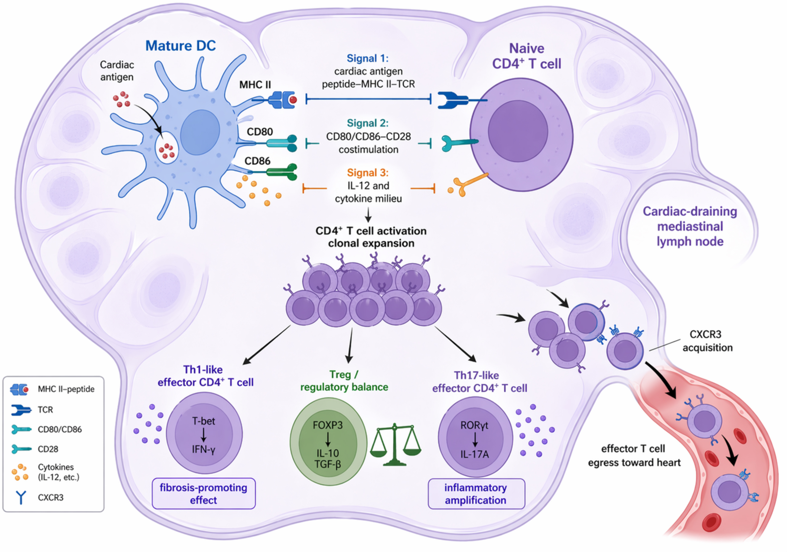
Figure 2. Candidate processes by which different DC subsets mediate CD4+ T cell priming and functional polarization under pressure overload. This figure summarizes the process by which cardiac antigens are processed by migratory DCs, cDC1, cDC2, and monocyte-derived DCs, and subsequently induce CD4+ T cell activation, clonal expansion, and differentiation toward Th1-like, Th17-like, or Treg phenotypes in the draining lymph nodes through MHC II-TCR recognition, CD80/CD86-CD28 costimulation, and cytokine signaling. TCR: T cell receptor; CD28: cluster of differentiation 28; IL-12: interleukin-12; Th1: T helper 1 cell; IFN-γ: interferon-gamma; FOXP3: forkhead box protein 3; IL-10: interleukin-10; TGF-β: transforming growth factor-beta; Th17: T helper 17 cell; RORγt: RAR-related orphan receptor gamma t; IL-17A: interleukin-17A; CXCR3: C-X-C chemokine receptor type 3; DC: dendritic cell; cDC1: type 1 conventional dendritic cell; cDC2: type 2 conventional dendritic cell; MHC II: major histocompatibility complex class II; CD80: cluster of differentiation 80; CD86: cluster of differentiation 86; Treg: regulatory T cell.



