


Sleep disorders in chronic heart failure: a comprehensive review of mechanisms, clinical evaluation, and management
 0
0 Abstract
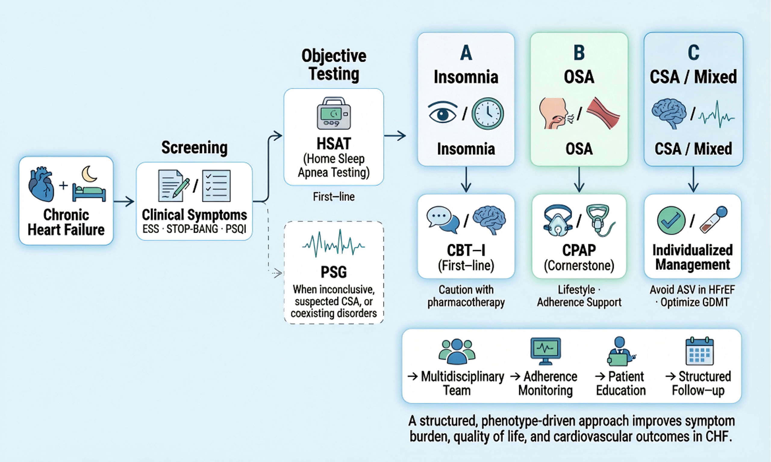
Sleep disorders are highly prevalent in patients with chronic heart failure (CHF) and represent an important yet underrecognized contributor to symptom burden, impaired quality of life, and adverse clinical outcomes. Insomnia and sleep-disordered breathing (SDB), including obstructive sleep apnea (OSA) and central sleep apnea (CSA), frequently coexist and interact with cardiac dysfunction through complex bidirectional mechanisms involving neurohormonal activation, fluid redistribution, and ventilatory instability. This review provides a comprehensive and clinically oriented overview of sleep disorders in CHF. We summarize current insights into pathophysiology and highlight the heterogeneity of sleep phenotypes across heart failure populations. A practical diagnostic framework is proposed, integrating clinical screening tools with objective testing modalities, including Home Sleep Apnea Testing (HSAT) and Polysomnography (PSG), with emphasis on appropriate patient selection. Management strategies are discussed using a phenotype-driven approach. Cognitive behavioral therapy for insomnia remains the first-line treatment for chronic insomnia, while continuous positive airway pressure (CPAP) is the cornerstone therapy for OSA. In contrast, management of CSA requires careful patient selection, with avoidance of adaptive servo-ventilation in patients with reduced ejection fraction. Optimization of Guideline-Directed Medical Therapy (GDMT) plays a complementary role in improving sleep-related outcomes. Finally, we address implementation in clinical practice, emphasizing multidisciplinary care models, patient education, adherence monitoring, and integration into heart failure management pathways. Emerging directions, including digital health technologies and precision medicine approaches, are also discussed. A structured, integrated approach to the recognition and management of sleep disorders in CHF has the potential to improve both patient-centered outcomes and long-term cardiovascular prognosis.
Keywords
INTRODUCTION
Chronic heart failure (CHF) remains a major contributor to global morbidity, mortality, and healthcare utilization[1,2]. It represents the final common pathway of diverse cardiac insults - including myocardial infarction, cardiomyopathies, and chronic pressure or volume overload - resulting in impaired ventricular systolic and/or diastolic function and a substantial, persistent symptom burden[1].
Within this clinical spectrum, sleep disturbances are highly prevalent yet frequently underrecognized comorbidities in CHF[3,4]. Sleep-disordered breathing (SDB) affects approximately 40%-80% of patients with CHF, making it one of the most common comorbidities in this population. Early studies reported SDB in nearly 50% of patients with stable CHF[5]. In addition, approximately one-third of patients with CHF meet diagnostic criteria for insomnia, frequently presenting with overlapping symptoms such as difficulty initiating or maintaining sleep, non-restorative sleep, nocturnal dyspnea, snoring, and excessive daytime fatigue[3-5]. Beyond their impact on sleep quality, these disorders impair self-care, reduce functional capacity, exacerbate psychological distress, and are associated with increased hospitalization rates, poorer quality of life, and adverse long-term cardiovascular outcomes.
Importantly, the relationship between CHF and sleep disorders is bidirectional[6-8]. CHF predisposes patients to sleep disturbances through pulmonary congestion, nocturnal dyspnea, nocturia, rostral fluid redistribution, neurohormonal activation, and ventilatory control instability[9-14]. Conversely, sleep disorders may accelerate CHF progression through recurrent arousals, sleep fragmentation, intermittent hypoxemia, sympathetic activation, blood pressure elevation, endothelial dysfunction, and increased cardiac afterload[15-21]. These pathophysiological mechanisms may contribute to adverse cardiac remodeling, arrhythmic events, recurrent hospitalization, and increased mortality[21-23]. Accordingly, sleep disorders in CHF should not be regarded merely as accompanying symptoms, but as clinically important and potentially modifiable contributors to disease progression.
Restorative sleep is essential for physiological recovery and chronic disease management. The National Sleep Foundation recommends 7-9 h of sleep per night for healthy adults[24]. However, patients with CHF - particularly during hospitalization - often experience fragmented sleep due to a combination of disease-related factors (e.g., dyspnea, nocturia), psychological stress, and environmental disruptions such as noise, light, and nighttime clinical interventions[25,26]. Experimental evidence indicates that even one night of sleep deprivation can increase resting blood pressure from 82 ± 8 to 86 ± 7 mmHg, suggesting an acute pressor effect that may impose additional cardiovascular stress[27]. In patients with CHF, such physiological responses may further burden an already compromised cardiovascular system.
Two major sleep disorder phenotypes predominate in CHF: insomnia and SDB, particularly obstructive sleep apnea (OSA) and central sleep apnea (CSA) with Cheyne-Stokes respiration[4,5]. Importantly, symptom overlap, atypical clinical presentations, and phenotype-specific therapeutic implications complicate recognition and management in routine care. Accordingly, a tailored, phenotype-oriented approach is required rather than reliance on generalized sleep disorder frameworks.
This review provides a clinically focused and integrative synthesis of current evidence and guideline-informed strategies. We aim to support (i) systematic identification of insomnia and SDB within CHF care pathways, (ii) pragmatic diagnostic differentiation using scalable approaches, and (iii) individualized, multidisciplinary management strategies that are feasible in real-world clinical practice.
EPIDEMIOLOGY AND CLINICAL SIGNIFICANCE
Sleep disturbances are highly prevalent in patients with CHF and represent a clinically significant yet frequently underrecognized comorbidity. Epidemiological studies consistently demonstrate a substantially higher burden of sleep disorders in CHF than in the general population, with many patients experiencing impaired sleep quality, daytime dysfunction, and reduced quality of life[3,5].
SDB is particularly common in CHF. Early landmark studies reported SDB in approximately 50% of patients with stable CHF, including both OSA and CSA[5]. Subsequent investigations have suggested an even greater burden, with SDB affecting approximately 40%-80% of patients depending on heart failure severity, diagnostic criteria, and study population[6,28,29]. By comparison, OSA prevalence in the general adult population has been estimated at 9%-38% when defined by an apnea-hypopnea index (AHI) ≥ 5 events/h and 6%-17% when defined by AHI ≥ 15 events/h[30].
Insomnia is also highly prevalent in CHF and frequently coexists with SDB. Reported prevalence estimates for insomnia symptoms range from 23% to 73%, reflecting differences in study populations and assessment methods[31]. Approximately one-third of patients with CHF meet diagnostic criteria for insomnia, often presenting with mixed phenotypes characterized by difficulty initiating sleep, difficulty maintaining sleep, early morning awakening, and non-restorative sleep[32]. In contrast, insomnia disorder affects approximately 10% of the general adult population, while occasional insomnia symptoms occur in approximately 20%[31]. Collectively, these findings indicate that both insomnia and SDB are substantially overrepresented in CHF and should be actively screened for rather than attributed solely to chronic illness.
The clinical significance of sleep disturbances extends beyond symptom burden. Poor sleep quality has been associated with impaired functional status, reduced self-care capacity, worse emotional well-being, and adverse cardiovascular outcomes[3,6]. In a prospective study of patients with Heart Failure (HF), poor sleep quality defined by a Pittsburgh Sleep Quality Index score > 5 was present in 63% of patients and was associated with a 2.5-fold higher risk of reduced cardiac event-free survival, including cardiac death, hospitalization, or emergency department visits for cardiac causes[33]. Similarly, untreated moderate-to-severe OSA in HF was associated with higher mortality than mild or no sleep apnea, with adjusted death rates of 8.7 versus 4.2 deaths per 100 patient-years[22].
These observations are clinically important because the relationship between CHF and sleep disorders is bidirectional. CHF predisposes patients to insomnia and SDB through dyspnea, orthopnea, nocturia, pulmonary congestion, rostral fluid redistribution, neurohormonal activation, and ventilatory instability. Conversely, insomnia and SDB may aggravate CHF through sleep fragmentation, intermittent hypoxemia, recurrent arousals, sympathetic activation, blood pressure surges, endothelial dysfunction, inflammation, and increased cardiac afterload[7,28,34]. These mechanisms may contribute to adverse cardiac remodeling, arrhythmias, recurrent hospitalization, and mortality. Although much of the available evidence remains observational, the consistency of epidemiological associations and mechanistic findings supports the concept that sleep disorders are clinically relevant and potentially modifiable contributors to CHF progression rather than merely accompanying symptoms.
The impact of sleep disturbances also extends beyond patients. CHF-related nocturnal symptoms, including dyspnea, apnea, cough, and nocturia, frequently disrupt caregivers’ sleep, contributing to fragmented sleep patterns and caregiver burden[35]. Therefore, systematic integration of sleep assessment into CHF care pathways may represent an important opportunity to improve patient-centered outcomes, caregiver well-being, and long-term cardiovascular prognosis.
PATHOPHYSIOLOGICAL MECHANISMS
The relationship between CHF and SDB is bidirectional and mediated by complex, overlapping mechanisms that mutually reinforce disease progression. Hemodynamic alterations and neurohumoral activation in CHF predispose to the development of SDB, while SDB - particularly OSA and CSA - imposes additional cardiovascular stress through intermittent hypoxia, sympathetic activation, and hemodynamic fluctuations, thereby accelerating heart failure progression[7,28,34]. Understanding this interaction is essential for targeted and effective management.
From heart failure to sleep-disordered breathing
CSA, frequently presenting as Cheyne-Stokes respiration (CSR), is highly prevalent in CHF and is primarily driven by instability of ventilatory control, characterized by increased loop gain[9,10]. During sleep, respiratory drive depends largely on chemoreceptor responsiveness to PaCO2. In CHF, pulmonary congestion activates pulmonary J receptors, leading to chronic hyperventilation and a reduction in PaCO2 toward or below the apneic threshold[11,15].
Consequently, even minor fluctuations in ventilation - such as post-arousal hyperventilation - can lower PaCO2 below the threshold required to sustain respiratory drive, triggering central apnea. During apnea, PaCO2 gradually rises until it exceeds the threshold, inducing compensatory hyperventilation. This feedback loop generates the characteristic periodic breathing pattern of CSA/CSR[9,36].
Reduced cardiac output further contributes by prolonging circulatory delay between the lungs and central chemoreceptors. This delay introduces a temporal mismatch between blood gas changes and ventilatory responses, thereby amplifying instability in respiratory control and determining the cycle length of CSR[37,38].
Nocturnal rostral fluid shift represents another key mechanism. Redistribution of fluid from the lower extremities to the thorax and neck in the supine position is accentuated in CHF[12]. Fluid accumulation in the neck increases peripharyngeal tissue pressure and upper airway collapsibility, promoting OSA[39,40]. Simultaneously, pulmonary fluid accumulation worsens congestion, stimulates vagal C-fibers, and induces hyperventilation with further reductions in PaCO2, thereby facilitating CSA[11,13]. This fluid shift provides a unifying pathophysiological link between OSA and CSA and may explain the dynamic transition between obstructive and central events observed in some patients during sleep[12,14].
From sleep-disordered breathing to heart failure progression
SDB contributes to CHF progression through interrelated mechanisms including neurohumoral activation, hemodynamic stress, intermittent hypoxia, oxidative stress, and systemic inflammation. These processes act synergistically to promote adverse cardiac remodeling and end-organ dysfunction.
Neurohumoral activation and autonomic dysregulation
Neurohumoral activation is a central mechanism linking SDB to heart failure progression. Reduced cardiac output in CHF activates the sympathetic nervous system and Renin-Angiotensin-Aldosterone System (RAAS), initially compensatory but ultimately maladaptive[41,42]. Sustained sympathetic overactivity promotes tachycardia, increased myocardial oxygen demand, and direct cardiotoxic effects, contributing to hypertrophy, apoptosis, and ventricular dysfunction[41].
SDB further amplifies this imbalance. Recurrent apnea and hypopnea induce intermittent hypoxia and arousal-related sympathetic surges. Hypoxemia activates peripheral chemoreceptors, leading to vasoconstriction and transient blood pressure elevations, while repeated arousals sustain sympathetic activation into daytime wakefulness[16,17].
Intermittent hypoxia also promotes endothelial dysfunction through increased endothelin-1 production, oxidative stress, and reduced nitric oxide bioavailability[18,19], contributing to increased vascular tone and afterload. These processes contribute to vascular dysfunction and increased afterload, further aggravating cardiac stress. Repetitive hypoxia-reoxygenation cycles may impair cerebral perfusion and oxygenation, contributing to neurocognitive dysfunction. In addition, SDB has been associated with an increased risk of cerebrovascular events.
In addition, cerebral hypoperfusion and chronic neurohumoral activation are associated with cognitive impairment in CHF[42]. Sleep disturbances further exacerbate deficits in memory and executive function and are linked to reduced treatment adherence. Depression is also highly prevalent and contributes to worse clinical outcomes, creating a reinforcing cycle of sleep disruption, neurohumoral activation, and functional decline[43].
Molecular and cellular pathways
Beyond organ-level hemodynamic stress, SDB may promote CHF progression through several molecular and cellular mechanisms. Recurrent cycles of hypoxia and reoxygenation resemble repeated ischemia-reperfusion injury and increase the generation of reactive oxygen and nitrogen species through mitochondrial dysfunction, Nicotinamide Adenine Dinucleotide Phosphate (NADPH) oxidase activation, xanthine oxidase activity, and endothelial nitric oxide synthase uncoupling[44]. The resulting oxidative stress reduces nitric oxide bioavailability, impairs endothelial function, and contributes to vascular stiffness and increased cardiac afterload.
Intermittent hypoxia also activates oxygen- and redox-sensitive signaling pathways, including hypoxia-inducible factor-1α (HIF-1α), nuclear factor-κB (NF-κB), and mitogen-activated protein kinase pathways[44-47]. These pathways promote inflammatory activation, endothelial injury, and microvascular dysfunction, while NLRP3 inflammasome activation may further amplify vascular and myocardial inflammation[48].
At the myocardial level, recurrent sympathetic activation, renin-angiotensin-aldosterone system activation, oxidative stress, and inflammation contribute to cardiomyocyte injury, fibroblast activation, extracellular matrix remodeling, and myocardial fibrosis[41,42,49,50]. These processes provide a mechanistic link between nocturnal respiratory instability and adverse cardiac remodeling, ventricular dysfunction, arrhythmogenesis, and recurrent hospitalization.
Hemodynamic stress and cardiac remodeling
OSA imposes substantial hemodynamic stress through repetitive upper airway obstruction and generation of markedly negative intrathoracic pressure (up to -60 to -80 cmH2O)[15,20]. These pressure swings increase left ventricular transmural pressure (afterload) and venous return, promoting right ventricular dilation and interventricular septal shift, which impairs left ventricular filling and reduces cardiac output[51].
Over time, these repetitive stresses contribute to adverse remodeling, including ventricular hypertrophy, chamber dilation, and progressive systolic dysfunction[49,50]. Although CSA does not generate large negative intrathoracic pressures, its cyclical hyperventilation and apnea lead to fluctuations in heart rate and blood pressure through chemoreflex activation and recurrent arousals, resulting in repetitive cardiovascular stress[13,52].
Intermittent hypoxia, oxidative stress, and systemic inflammation
Intermittent hypoxia with subsequent reoxygenation is a hallmark of SDB and resembles ischemia-reperfusion injury[45]. This process drives the generation of reactive oxygen species, leading to oxidative stress and endothelial dysfunction[19]. Oxidative stress activates pro-inflammatory pathways, including NF-κB, resulting in elevated levels of inflammatory mediators such as C-reactive protein, interleukin-6, and tumor necrosis factor-α[46,47]. These mediators exert negative inotropic effects, promote myocardial fibrosis, and contribute to metabolic dysregulation. Together, intermittent hypoxia, oxidative stress, and inflammation form a self-reinforcing cascade that exacerbates myocardial injury and accelerates heart failure progression[19,25].
The interaction between CHF and SDB is therefore self-perpetuating. CHF promotes the development of SDB through hemodynamic and ventilatory control abnormalities, while SDB accelerates cardiac dysfunction via autonomic imbalance, mechanical stress, and inflammatory pathways. This bidirectional relationship contributes to worse clinical outcomes, including increased mortality, rehospitalization, and arrhythmic risk[22,53].
MECHANISMS OF INSOMNIA IN CHF
Insomnia in CHF is multifactorial and should not be viewed simply as a subjective sleep complaint. Disease-related symptoms, including orthopnea, paroxysmal nocturnal dyspnea, cough, nocturia, palpitations, pain, and medication effects, may repeatedly disrupt sleep and increase difficulty initiating or maintaining sleep[3,4]. Over time, recurrent nighttime symptoms may promote sleep-related worry, maladaptive sleep behaviors, and conditioned wakefulness, contributing to persistent insomnia even when cardiac symptoms are partially controlled[54].
At the neurobiological level, insomnia is characterized by cognitive, emotional, autonomic, and neuroendocrine hyperarousal[54,55]. In patients with CHF, illness-related anxiety, fear of nocturnal symptoms, depression, and reduced perceived control may increase sympathetic activity and hypothalamic-pituitary-adrenal axis activation, delaying sleep onset and impairing sleep quality[55,56]. Inflammatory mediators may further contribute to fatigue, poor sleep quality, and impaired daytime function[46,47].
Circadian disruption represents an additional mechanism. Reduced daytime activity, frequent daytime napping, hospitalization-related environmental disturbances, and irregular sleep-wake schedules may weaken circadian entrainment and reduce sleep efficiency[25,26]. Insomnia and SDB frequently coexist in CHF, creating a reinforcing cycle in which respiratory-event-related arousals worsen insomnia, while chronic sleep fragmentation may further increase sympathetic activation and cardiovascular stress.
These mechanistic insights have direct therapeutic implications. Optimization of guideline-directed medical therapy and control of nocturnal symptoms may reduce both ventilatory instability and insomnia-provoking symptoms[1,57,58]. Cognitive behavioral therapy for insomnia (CBT-I) is particularly relevant because it targets conditioned arousal, maladaptive sleep behaviors, cognitive hyperarousal, and circadian dysregulation[54,59-61]. Cardiac rehabilitation and structured daytime activity may further improve sleep quality by stabilizing circadian rhythms, improving functional capacity, and reducing psychological distress[62,63]. Together, these observations support a more individualized and mechanism-based approach to management.
SCREENING AND RISK STRATIFICATION IN CHF
Based on these diagnostic distinctions, screening strategies in CHF should aim not only to identify sleep disturbance but also to distinguish among insomnia, OSA, CSA, and mixed phenotypes, each requiring different diagnostic tests and management approaches. SDB is highly prevalent in patients with CHF, affecting approximately 40%-80% of this population, and is associated with adverse cardiovascular outcomes, impaired cognitive function, and reduced quality of life[28,29,34]. Despite this substantial burden, sleep disorders remain underrecognized in routine cardiology practice, and many affected patients do not undergo formal diagnostic evaluation or receive targeted treatment[28,64]. Effective screening strategies must therefore balance diagnostic sensitivity with clinical feasibility to enable integration into routine heart failure care pathways.
Screening questionnaires and clinical tools
Several validated tools may be used for initial risk stratification in ambulatory patients with CHF. The Epworth Sleepiness Scale (ESS) assesses subjective daytime sleepiness, with scores ≥ 10 suggesting excessive sleepiness and the need for further evaluation[65]. However, its sensitivity may be limited in CHF because fatigue often predominates over overt sleepiness.
The STOP-BANG (Snoring, Tiredness, Observed apnea, high blood Pressure, Body mass index, Age, Neck circumference, and Gender) questionnaire evaluates snoring, tiredness, observed apnea, high blood pressure, body mass index, age, neck circumference, and male sex. A score ≥ 3 indicates increased risk of OSA, although specificity may be reduced in CHF because several risk factors are common in this population[66].
Sleep quality and insomnia can be assessed using the Pittsburgh Sleep Quality Index (PSQI) and Insomnia Severity Index (ISI). A PSQI score > 5 indicates poor sleep quality, whereas an ISI score ≥ 15 suggests at least moderate insomnia[67,68]. Sleep diaries recorded over 1-2 weeks may provide additional information on sleep patterns, nocturnal awakenings, and sleep efficiency, particularly when insomnia or circadian rhythm disturbances are suspected[69,70].
In clinical practice, “high risk” should be determined using a combination of questionnaire results and clinical features rather than a single screening tool. Patients with CHF may be considered high risk if they have STOP-BANG ≥ 3, ESS score ≥ 10, PSQI > 5, ISI ≥ 15, or clinical features such as witnessed apnea, habitual snoring, nocturnal choking, refractory fatigue, recurrent nocturnal dyspnea, atrial fibrillation, resistant hypertension, pulmonary hypertension, obesity, or suspected CSA[1,64-68]. Because symptoms may be atypical or absent in CHF, clinicians should maintain a low threshold for objective sleep testing[64].
Objective screening: respiratory polygraphy and overnight monitoring
Given the limitations of symptom-based screening, objective testing is essential. Portable respiratory polygraphy represents a pragmatic and cost-effective option for both outpatient and inpatient assessment[64,71]. Standard measurements include airflow, respiratory effort, oxygen saturation, heart rate or rhythm, body position, and snoring.
Clinical studies highlight the high burden of SDB in CHF. In a cohort of patients with non-ischemic CHF, Damy et al. reported SDB in 85% of patients (AHI ≥ 5 events/h), with predominantly obstructive events. AHI was independently associated with left ventricular hypertrophy, underscoring the structural impact of untreated SDB[71]. Similarly, increasing SDB severity has been associated with biomarkers of myocardial injury and metabolic dysregulation, supporting the role of objective testing in risk stratification[72].
Home respiratory polygraphy demonstrates reasonable diagnostic agreement with in-laboratory Polysomnography (PSG) and offers advantages in accessibility, cost, and ecological validity[73]. However, the absence of electroencephalographic data limits accurate assessment of sleep stages and total sleep time, potentially leading to underestimation of AHI, particularly in patients with fragmented sleep[73].
Role of polysomnography
PSG remains the gold standard for diagnosing SDB. It provides a comprehensive evaluation of sleep architecture, respiratory events, oxygen desaturation, cardiac rhythm, body position, and limb movements[74]. Compared with respiratory polygraphy, PSG offers several advantages in CHF. Electroencephalographic monitoring enables precise sleep staging and detection of arousal-related events. It also allows reliable differentiation between OSA and CSA, which is critical given their distinct therapeutic implications. In addition, PSG can identify coexisting sleep disorders, such as periodic limb movement disorder or Rapid Eye Movement (REM) sleep behavior disorder, which may contribute to sleep fragmentation[74,75]. Despite these advantages, PSG is resource-intensive, requires specialized infrastructure, and is often associated with long waiting times, which may delay diagnosis and prolong exposure to untreated SDB.
Clinical application
Available evidence supports a stepwise screening approach in CHF. Questionnaires such as ESS, STOP-BANG, and PSQI may assist with initial risk stratification, but their limitations necessitate a low threshold for objective testing[3,64-67]. Portable respiratory polygraphy is a practical first-line modality in many patients, while PSG should be reserved for diagnostically complex cases, suspected CSA, inconclusive results, or suspected coexisting sleep disorders. A pragmatic pathway combining feasibility and diagnostic accuracy is essential for implementation in routine care. For ease of clinical application, the principal screening questionnaires, diagnostic tools, and emerging technologies discussed in this review are summarized in Table 1.
Comparison of screening and diagnostic tools for sleep disorders in chronic heart failure
| Tool | Main purpose | Key protocol or parameters | Advantages | Limitations in CHF | Suggested clinical use |
| ESS[65] | Subjective daytime sleepiness | 8 items; score ≥ 10 suggests excessive sleepiness | Simple and rapid | Low sensitivity because CHF patients often report fatigue rather than sleepiness | Initial screening only |
| STOP-BANG[66] | OSA risk stratification | 8 items; score ≥ 3 indicates high risk | High sensitivity; easy to administer | Low specificity in CHF due to age, hypertension, and male sex | Identifying patients requiring objective testing |
| PSQI[67] | Global sleep quality | 7 domains; score > 5 indicates poor sleep quality | Captures insomnia-like symptoms and sleep quality | Does not distinguish insomnia from SDB | Assessing sleep quality burden |
| ISI[68] | Insomnia severity | 7 items; score ≥ 15 suggests moderate insomnia | Useful for insomnia severity and follow-up | Does not identify respiratory events | Insomnia assessment and monitoring |
| Sleep diary[68] | Longitudinal sleep pattern | 1-2 weeks of bedtime, wake time, awakenings, naps | Useful for insomnia and circadian rhythm assessment | Requires patient adherence | Complementary tool for insomnia |
| HSAT/respiratory polygraphy[76] | Objective SDB assessment | Airflow, respiratory effort, oxygen saturation, body position, heart rate | Accessible and home-based | No EEG; may underestimate AHI; limited CSA differentiation | First-line test for suspected moderate-to-severe OSA |
| PSG[75] | Definitive diagnosis and phenotyping | Sleep stages, respiratory events, oxygenation, rhythm, limb movements | Gold standard; differentiates OSA, CSA, and mixed apnea | Resource-intensive | Suspected CSA, mixed apnea, inconclusive HSAT, complex cases |
| Wearable/smartphone/radar tools[77] | Preliminary or longitudinal monitoring | PPG, accelerometry, sound, motion, radar signals | Convenient and scalable | Limited validation in CHF; not diagnostic | Adjunctive screening or follow-up |
Emerging screening modalities
Several emerging technologies may facilitate accessible and longitudinal screening for sleep disorders in CHF, although each has specific strengths and limitations. Home Sleep Apnea Testing (HSAT) and portable respiratory polygraphy provide lower-cost, home-based assessment and have acceptable diagnostic performance for moderate-to-severe OSA[76]. However, the absence of electroencephalography limits sleep staging, may underestimate AHI, and reduces accuracy in distinguishing OSA from CSA[73,75].
Wearable devices and patch-based systems use physiological signals such as photoplethysmography, accelerometry, heart rate variability, electrocardiography, and respiratory monitoring to estimate sleep and respiratory parameters[77-79]. Their advantages include convenience, patient acceptability, and the ability to support repeated longitudinal assessment. However, performance varies among devices, validation in CHF remains limited, and diagnostic accuracy is generally lower than PSG[75,77,79].
Smartphone-based applications and non-contact radar systems offer inexpensive and unobtrusive monitoring without specialized equipment. However, these technologies remain primarily screening tools because their performance may be affected by environmental factors, motion artifacts, and limited validation data[80-82].
Overall, emerging technologies may support preliminary screening and follow-up, but PSG remains the reference standard when CSA, mixed sleep apnea, inconclusive HSAT findings, or coexisting sleep disorders are suspected[75,76].
Integrated screening approach in heart failure care
Given the high prevalence and clinical impact of SDB in CHF, a structured and pragmatic screening pathway is essential.
Step 1: initial risk assessment and subjective screening
All patients with CHF should undergo systematic evaluation, including targeted history (snoring, witnessed apneas, nocturnal dyspnea, non-restorative sleep, fatigue, atrial fibrillation, resistant hypertension) and standardized questionnaires (ESS, STOP-BANG, PSQI). Absence of typical symptoms should not exclude further assessment[64].
Step 2: objective testing in high-risk patients
Patients identified as high risk or presenting with suggestive clinical features should undergo objective sleep assessment. In stable patients with suspected OSA, HSAT or portable respiratory monitoring represents an appropriate first-line approach. Key parameters obtained from these studies include the apnea-hypopnea index, oxygen desaturation index, and minimum oxygen saturation, which together provide an estimate of disease severity and physiological impact.
PSG should be considered when initial testing is negative or inconclusive despite high clinical suspicion, or when central sleep apnea is suspected, particularly in patients with reduced ejection fraction. It is also recommended in cases where coexisting sleep disorders are likely or when significant comorbidities may affect diagnostic accuracy. In these contexts, PSG remains essential due to its ability to differentiate obstructive from central events and to provide comprehensive assessment of sleep architecture[75].
Step 3: multidisciplinary management
Once SDB is diagnosed, management should involve a multidisciplinary approach integrating cardiology and sleep medicine. Treatment strategies - including continuous positive airway pressure (CPAP) and, where appropriate, Adaptive servo-ventilation (ASV) - should be individualized. Follow-up should include assessment of adherence, symptom improvement, and cardiac function.
DIAGNOSTIC DIFFERENTIATION AND PHENOTYPING
In patients with CHF, differentiation between insomnia and SDB is often challenging because symptoms such as fatigue, non-restorative sleep, nocturnal awakenings, reduced exercise tolerance, nocturnal dyspnea, orthopnea, anxiety, and depression frequently overlap[1,3,4,44]. Furthermore, insomnia, OSA, and CSA may coexist in the same patient. Accurate phenotyping is therefore essential because these disorders differ in their underlying mechanisms, diagnostic evaluation, and management strategies.
Clinical features and symptom patterns
Insomnia is characterized by persistent dissatisfaction with sleep quantity or quality, including difficulty initiating or maintaining sleep or early morning awakening, accompanied by daytime impairment such as fatigue, impaired concentration, and mood disturbances[68]. In CHF, insomnia is often associated with anxiety, depression, and disease-related psychological stress, and patients are typically able to describe their sleep difficulties clearly[3].
In contrast, SDB is characterized by abnormal respiratory events during sleep. OSA typically presents with loud snoring, witnessed apneas, nocturnal choking, and excessive daytime sleepiness[13,15]. However, these classic symptoms are less predictive in CHF populations and may contribute to under-recognition[64]. CSA, particularly in the form of Cheyne-Stokes respiration, is more common in advanced CHF and often presents with periodic breathing, nocturnal dyspnea, and recurrent awakenings rather than prominent snoring or hypersomnolence[52]. A key distinguishing feature is that insomnia reflects intrinsic hyperarousal, whereas sleep fragmentation in SDB is secondary to recurrent respiratory events and arousals. Information from bed partners regarding snoring, apneas, or abnormal breathing patterns may therefore be particularly valuable.
Diagnostic assessment
Standardized questionnaires may support initial evaluation but have limited diagnostic accuracy in CHF. The ESS evaluates daytime sleepiness, while the PSQI and ISI assess sleep quality and insomnia severity. STOP-BANG and Berlin questionnaires may help identify patients at increased risk of OSA, although specificity is reduced in CHF populations[65-68].
PSG remains the gold standard for diagnosing SDB and distinguishing OSA from CSA. It provides comprehensive assessment of sleep architecture, respiratory events, oxygen saturation, and cardiac rhythm, enabling precise characterization of apnea type and severity[75]. In CSA, PSG typically demonstrates absent respiratory effort during apneic episodes and Cheyne-Stokes respiration patterns[13,15]. PSG also facilitates identification of alternative causes of sleep fragmentation, such as periodic limb movement disorder or REM sleep behavior disorder[75].
Portable respiratory polygraphy and HSAT are increasingly used because of their accessibility and feasibility in outpatient settings. In patients with a high pre-test probability of moderate-to-severe OSA, positive HSAT findings may be sufficient to establish the diagnosis and initiate treatment[76]. However, the absence of electroencephalographic monitoring limits sleep stage assessment, may underestimate AHI, and reduces accuracy in differentiating OSA from CSA. Emerging technologies, including wearable devices, smartphone-based applications, and radar-based systems, may support preliminary screening and longitudinal monitoring but currently lack sufficient validation for definitive diagnosis[77,80,81].
A practical diagnostic pathway is summarized in Figure 1. Initial evaluation should include targeted clinical history, ideally incorporating bed partner observations, together with standardized questionnaires. Patients with suspected SDB should undergo objective testing, with HSAT serving as an appropriate first-line modality in selected patients and PSG reserved for inconclusive results, suspected CSA, mixed sleep apnea, or coexisting sleep disorders[65-67].
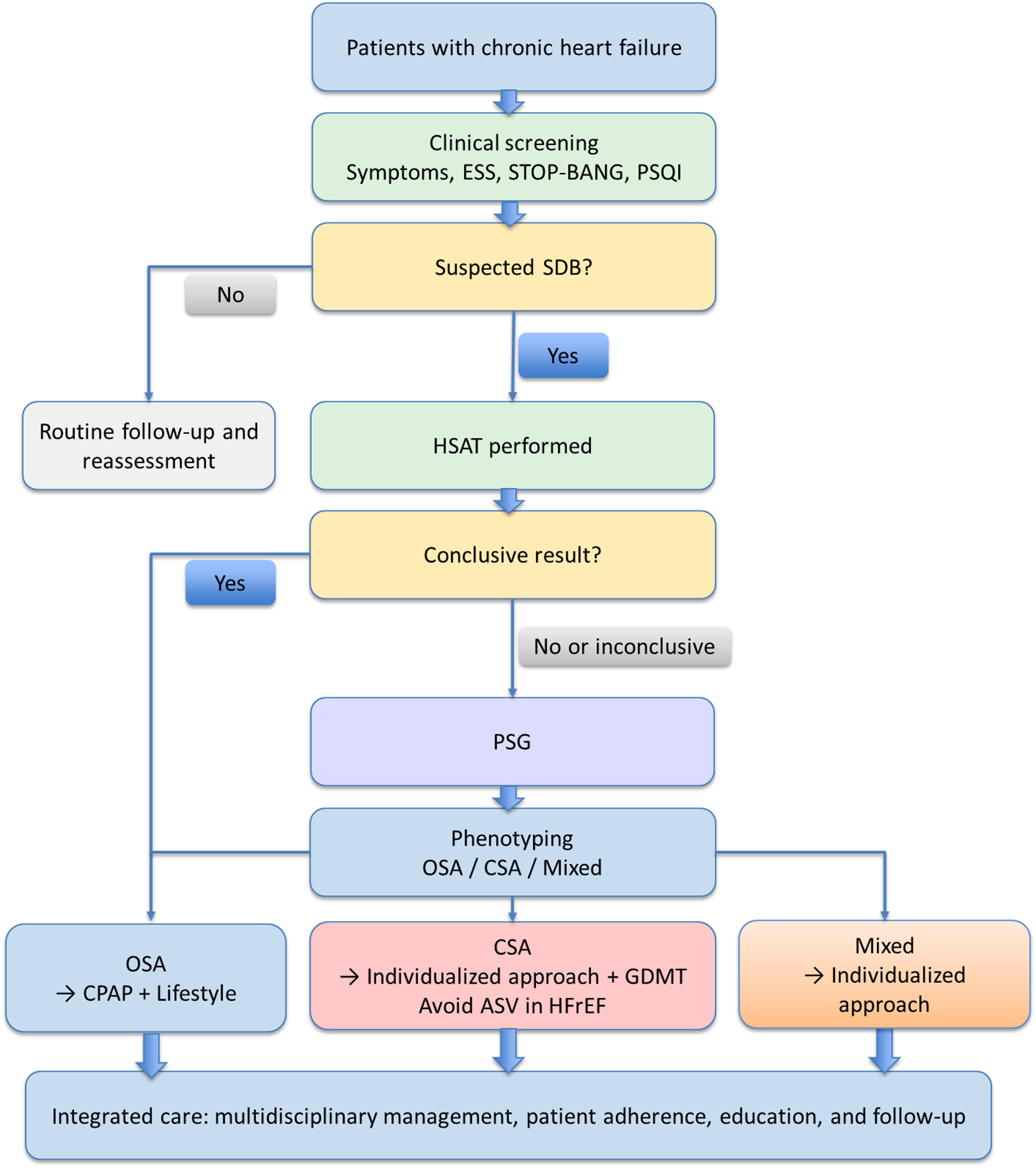
Figure 1. Integrated diagnostic and management pathway for sleep disorders in chronic heart failure. A stepwise approach for screening, diagnostic evaluation, phenotyping, and management of sleep disorders in patients with chronic heart failure, integrating clinical assessment, home sleep apnea testing, polysomnography, phenotype-specific treatment, and longitudinal multidisciplinary care. CHF: Chronic heart failure; ESS: epworth sleepiness scale; STOP-BANG: snoring, tiredness, observed apnea, high blood pressure, body mass index, age, neck circumference, and gender; PSQI: pittsburgh sleep quality index; SDB: sleep-disordered breathing; HSAT: home sleep apnea testing; PSG: polysomnography; OSA: obstructive sleep apnea; CSA: central sleep apnea; CPAP: continuous positive airway pressure; GDMT: guideline-directed medical therapy; ASV: adaptive servo-ventilation; HFrEF: heart failure with reduced ejection fraction.
Special considerations in heart failure
Mixed sleep apnea is common in CHF, with patients exhibiting both obstructive and central respiratory events or transitioning between patterns over time. This likely reflects dynamic changes in fluid distribution, sleep stage, and chemoreceptor sensitivity and often requires full-night PSG for accurate characterization[12].
Coexistence of insomnia and SDB is also frequent. Therefore, diagnosis should integrate subjective symptoms with objective findings. Patients with insomnia complaints but elevated AHI should be considered to have SDB with comorbid insomnia and may require combined management strategies[3].
Assessment of cognition and mood may further aid differentiation. SDB is more strongly associated with impaired attention and executive function, whereas insomnia is often linked to subjective memory complaints and emotional dysregulation[3].
A structured, stepwise diagnostic approach is recommended to facilitate clinical implementation and is illustrated in Figure 1. Initial evaluation should include targeted clinical history - ideally incorporating bed partner observations - together with standardized questionnaires such as the ESS, PSQI, and STOP-BANG. An ESS score ≥ 10 or STOP-BANG score ≥ 3 increases the likelihood of SDB, while a PSQI score > 5 indicates clinically significant sleep disturbance warranting further assessment.
Patients with suspected SDB should undergo objective testing. In those with a high pre-test probability of moderate-to-severe OSA, HSAT represents an appropriate first-line modality. However, referral for PSG is recommended when HSAT results are negative or inconclusive despite persistent clinical suspicion, when CSA is suspected - particularly in patients with reduced ejection fraction or atrial fibrillation - or when coexisting sleep disorders are likely.
PSG should be used to confirm the diagnosis and define the SDB phenotype. Key parameters include AHI, the type and distribution of respiratory events, their relationship to sleep stages, patterns of oxygen desaturation, and the presence of Cheyne-Stokes respiration. In addition, PSG enables exclusion of alternative causes of sleep disruption, which is essential for guiding appropriate management.
MANAGEMENT STRATEGIES
Management of sleep disorders in patients with CHF requires accurate phenotyping and a multidisciplinary approach. Treatment selection should be guided by the dominant underlying mechanism. Insomnia is primarily driven by hyperarousal, maladaptive sleep behaviors, circadian disruption, and CHF-related nocturnal symptoms, whereas OSA results from recurrent upper airway obstruction and CSA reflects ventilatory instability associated with cardiac dysfunction. Accordingly, management should be phenotype-specific and mechanism-based.
Management of insomnia
Non-pharmacological therapy: cognitive behavioral therapy
CBT-I is recommended as first-line therapy for chronic insomnia and has demonstrated both safety and efficacy in patients with CHF[59]. CBT-I is particularly relevant in CHF because it targets conditioned arousal, sleep-related worry, maladaptive sleep behaviors, excessive time in bed, and circadian dysregulation[59-61]. In parallel, optimization of nocturnal CHF symptoms - including orthopnea, paroxysmal nocturnal dyspnea, cough, palpitations, and nocturia - should be incorporated into treatment because these symptoms frequently trigger sleep disruption and perpetuate insomnia[1,3,44]. Core components include stimulus control, sleep restriction, cognitive restructuring, relaxation techniques, and sleep hygiene education.
The HeartSleep randomized controlled trial demonstrated that an 8-week CBT-I intervention significantly improved insomnia severity, sleep quality, sleep efficiency, fatigue, daytime sleepiness, and psychomotor vigilance, with benefits maintained at 12-month follow-up[60]. Subsequent analyses also reported improvements in cognitive function and functional capacity[61]. These findings support CBT-I as a central component of insomnia management in CHF.
Pharmacological therapy
Pharmacological treatment may be considered when CBT-I is unavailable or insufficient but requires careful risk-benefit assessment in CHF. Benzodiazepines and non-benzodiazepine hypnotics should be used cautiously because of potential respiratory depression, residual sedation, and adverse clinical outcomes, including rehospitalization in older adults[83,84]. Orexin receptor antagonists (e.g., suvorexant) may represent a safer alternative by promoting sleep through wakefulness pathways rather than respiratory suppression, although evidence in CHF remains limited[85]. Sedating antidepressants (e.g., trazodone and mirtazapine) may be considered in patients with comorbid depression but require caution regarding hemodynamic effects and drug interactions[86]. Overall, hypnotics should be prescribed at the lowest effective dose for the shortest possible duration.
Management of obstructive sleep apnea
CPAP remains the cornerstone therapy for OSA. By preventing upper airway collapse, CPAP reduces respiratory events, intermittent hypoxemia, arousal-related sympathetic activation, and negative intrathoracic pressure swings[13,15,87,88]. These effects may improve oxygenation, reduce cardiac afterload, and attenuate neurohumoral activation. Meta-analyses have demonstrated improvements in AHI, oxygenation, and Left Ventricular Ejection Fraction (LVEF) in selected patients with CHF and OSA[87]. Clinical studies have also reported improvements in symptoms, functional status, and daytime sleepiness[88]. Adherence remains a major determinant of treatment effectiveness, highlighting the importance of patient education, follow-up, and individualized device adjustment. Alternative modalities such as APAP and Bilevel Positive Airway Pressure (BiPAP) may be considered in selected patients with CPAP intolerance[87].
Management of central sleep apnea
CSA in CHF is often a manifestation of underlying cardiac dysfunction and is driven by ventilatory instability, hypocapnia, pulmonary congestion, and prolonged circulatory delay[52]. Accordingly, management should target these underlying mechanisms. Because CSA frequently reflects the severity of heart failure, optimization of heart failure therapy, decongestion, and improvement of cardiac output remain the central therapeutic priorities[1,52,57].
Although ASV effectively suppresses central respiratory events, the SERVE-HF (Treatment of Sleep-Disordered Breathing with Predominant Central Sleep Apnoea by Adaptive Servo Ventilation in Patients with Heart Failure) trial demonstrated increased all-cause and cardiovascular mortality in patients with Heart Failure with reduced Ejection Fraction (HFrEF) (LVEF ≤ 45%), leading to a contraindication in this population[89,90]. In patients with Heart Failure with preserved Ejection Fraction (HFpEF), ASV may improve respiratory parameters such as AHI and oxygenation, although evidence for improvement in clinical outcomes remains limited[91]. Supplemental oxygen and selected respiratory stimulants may improve ventilatory stability in selected patients, but their role remains uncertain because outcome data are limited[52]. Close collaboration between cardiology and sleep medicine specialists is therefore essential.
Optimization of heart failure therapy and its impact on SDB
Guideline-directed medical therapy (GDMT) remains the foundation of CHF management and may indirectly improve SDB through optimization of cardiac function and hemodynamics[57]. Improved cardiac output, reduced pulmonary congestion, attenuation of neurohormonal activation, and reduced rostral fluid redistribution may lessen both CSA-promoting ventilatory instability and OSA-promoting upper airway edema[1,12,13,39,57]. Therapies targeting the renin-angiotensin system, including Angiotensin Converting Enzyme (ACE) inhibitors, Angiotensin Receptor Blockers (ARBs), and angiotensin receptor-neprilysin inhibitors, may improve cardiac loading conditions and reduce pulmonary congestion[1]. In addition, sodium-glucose cotransporter-2 inhibitors (SGLT2i) have been associated with improvements in cardiac performance, metabolic status, and body weight, which may contribute to reductions in OSA severity[58]. Because polypharmacy is common in CHF, careful medication review, individualized titration, and regular reassessment are important to maximize both cardiovascular and sleep-related benefits.
In summary, management of sleep disorders in CHF requires a phenotype-driven and multidisciplinary approach. CBT-I is recommended as first-line therapy for insomnia, CPAP remains the cornerstone treatment for OSA, and CSA management should focus on optimization of heart failure therapy with careful patient selection for advanced interventions. Integration of sleep and heart failure management may improve both quality of life and long-term outcomes. A summary of phenotype-specific treatment strategies, mechanistic targets, and key clinical considerations is provided in Table 2.
Phenotype-specific and mechanism-based treatment strategies for sleep disorders in chronic heart failure
| Phenotype | Dominant mechanisms | Main treatment strategy | Mechanistic target | Clinical considerations |
| Insomnia[59] | Hyperarousal, conditioned wakefulness, circadian disruption, nocturnal CHF symptoms | CBT-I | Reduces maladaptive sleep behaviors, sleep-related worry, and conditioned arousal | First-line treatment; can be integrated with cardiac rehabilitation |
| Insomnia with nocturnal CHF symptoms[1] | Orthopnea, nocturia, dyspnea, cough, palpitations | Optimization of CHF symptoms and medication timing | Reduces nighttime awakenings and symptom-triggered arousal | Consider diuretic timing and congestion control |
| OSA[87,88] | Upper airway collapse, intermittent hypoxemia, negative intrathoracic pressure swings | CPAP | Maintains airway patency, reduces hypoxemia and afterload | Monitor adherence, mask leak, residual AHI |
| CSA[1,57] | Ventilatory instability, hypocapnia, prolonged circulatory delay, pulmonary congestion | GDMT optimization and decongestion | Improves cardiac function and reduces ventilatory instability | Individualized management with sleep specialist input |
| CSA in HFrEF[89] | Central events with reduced ejection fraction | Avoid ASV when contraindicated | Safety-focused approach | Avoid ASV in HFrEF patients where contraindicated |
| Mixed apnea[75] | Dynamic obstructive and central events | PSG-guided individualized treatment | Phenotype-specific treatment selection | Requires full-night phenotyping |
| Poor PAP adherence[94] | Mask discomfort, leak, residual events, low perceived benefit | Education, telemonitoring, device optimization | Improves sustained treatment exposure | Early follow-up is essential |
IMPLEMENTATION IN CLINICAL PRACTICE
Effective management of sleep disorders in CHF requires integration into routine care pathways with structured long-term follow-up. Multidisciplinary collaboration, adherence monitoring, and patient education are essential to shift care toward proactive management[1,92,93].
Multidisciplinary care model
Cardiologists should screen for sleep disorders, recognize HF-specific patterns (e.g., CSA), optimize GDMT, and guide treatment decisions. Sleep specialists confirm diagnosis and manage Positive Airway Pressure (PAP) therapy, while respiratory therapists support device initiation and adherence. Nursing staff contribute to screening, non-pharmacological interventions, and patient education. Clinical pharmacists assist with medication optimization and safety[1,34,62,94-96].
Patient education and self-management
Patient and caregiver engagement is essential for successful implementation of sleep disorder management in CHF. Sleep disturbances frequently affect both patients and their caregivers, and a dyadic care approach may improve adherence and clinical outcomes[35].
Effective education should encompass disease awareness, engagement in screening processes, realistic treatment expectations, and the development of self-monitoring skills, while actively involving caregivers in the management process. Improved understanding has been associated with greater diagnostic uptake and enhanced long-term adherence to therapy[2].
Long-term management of PAP adherence
The clinical effectiveness of PAP therapy is highly dependent on sustained adherence. Suboptimal adherence is associated with limited improvement in cardiac function and clinical outcomes[97-99]. Contemporary PAP devices allow remote monitoring of usage patterns, mask leak, and residual AHI, enabling early identification of adherence barriers and timely intervention[94].
Adherence is commonly defined as use for at least 4 hours per night on 70% or more of nights[99]. Strategies to improve adherence should include early and structured follow-up, proactive management of side effects, telemonitoring, patient education, individualized device optimization, and psychological support. Implementation of such comprehensive approaches has been shown to improve both adherence and clinical outcomes[94,99].
Integration with cardiac rehabilitation
Cardiac rehabilitation provides an effective platform for integrating sleep disorder management into routine CHF care[63]. Regular physical activity has been associated with improved sleep quality in CHF, with higher activity levels correlating with better sleep outcomes[99]. Moreover, improved daytime activity may reinforce circadian regulation, creating a beneficial feedback loop between sleep quality and functional capacity[63].
Psychological support components of rehabilitation programs offer an opportunity to incorporate CBT-I, which has been shown to improve insomnia symptoms, fatigue, mood, and functional status in CHF populations[62]. In addition, dietary patterns such as the Mediterranean diet have been associated with improved sleep quality and reduced insomnia symptoms, suggesting a broader role for lifestyle interventions in sleep management[100].
Longitudinal follow-up pathway
Structured, longitudinal follow-up is critical to ensure sustained treatment effects. Drawing on established frameworks for chronic disease management and PAP adherence monitoring[73], a phased approach may be conceptualized, including an early baseline phase (0-1 month) focused on treatment initiation and education, an adaptation phase (1-3 months) addressing adherence and early response, a consolidation phase (3-12 months) reinforcing behavioral and therapeutic stability, and a long-term maintenance phase beyond 12 months. Such structured follow-up models have been associated with improved adherence and better clinical outcomes in patients with SDB[94,99].
Quality indicators and outcome assessment
To ensure effective implementation, standardized quality indicators should be incorporated into clinical practice. Consistent with established quality improvement frameworks in cardiovascular care1, these include process measures (e.g., screening rates, diagnostic completion), intermediate outcomes (e.g., adherence, symptom improvement), and clinical outcomes (e.g., hospitalization, functional status, mortality). Integration of these metrics into existing heart failure quality frameworks may enhance care delivery and support continuous quality improvement[1,93].
FUTURE DIRECTIONS
Evidence supporting sleep interventions in HF remains limited. Larger multicenter trials are needed to evaluate CBT-I across HF phenotypes and to determine whether CPAP improves hard clinical endpoints beyond surrogate markers[60,88]. The SERVE-HF findings highlight the need for safer and phenotype-specific approaches to CSA management[89]. Future research should focus on pathophysiology-driven phenotyping and personalized treatment strategies, including targeted approaches for fluid redistribution-related OSA and chemoreflex-driven CSA[101,102]. Integration of multi-omics and digital health technologies may further enable precision medicine, although validation in HF populations remains necessary[103]. Implementation science and health economic evaluation will be essential to support scalable, cost-effective integration into routine care[92,104].
CONCLUSIONS
Sleep disturbances, including insomnia and SDB, are highly prevalent in patients with chronic heart failure yet remain underrecognized and insufficiently integrated into routine clinical care. Beyond being comorbid conditions, accumulating evidence suggests that sleep disorders may act as modifiable contributors to symptom burden, functional impairment, and adverse outcomes.
This review highlights the need for a structured, multidisciplinary approach incorporating systematic screening, accurate phenotyping, and individualized, mechanism-based treatment strategies. Importantly, integrating sleep management into established heart failure care pathways represents a clinically actionable opportunity to improve patient-centered outcomes.
Bridging current gaps will require not only high-quality randomized evidence but also effective implementation strategies. Addressing sleep disturbances may represent a critical and underutilized avenue for optimizing long-term management in heart failure.
DECLARATIONS
Acknowledgments
Special thanks to Ms. Juan Zhang for her assistance during the manuscript revision process.
Authors’ contributions
Made substantial contributions to conception and design of the study: Xiao Y, Li F, Long W
Performed data acquisition and conducted literature search with appraisal of study quality: Wang Y, Hu L, Tan X, Qiu J
Performed data analysis and interpretation, and wrote the initial draft: Xiao Y
Revised the manuscript critically for important intellectual content: Zhu Y, Li F, Long W
Availability of data and materials
Not applicable.
AI and AI-assisted tools statement
During the preparation of this manuscript, the AI tool ChatGPT (OpenAI; model: GPT-5.5; released 2026-03-26) was used solely for language editing and improving readability. The tool did not influence the study design, data collection, analysis, interpretation, or the scientific content of the work. All authors take full responsibility for the accuracy, integrity, and final content of the manuscript.
Financial support and sponsorship
None.
Conflicts of interest
All authors declared that there are no conflicts of interest.
Ethical approval and consent to participate
Not applicable.
Consent for publication
Not applicable.
Copyright
© The Author(s) 2026.
REFERENCES
1. McDonagh TA, Metra M, Adamo M, et al. 2021 ESC Guidelines for the diagnosis and treatment of acute and chronic heart failure: developed by the Task Force for the diagnosis and treatment of acute and chronic heart failure of the European Society of Cardiology (ESC) With the special contribution of the Heart Failure Association (HFA) of the ESC. Eur Heart J. 2021;42:3599-726.
2. Riegel B, Lee CS, Dickson VV. Self care in patients with chronic heart failure. Nat Rev Cardiol. 2011;8:644-54.
3. Redeker NS, Stein S. Characteristics of sleep in patients with stable heart failure versus a comparison group. Heart Lung. 2006;35:252-61.
4. Atay ME, Çiftçi B. Hospital-acquired insomnia in patients with heart failure: a mixed methods study. BMC Nurs. 2026;25:151.
5. Javaheri S, Parker TJ, Liming JD, et al. Sleep apnea in 81 ambulatory male patients with stable heart failure: types and their prevalences, consequences, and presentations. Circulation. 1998;97:2154-9.
6. Arzt M, Woehrle H, Oldenburg O, et al. Prevalence and predictors of sleep-disordered breathing in patients with stable chronic heart failure: the SchlaHF registry. JACC Heart Fail. 2016;4:116-25.
7. Javaheri S, Germany R. Sleep and breathing disorders in heart failure. Handb Clin Neurol. 2022;189:295-307.
8. Lin Y, Chen Y, Tu W, et al. Comparative effectiveness of therapies for sleep-disordered breathing in heart failure patients: a comprehensive systematic review and network meta-analysis. Respir Med. 2025;236:107907.
11. Solin P, Bergin P, Richardson M, Kaye DM, Walters EH, Naughton MT. Influence of pulmonary capillary wedge pressure on central apnea in heart failure. Circulation. 1999;99:1574-9.
12. Yumino D, Bradley TD. Central sleep apnea and Cheyne-Stokes respiration. Proc Am Thorac Soc. 2008;5:226-36.
13. Bradley TD, Floras JS. Sleep apnea and heart failure: part II: central sleep apnea. Circulation. 2003;107:1822-6.
14. Yumino D, Redolfi S, Ruttanaumpawan P, et al. Nocturnal rostral fluid shift: a unifying concept for the pathogenesis of obstructive and central sleep apnea in men with heart failure. Circulation. 2010;121:1598-605.
15. Bradley TD, Floras JS. Sleep apnea and heart failure: part I: obstructive sleep apnea. Circulation. 2003;107:1671-8.
16. Somers VK, Dyken ME, Clary MP, Abboud FM. Sympathetic neural mechanisms in obstructive sleep apnea. J Clin Invest. 1995;96:1897-904.
17. Narkiewicz K, Somers VK. Sympathetic nerve activity in obstructive sleep apnoea. Acta Physiol Scand. 2003;177:385-90.
18. Schulz R, Schmidt D, Blum A, et al. Decreased plasma levels of nitric oxide derivatives in obstructive sleep apnoea: response to CPAP therapy. Thorax. 2000;55:1046-51.
19. Lavie L. Oxidative stress in obstructive sleep apnea and intermittent hypoxia-revisited-the bad ugly and good: implications to the heart and brain. Sleep Med Rev. 2015;20:27-45.
20. Somers VK, White DP, Amin R, et al. Sleep apnea and cardiovascular disease: an American Heart Association/American College of Cardiology Foundation Scientific Statement From the American Heart Association Council for High Blood Pressure Research Professional Education Committee, Council on Clinical Cardiology, Stroke Council, and Council on Cardiovascular Nursing In Collaboration With the National Heart, Lung, and Blood Institute National Center on Sleep Disorders Research (National Institutes of Health). Circulation. 2008;118:1080-111.
21. Javaheri S, Barbe F, Campos-Rodriguez F, et al. Sleep apnea: types, mechanisms, and clinical cardiovascular consequences. J Am Coll Cardiol. 2017;69:841-58.
22. Wang H, Parker JD, Newton GE, et al. Influence of obstructive sleep apnea on mortality in patients with heart failure. J Am Coll Cardiol. 2007;49:1625-31.
23. Oldenburg O, Wellmann B, Buchholz A, et al. Nocturnal hypoxaemia is associated with increased mortality in stable heart failure patients. Eur Heart J. 2016;37:1695-703.
24. Hirshkowitz M, Whiton K, Albert SM, et al. National sleep foundation’s sleep time duration recommendations: methodology and results summary. Sleep Health. 2015;1:40-3.
25. Lévy P, Naughton MT, Tamisier R, Cowie MR, Bradley TD. Sleep apnoea and heart failure. Eur Respir J. 2022;59:2101640.
26. Holleck ME, Tikkanen K, Holleck JL, et al. Reducing nighttime interruptions and improving sleep for hospitalized patients by restructuring nighttime clinical workflow. J Gen Intern Med. 2023;38:2091-7.
27. Kato M, Phillips BG, Sigurdsson G, Narkiewicz K, Pesek CA, Somers VK. Effects of sleep deprivation on neural circulatory control. Hypertension. 2000;35:1173-5.
28. Kazimierczak A, Krzesiński P, Krzyżanowski K, Gielerak G. Sleep-disordered breathing in patients with heart failure: new trends in therapy. Biomed Res Int. 2013;2013:459613.
29. Khayat R, Jarjoura D, Porter K, et al. Sleep disordered breathing and post-discharge mortality in patients with acute heart failure. Eur Heart J. 2015;36:1463-9.
30. Senaratna CV, Perret JL, Lodge CJ, et al. Prevalence of obstructive sleep apnea in the general population: a systematic review. Sleep Med Rev. 2017;34:70-81.
31. Morin CM, Jarrin DC. Epidemiology of insomnia: prevalence, course, risk factors, and public health burden. Sleep Med Clin. 2022;17:173-91.
32. Klompstra L, Jaarsma T, Stromberg A. Factors associated with sleeping difficulties in patients with heart failure. Eur Heart J. 2024;45:ehae666.3409.
33. Lee KS, Lennie TA, Heo S, Song EK, Moser DK. Prognostic importance of sleep quality in patients with heart failure. Am J Crit Care. 2016;25:516-25.
34. Oldenburg O, Lamp B, Faber L, Teschler H, Horstkotte D, Töpfer V. Sleep-disordered breathing in patients with symptomatic heart failure: a contemporary study of prevalence in and characteristics of 700 patients. Eur J Heart Fail. 2007;9:251-7.
35. Al-Rawashdeh SY, Lennie TA, Chung ML. The association of sleep disturbances with quality of life in heart failure patient-caregiver dyads. West J Nurs Res. 2017;39:492-506.
36. Javaheri S. A mechanism of central sleep apnea in patients with heart failure. N Engl J Med. 1999;341:949-54.
37. Francis DP, Willson K, Davies LC, Coats AJ, Piepoli M. Quantitative general theory for periodic breathing in chronic heart failure and its clinical implications. Circulation. 2000;102:2214-21.
38. Lorenzi-Filho G, Rankin F, Bies I, Douglas Bradley T. Effects of inhaled carbon dioxide and oxygen on Cheyne-Stokes respiration in patients with heart failure. Am J Respir Crit Care Med. 1999;159:1490-8.
39. White LH, Bradley TD. Role of nocturnal rostral fluid shift in the pathogenesis of obstructive and central sleep apnoea. J Physiol. 2013;591:1179-93.
40. Redolfi S, Yumino D, Ruttanaumpawan P, et al. Relationship between overnight rostral fluid shift and obstructive sleep apnea in nonobese men. Am J Respir Crit Care Med. 2009;179:241-6.
41. Mann DL, Bristow MR. Mechanisms and models in heart failure: the biomechanical model and beyond. Circulation. 2005;111:2837-49.
42. Triposkiadis F, Karayannis G, Giamouzis G, Skoularigis J, Louridas G, Butler J. The sympathetic nervous system in heart failure physiology, pathophysiology, and clinical implications. J Am Coll Cardiol. 2009;54:1747-62.
43. Celano CM, Villegas AC, Albanese AM, Gaggin HK, Huffman JC. Depression and anxiety in heart failure: a review. Harv Rev Psychiatry. 2018;26:175-84.
44. Nanduri J, Yuan G, Kumar GK, Semenza GL, Prabhakar NR. Transcriptional responses to intermittent hypoxia. Respir Physiol Neurobiol. 2008;164:277-81.
45. Ryan S, Taylor CT, McNicholas WT. Selective activation of inflammatory pathways by intermittent hypoxia in obstructive sleep apnea syndrome. Circulation. 2005;112:2660-7.
46. Ghiasi M. Investigating the NF-κB signaling pathway in heart failure: exploring potential therapeutic approaches. Heliyon. 2024;10:e40812.
47. Fan H, Niu H, Zhao B, Gao X. Inflammation in obstructive sleep apnea: a global bibliometric perspective. Nat Sci Sleep. 2025;17:2275-95.
48. Wei M, Teske JA, Mashaqi S, Combs D. Obstructive sleep apnea, the NLRP3 inflammasome and the potential effects of incretin therapies. Front Sleep. 2024;3:1524593.
49. Anker SD, von Haehling S, Germany R. Sleep-disordered breathing and cardiovascular disease. Indian Heart J. 2016;68 Suppl 1:S69-76.
50. Kasai T, Bradley TD. Obstructive sleep apnea and heart failure: pathophysiologic and therapeutic implications. J Am Coll Cardiol. 2011;57:119-27.
51. Naughton MT. The link between obstructive sleep apnea and heart failure: underappreciated opportunity for treatment. Curr Heart Fail Rep. 2006;3:183-8.
52. Javaheri S. Central sleep apnea in congestive heart failure: prevalence, mechanisms, impact, and therapeutic options. Semin Respir Crit Care Med. 2005;26:44-55.
53. Nakamura S, Asai K, Kubota Y, et al. Impact of sleep-disordered breathing and efficacy of positive airway pressure on mortality in patients with chronic heart failure and sleep-disordered breathing: a meta-analysis. Clin Res Cardiol. 2015;104:208-16.
54. Riemann D, Spiegelhalder K, Feige B, et al. The hyperarousal model of insomnia: a review of the concept and its evidence. Sleep Med Rev. 2010;14:19-31.
55. Kalmbach DA, Cuamatzi-Castelan AS, Tonnu CV, et al. Hyperarousal and sleep reactivity in insomnia: current insights. Nat Sci Sleep. 2018;10:193-201.
56. Vgontzas AN, Bixler EO, Lin HM, et al. Chronic insomnia is associated with nyctohemeral activation of the hypothalamic-pituitary-adrenal axis: clinical implications. J Clin Endocrinol Metab. 2001;86:3787-94.
57. Tamura A, Kawano Y, Naono S, Kotoku M, Kadota J. Relationship between beta-blocker treatment and the severity of central sleep apnea in chronic heart failure. Chest. 2007;131:130-5.
58. McMurray JJV, Solomon SD, Inzucchi SE, et al. Dapagliflozin in patients with heart failure and reduced ejection fraction. N Engl J Med. 2019;381:1995-2008.
59. Qaseem A, Kansagara D, Forciea MA, Cooke M, Denberg TD. Management of chronic insomnia disorder in adults: a clinical practice guideline from the American College of Physicians. Ann Intern Med. 2016;165:125-33.
60. Redeker NS, Jeon S, Andrews L, Cline J, Mohsenin V, Jacoby D. Effects of cognitive behavioral therapy for insomnia on sleep-related cognitions among patients with stable heart failure. Behav Sleep Med. 2019;17:342-54.
61. Redeker NS, Conley S, Anderson G, et al. Effects of cognitive behavioral therapy for insomnia on sleep, symptoms, stress, and autonomic function among patients with heart failure. Behav Sleep Med. 2020;18:190-202.
62. Redeker NS, Yaggi HK, Jacoby D, et al. Cognitive behavioral therapy for insomnia has sustained effects on insomnia, fatigue, and function among people with chronic heart failure and insomnia: the HeartSleep Study. Sleep. 2022:45.
63. Ash G, Jeon S, Conley S, et al. Day-to-day relationships between physical activity and sleep characteristics among people with heart failure and insomnia. Behav Sleep Med. 2021;19:602-14.
64. Paulino A, Damy T, Margarit L, et al. Prevalence of sleep-disordered breathing in a 316-patient French cohort of stable congestive heart failure. Arch Cardiovasc Dis. 2009;102:169-75.
65. Johns MW. Reliability and factor analysis of the Epworth Sleepiness Scale. Sleep. 1992;15:376-81.
66. Chung F, Abdullah HR, Liao P. STOP-bang questionnaire: a practical approach to screen for obstructive sleep apnea. Chest. 2016;149:631-8.
67. Buysse DJ, Reynolds CF 3rd, Monk TH, Berman SR, Kupfer DJ. The Pittsburgh Sleep Quality Index: a new instrument for psychiatric practice and research. Psychiatry Res. 1989;28:193-213.
68. Bastien CH, Vallières A, Morin CM. Validation of the insomnia severity index as an outcome measure for insomnia research. Sleep Med. 2001;2:297-307.
69. Carney CE, Buysse DJ, Ancoli-Israel S, et al. The consensus sleep diary: standardizing prospective sleep self-monitoring. Sleep. 2012;35:287-302.
70. Hartmann JA, Carney CE, Lachowski A, Edinger JD. Exploring the construct of subjective sleep quality in patients with insomnia. J Clin Psychiatry. 2015;76:e768-73.
71. Damy T, Paulino A, Margarit L, et al. Left ventricle remodelling is associated with sleep-disordered breathing in non-ischaemic cardiopathy with systolic dysfunction. J Sleep Res. 2011;20:101-9.
72. Miyata M, Yoshihisa A, Yamauchi H, et al. Impact of sleep-disordered breathing on myocardial damage and metabolism in patients with chronic heart failure. Heart Vessels. 2015;30:318-24.
73. Quintana-Gallego E, Villa-Gil M, Carmona-Bernal C, et al. Home respiratory polygraphy for diagnosis of sleep-disordered breathing in heart failure. Eur Respir J. 2004;24:443-8.
74. Markun LC, Sampat A. Clinician-Focused overview and developments in polysomnography. Curr Sleep Med Rep. 2020;6:309-21.
75. Sateia MJ. International classification of sleep disorders-third edition: highlights and modifications. Chest. 2014;146:1387-94.
76. Kapur VK, Auckley DH, Chowdhuri S, et al. Clinical practice guideline for diagnostic testing for adult obstructive sleep apnea: an American Academy of sleep medicine clinical practice guideline. J Clin Sleep Med. 2017;13:479-504.
77. Zambotti M, Rosas L, Colrain IM, Baker FC. The sleep of the ring: comparison of the OURA sleep tracker against polysomnography. Behav Sleep Med. 2019;17:124-36.
78. Abdelrazik A, Eldesouky M, Antoun I, et al. Wearable devices for arrhythmia detection: advancements and clinical implications. Sensors. 2025;25:2848.
79. Zambotti M, Cellini N, Goldstone A, Colrain IM, Baker FC. Wearable sleep technology in clinical and research settings. Med Sci Sports Exerc. 2019;51:1538-57.
80. Nakano H, Hirayama K, Sadamitsu Y, et al. Monitoring sound to quantify snoring and sleep apnea severity using a smartphone: proof of concept. J Clin Sleep Med. 2014;10:73-8.
81. Massagram W, Lubecke V, Host-madsen A, Boric-lubecke O. Assessment of heart rate variability and respiratory sinus arrhythmia via doppler radar. IEEE Trans Microwave Theory Techn. 2009;57:2542-9.
82. Wang W, Li C, Chen Z, et al. Detection of sleep apnea-hypopnea events using millimeter-wave radar and pulse oximeter. Annu Int Conf IEEE Eng Med Biol Soc. 2024;2024:1-5.
83. Brandt J, Leong C. Benzodiazepines and Z-drugs: an updated review of major adverse outcomes reported on in epidemiologic research. Drugs R D. 2017;17:493-507.
84. Gerlach LB, Wiechers IR, Maust DT. Prescription benzodiazepine use among older adults: a critical review. Harv Rev Psychiatry. 2018;26:264-73.
85. Herring WJ, Connor KM, Snyder E, et al. Suvorexant in patients with insomnia: pooled analyses of three-month data from phase-3 randomized controlled clinical trials. J Clin Sleep Med. 2016;12:1215-25.
86. Wichniak A, Wierzbicka A, Walęcka M, Jernajczyk W. Effects of antidepressants on sleep. Curr Psychiatry Rep. 2017;19:63.
87. Abuzaid AS, Al Ashry HS, Elbadawi A, et al. Meta-analysis of cardiovascular outcomes with continuous positive airway pressure therapy in patients with obstructive sleep apnea. Am J Cardiol. 2017;120:693-9.
88. Kaneko Y, Floras JS, Usui K, et al. Cardiovascular effects of continuous positive airway pressure in patients with heart failure and obstructive sleep apnea. N Engl J Med. 2003;348:1233-41.
89. Cowie MR, Woehrle H, Wegscheider K, et al. Adaptive servo-ventilation for central sleep apnoea in systolic heart failure: results of the major substudy of SERVE-HF. Eur J Heart Fail. 2018;20:536-44.
90. Oldenburg O, Wellmann B, Bitter T, et al. Adaptive servo-ventilation to treat central sleep apnea in heart failure with reduced ejection fraction: the Bad Oeynhausen prospective ASV registry. Clin Res Cardiol. 2018;107:719-28.
91. Yoshihisa A, Suzuki S, Yamaki T, et al. Impact of adaptive servo-ventilation on cardiovascular function and prognosis in heart failure patients with preserved left ventricular ejection fraction and sleep-disordered breathing. Eur J Heart Fail. 2013;15:543-50.
92. Heidenreich PA, Bozkurt B, Aguilar D, et al. 2022 AHA/ACC/HFSA guideline for the management of heart failure: a report of the American College of Cardiology/American Heart Association Joint Committee on clinical practice guidelines. J Am Coll Cardiol. 2022;79:e263-421.
93. Ponikowski P, Anker SD, AlHabib KF, et al. Heart failure: preventing disease and death worldwide. ESC Heart Fail. 2014;1:4-25.
94. Cowie MR, Linz D, Redline S, Somers VK, Simonds AK. Sleep disordered breathing and cardiovascular disease: JACC state-of-the-art review. J Am Coll Cardiol. 2021;78:608-24.
95. Erickson VS, Westlake CA, Dracup KA, Woo MA, Hage A. Sleep disturbance symptoms in patients with heart failure. AACN Clin Issues. 2003;14:477-87.
96. Rissardo JP, Fornari Caprara AL, Bhal N, Repudi R, Zlatin L, Walker IM. Drug-induced myoclonus: a systematic review. Medicina. 2025;61:131.
97. Trupp RJ, Corwin EJ. Sleep-disordered breathing, cognitive functioning, and adherence in heart failure: linked through pathology? Prog Cardiovasc Nurs. 2008;23:32-6.
98. Weaver TE, Grunstein RR. Adherence to continuous positive airway pressure therapy: the challenge to effective treatment. Proc Am Thorac Soc. 2008;5:173-8.
99. Bakker JP, Weaver TE, Parthasarathy S, Aloia MS. Adherence to CPAP: what should we be aiming for, and how can we get there? Chest. 2019;155:1272-87.
100. Zuraikat FM, Wood RA, Barragán R, St-Onge MP. Sleep and diet: mounting evidence of a cyclical relationship. Annu Rev Nutr. 2021;41:309-32.
101. Arzt M, Oldenburg O, Graml A, et al. Phenotyping of sleep-disordered breathing in patients with chronic heart failure with reduced ejection fraction-the SchlaHF registry. J Am Heart Assoc. 2017:6.
102. Schmickl CN, Landry SA, Orr JE, et al. Acetazolamide for OSA and central sleep apnea: a comprehensive systematic review and meta-analysis. Chest. 2020;158:2632-45.
103. Shah SJ, Katz DH, Selvaraj S, et al. Phenomapping for novel classification of heart failure with preserved ejection fraction. Circulation. 2015;131:269-79.
Cite This Article
 Open Access
How to Cite
Download Citation
Export Citation File:
Type of Import
Tips on Downloading Citation
Citation Manager File Format
Type of Import
Direct Import: When the Direct Import option is selected (the default state), a dialogue box will give you the option to Save or Open the downloaded citation data. Choosing Open will either launch your citation manager or give you a choice of applications with which to use the metadata. The Save option saves the file locally for later use.
Indirect Import: When the Indirect Import option is selected, the metadata is displayed and may be copied and pasted as needed.
About This Article
Copyright

Data & Comments
Data
0









Comments
Comments must be written in English. Spam, offensive content, impersonation, and private information will not be permitted. If any comment is reported and identified as inappropriate content by OAE staff, the comment will be removed without notice. If you have any queries or need any help, please contact us at [email protected].