

YAP/TAZ in vascular homeostasis and disease: context-dependent regulation and therapeutic opportunities
 0
0 Graphical Abstract
Abstract
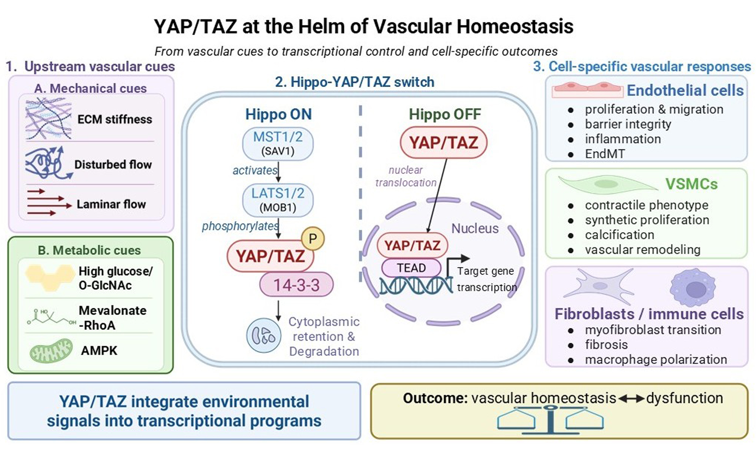
Vascular homeostasis depends on coordinated responses among endothelial cells, vascular smooth muscle cells, and perivascular cells. The Hippo pathway effectors Yes-associated protein (YAP) and transcriptional coactivator with PDZ-binding motif (TAZ) integrate mechanical, metabolic, and biochemical cues within these cell types. Their effects are highly context-dependent, supporting vascular development and integrity under physiological conditions while promoting inflammation, maladaptive remodeling, or calcification in specific disease settings. This review synthesizes the regulatory networks that control YAP/TAZ across vascular cell types and examines their roles in atherosclerosis, angiogenesis, vascular calcification, and related disorders. We further assess emerging YAP/TAZ-directed therapies and define the central translational challenge: achieving cell-, site-, and stage-specific modulation without disrupting essential homeostatic and regenerative functions.
Keywords


 Download PDF
INTRODUCTION
The vasculature is a dynamic network that regulates blood pressure, supports gas and nutrient exchange, enables immune surveillance, and maintains fluid homeostasis[1-3]. Yes-associated protein (YAP) was first identified by Sudol et al. in 1994 as a novel SH3 domain-binding protein[4]. Its paralog, transcriptional coactivator with PDZ-binding motif (TAZ), was subsequently identified by Kanai et al. in 2000 as a 14-3-3-binding protein[5]. Both proteins were subsequently recognized as downstream effectors of the Hippo pathway and as context-dependent regulators of tissue development, homeostasis, and disease. Here, we synthesize YAP/TAZ regulatory networks in endothelial, smooth muscle, perivascular, and immune cells, with emphasis on atherosclerosis, angiogenesis, and vascular calcification. We focus on how YAP/TAZ integrate mechanical and metabolic signals and why their vascular effects vary across cell types and disease stages. This framework clarifies both the therapeutic potential and the risks of targeting YAP/TAZ in cardiovascular disease.
AN OVERVIEW OF YAP/TAZ
YAP and TAZ are transcriptional co-regulators that lack DNA-binding domains but act primarily through transcription factors, most notably the Transcriptional Enhancer Associate Domain (TEAD) family[6]. Structurally, YAP isoforms (such as YAP1 with one WW domain and YAP2 with two) and TAZ share several characteristic domains, including the TEAD-binding domain, WW domains, and a C-terminal PDZ-binding motif, which are essential for their protein interactions and transcriptional activities[7]. Although YAP and TAZ share structural and functional similarities, they are not functionally interchangeable. TAZ shows distinct regulatory features, including differential sensitivity to mechanical cues and unique interaction partners, which may explain their context-specific functions in vascular biology.
YAP and TAZ integrate diverse upstream signals, with the canonical Hippo kinase cascade serving as a core regulatory module[8]. Hippo pathway activity is strongly shaped by the tissue microenvironment. YAP/TAZ respond to mechanical forces, including tension, compression, and shear stress[9], extracellular matrix stiffness[10], metabolic status[11], G protein-coupled receptor signaling[12], and soluble factors. This responsiveness positions YAP/TAZ as integrators of cardiovascular microenvironmental signals.
The Hippo pathway centers on a core phosphorylation cascade: MST1/2 kinases, in concert with their adaptors SAV1 and MOB1, phosphorylate and activate LATS1/2 kinases, which then phosphorylate YAP/TAZ[13]. This phosphorylation promotes 14-3-3 protein binding, leading to YAP/TAZ cytoplasmic sequestration and degradation, thereby inhibiting their transcriptional activity[14]. Conversely, when the Hippo pathway is inactivated, dephosphorylated YAP/TAZ translocate into the nucleus. There, they associate with TEAD transcription factors on chromatin and induce genes that regulate processes including cell proliferation and survival[6,9]. In addition to this established pathway, evidence also indicates that diverse post-translational modifications dynamically regulate YAP/TAZ stability, localization, and function in response to microenvironmental cues[15,16].
Beyond their initial characterization as regulators of organ size[17], YAP/TAZ are now known to be important regulators of cellular homeostasis and disease across diverse contexts[18,19]. This versatility enables YAP/TAZ to coordinate structural and functional responses across cardiovascular cell types, including endothelial cells, vascular smooth muscle cells, and cardiac myocytes. Their vascular effects therefore depend on how each cell type interprets mechanical and metabolic inputs through distinct YAP/TAZ-dependent transcriptional programs [Figure 1].
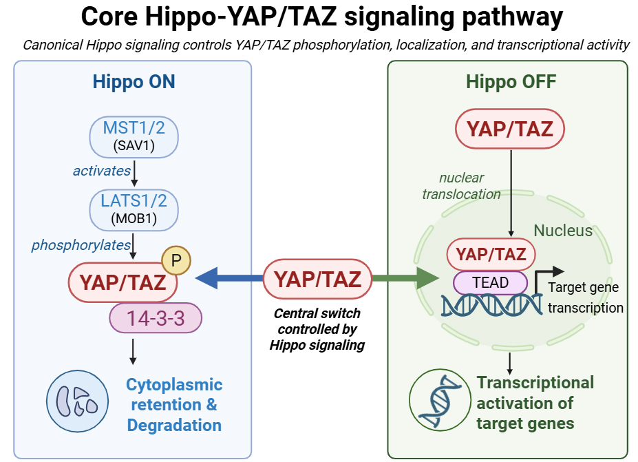
Figure 1. Core Hippo-YAP/TAZ signaling pathway. Created in BioRender. Liu, Y. (2026) https://BioRender.com/t01kqrd. YAP: Yes-associated protein; TAZ: transcriptional coactivator with PDZ-binding motif; TEAD: TEA domain.
BIOLOGICAL REGULATION OF YAP/TAZ IN THE VASCULAR SYSTEM
Cells sense the physical properties of their environment, convert them into biochemical signals, and adapt their behavior accordingly. In vascular cells, fluid shear stress engages several mechanosensors, including integrins, the glycocalyx, primary cilia, G-protein-coupled receptors, and ion channels (K+, Ca2+)[20]. YAP/TAZ act downstream of these sensors as mechanosensitive transcriptional co-regulators that couple physical cues to genes involved in vascular homeostasis and remodeling [Figure 2].
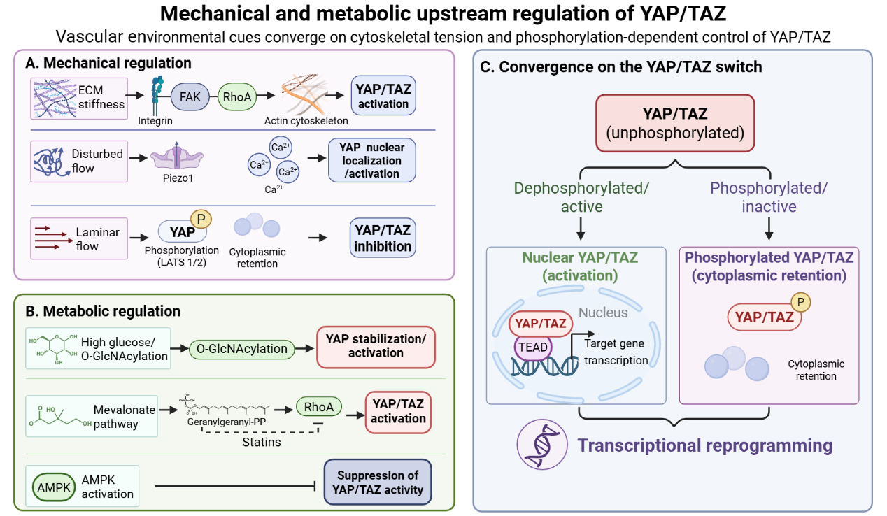
Figure 2. Mechanical and metabolic upstream regulation of YAP/TAZ. (A) Mechanical cues regulate YAP/TAZ through integrin-FAK-RhoA signaling, Piezo1-mediated Ca2+ influx, and LATS1/2-dependent phosphorylation; (B) Metabolic cues regulate YAP/TAZ through O-GlcNAcylation, the mevalonate-RhoA pathway, and AMPK signaling; (C) Dephosphorylated YAP/TAZ enters the nucleus and binds TEAD to activate transcription, whereas phosphorylated YAP/TAZ is retained in the cytoplasm. Created in BioRender. Liu Y (2026) https://BioRender.com/ro500te. YAP: Yes-associated protein; TAZ: transcriptional coactivator with PDZ-binding motif; TEAD: TEA domain; ECM: enabling extracellular matrix; FAK: focal adhesion kinase; AMPK: AMP-activated protein kinase.
Mechanotransduction: from microenvironment to nucleus
Vascular cells are continuously exposed to mechanical forces, including matrix stiffness, shear stress from blood flow, and cyclic stretch from pulsatile pressure during the cardiac cycle[21-24]. YAP/TAZ integrate these diverse mechanical cues to coordinate vascular homeostasis and remodeling. For example, cyclic stretch induces YAP nuclear translocation via the Thbs1/integrin αvβ1-Rap2-Hippo pathway in vascular smooth muscle cells (VSMCs), and YAP/TAZ are required for stretch-induced proliferative and pro-inflammatory responses[21,22,24].
Integrin-mediated mechanosensing of matrix stiffness
The cytoskeleton acts as a central conduit for mechanical signal transduction, translating extracellular forces into biochemical cues that regulate YAP/TAZ activity. This process relies on force-sensitive adhesion and cytoskeletal proteins, including integrins, talin, vinculin, and actin, that transmit tension from the cell surface to the nucleus[20,25]. At the subcellular level, substrate stiffness enhances αTAT1 recruitment to focal adhesions (FAs), increasing microtubule acetylation and releasing GEF-H1, which activates RhoA and actomyosin contractility, thereby reinforcing YAP/TAZ nuclear translocation and mechanosensitivity[26]. FAs regulate YAP localization through complementary mechanical and biochemical mechanisms: vinculin-talin binding mediates tension-dependent nuclear regulation, while talin-focal adhesion kinase (FAK) interaction provides biochemical control[27]. In vascular remodeling models such as transverse aortic constriction or arterial ligation, mechanical stress induces Thbs1, which binds integrin αvβ1 to activate YAP/TAZ, promote focal adhesion maturation, and inactivate the small GTPase Rap2[22]. Outside the vasculature, the integrin α1β1-F-actin-YAP pathway regulates COL1A1 expression during mechanically induced scleral remodeling, illustrating a broader matrix-responsive mechanism[28].
Shear stress sensing in endothelial cells
Beyond matrix stiffness, endothelial cells continuously sense hemodynamic forces, particularly shear stress, which is central to vascular physiology and atherosclerosis susceptibility[29,30]. Endothelial YAP/TAZ respond differently to flow patterns: under unidirectional shear stress, integrin activation and Gα13 signaling inhibit RhoA, leading to YAP phosphorylation at Ser127 and cytoplasmic retention; in contrast, disturbed flow promotes YAP nuclear translocation and pro-atherogenic gene expression[31]. Recent findings have expanded our understanding of the upstream mechanosensing mechanisms that regulate YAP activity. Discoidin domain receptor 1 (DDR1) acts as a direct endothelial mechanosensor; upon exposure to shear stress, it undergoes oligomerization and liquid-liquid phase separation with 14-3-3 protein, thereby facilitating YAP nuclear translocation and promoting atherogenesis[32]. Similarly, the endothelial serotonin receptor 5-HT1B has emerged as a novel mechanosensor that activates YAP through a β-arrestin/RhoA signaling axis under disturbed flow, amplifying atherosclerotic progression[33]. Increased 5-HT1B expression, in turn, promotes YAP nuclear localization through the β-arrestin/RhoA axis, establishing a self-amplifying loop that maintains YAP activity and endothelial inflammation[34]. This positive-feedback circuit may convert transient hemodynamic disturbance into sustained pro-atherogenic signaling.
Functional studies confirm that YAP/TAZ are indispensable for transducing disturbed flow into inflammatory phenotypes, as their depletion suppresses endothelial cell (EC) proliferation and inflammatory activation[31]. Piezo1, a mechanosensitive cation channel[25], responds acutely to tensile changes and modulates nuclear morphology under shear stress[35]. Piezo1-mediated nuclear flattening enhances nucleocytoplasmic transport, promoting YAP nuclear localization[36]. The YAP target angiomotin-like 2 (AmotL2) links junctional mechanical forces to chromatin remodeling by modulating EZH2 activity, establishing a mechano-regulated transcriptional feedback loop critical for endothelial homeostasis[24]. In addition, oscillatory shear stress activates an integrin α5β1/c-Abl signaling axis that induces YAP tyrosine phosphorylation at Y357, driving its nuclear translocation and pro-atherogenic activity[37]. Through junction-cytoskeletal-nuclear coupling, AmotL2 preserves nuclear morphology and lamin A integrity, maintaining chromatin accessibility at the YAP locus. Loss of AmotL2 leads to repressive H3K27me3 marks at the YAP promoter, thereby suppressing its expression and disrupting vascular homeostasis[24].
Mechanical and metabolic inputs converge upstream of YAP/TAZ. Matrix stiffness modulates cellular metabolism by altering nutrient transporter expression and mitochondrial function[38], while metabolic states in turn influence cytoskeletal organization and mechanosensitivity[39]. This bidirectional crosstalk allows YAP/TAZ to coordinate adaptive responses to physical and nutritional cues.
Metabolic regulation: a bidirectional circuit
Vessels play vital roles in supplying oxygen and nutrients to tissues throughout the body, making vascular cells sensitive sentinels of metabolic stress. Their metabolic state reflects local nutrient and oxygen availability and can signal tissue hypoxia. YAP/TAZ have emerged as important integrators that couple these metabolic cues to vascular adaptation: they are regulated by nutrients (glucose, fatty acids, insulin) and, in turn, regulate key metabolic pathways including glycolysis, lipogenesis, and glutaminolysis. Through this bidirectional circuit, YAP/TAZ align metabolic activity with vascular homeostasis and tissue repair[11].
YAP/TAZ as metabolic integrators in vascular homeostasis
YAP/TAZ activity is dynamically modulated by the metabolic status of the cell and its microenvironment. In endothelial cells (ECs), hyperglycemia in diabetic retinopathy induces O-GlcNAcylation of YAP at T383. This modification stabilizes YAP by inhibiting phosphorylation at S397. Activated YAP/TAZ enhance the hexosamine biosynthetic pathway, establishing a self-reinforcing loop that amplifies global O-GlcNAcylation, drives pro-angiogenic transcription, and disrupts vascular homeostasis[40]. In a distinct context, endothelial H₂S deficiency during pulmonary fibrosis inactivates AMP-activated protein kinase (AMPK), leading to YAP-mediated upregulation of the angiocrine factor plasminogen activator inhibitor-1 (PAI-1) and promoting fibrotic remodeling[41].
In VSMCs, YAP also responds to dietary stress, particularly through its regulation of transforming growth factor-β (TGF-β)-Smad signaling. The phosphatase PPM1B serves as a key negative regulator of this pathway by dephosphorylating p-Smad2/3. In VSMCs, YAP sustains TGF-β signaling by promoting removal of K63-linked ubiquitin chains from PPM1B at K326, thereby limiting PPM1B nuclear translocation and Smad2/3 dephosphorylation[42]. Further work identified the BRISC complex as a mediator of this process, with YAP-dependent assembly of ABRO1, YAP, and PPM1B promoting PPM1B deubiquitination and diet-induced arterial stiffness[43].
Metabolic feedback regulation of YAP/TAZ activity
Once activated, YAP/TAZ orchestrate metabolic programs that support vascular cell proliferation and remodeling. In ECs, the YAP/TAZ-TEAD axis promotes angiogenesis by inducing amino acid transporters such as SLC7A5. Subsequent amino acid uptake activates mechanistic target of rapamycin complex 1 (mTORC1) via Rag GTPases, driving endothelial proliferation and vascular growth[44]. The mevalonate pathway, crucial for cholesterol biosynthesis[45], also activates YAP/TAZ through geranylgeranylation-dependent Rho GTPase signaling, thereby linking metabolic flux to YAP-driven proliferation[46,47]. In pulmonary hypertension, YAP/TAZ couple mechanical and metabolic signals to transcriptional control of glutamine-to-proline[48] and glutaminolysis pathways[49], enabling extracellular matrix (ECM) stiffness-induced collagen biosynthesis and aspartate-dependent proliferation.
YAP/TAZ in classical cardiovascular risk factors
Classical cardiovascular risk factors - including hypertension, diabetes mellitus, dyslipidemia, and obesity - intersect with YAP/TAZ signaling through cell-type-specific mechanisms. In hypertension, YAP/TAZ are essential for maintaining vascular tone, and their dysregulation promotes hypertensive vascular remodeling via Foxm1 upregulation[50]. In diabetes, hyperglycemia activates YAP/TAZ signaling, promoting endothelial inflammation and monocyte-EC attachment[51]. The mevalonate pathway activates YAP/TAZ through geranylgeranylation-dependent Rho GTPase signaling[46,47]. Separately, macrophage YAP/TAZ-BRD4 signaling promotes inflammatory activation and oxidized Low-density Lipoprotein (oxLDL) uptake/lipid accumulation, contributing to atherogenesis[52]. In obesity, a high-fat, high-sucrose diet sustains YAP-mediated TGF-β signaling, exacerbating arterial stiffness[42]. Notably, despite their well-established roles as cardiovascular risk factors, direct mechanistic evidence linking lipoprotein(a) or homocysteine to YAP/TAZ signaling remains limited and warrants direct investigation. These associations are summarized in Table 1.
Modulation of YAP/TAZ signaling by classical cardiovascular risk factors
| Risk factor | Effect on YAP/TAZ | Vascular consequence | References |
| Hypertension | YAP/TAZ nuclear translocation increased; Foxm1 upregulation | VSMC proliferation, vascular fibrosis | [50] |
| Diabetes/hyperglycemia | O-GlcNAcylation stabilizes YAP/TAZ; ER stress activates YAP/TAZ-SMAD1/5 | Endothelial dysfunction, angiogenesis, accelerated atherosclerosis | [51] |
| Dyslipidemia/ox-LDL | Mevalonate-Rho signaling activates YAP/TAZ; macrophage YAP/TAZ–BRD4 signaling increases oxLDL uptake | Macrophage inflammation, lipid accumulation, atherogenesis | [46,47,52] |
| Obesity/metabolic syndrome | YAP-PPM1B-TGF-β axis sustained | Arterial stiffness, fibrotic remodeling | [107] |
CELL-TYPE-SPECIFIC ROLES IN VASCULAR PATHOPHYSIOLOGY
As discussed above, YAP/TAZ integrate mechanical and metabolic signals. How these signals translate into specific cellular outcomes, however, depends on cell type and vascular context. Endothelial cells, smooth muscle cells, and perivascular cells each respond through distinct transcriptional programs that collectively shape vascular function and remodeling. The following sections compare these cell-type-specific functions, from endothelial barrier regulation to smooth muscle phenotypic switching and immune-vascular crosstalk [Figure 3].
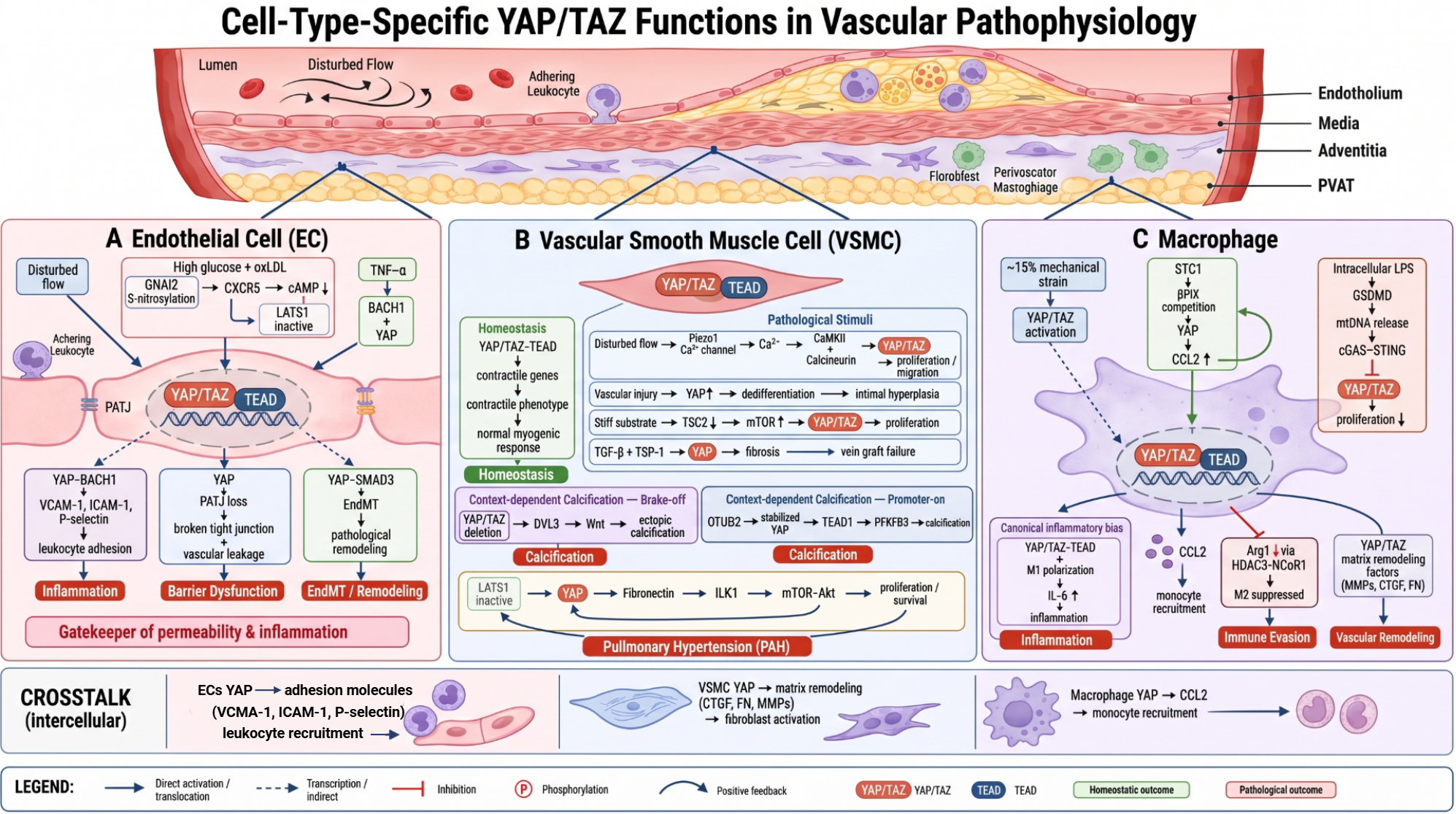
Figure 3. Cell-type-specific effects of YAP/TAZ in endothelial cells, smooth muscle cells, and macrophages within the context of a diseased vessel wall. (A) In endothelial cells, YAP/TAZ activation promotes leukocyte adhesion, barrier dysfunction, and EndMT-mediated vascular remodeling; (B) In vascular smooth muscle cells, YAP/TAZ maintain the contractile phenotype under homeostatic conditions but contribute to proliferation, phenotypic switching, calcification, and pulmonary hypertension in a context-dependent manner; (C) In macrophages, YAP/TAZ regulate inflammatory polarization, monocyte recruitment, immune evasion, and vascular remodeling in response to mechanical and inflammatory signals. Created in BioRender. Liu Y (2026) https://BioRender.com/jw33kg0. YAP: Yes-associated protein; TAZ: transcriptional coactivator with PDZ-binding motif; TEAD: TEA domain; ECM: enabling extracellular matrix; TNF-α: tumor necrosis factor-alpha; EndMT: endothelial-mesenchymal transition; SMAD3: SMAD family member 3; VCAM-1: vascular cell adhesion molecule-1; ICAM-1: intercellular adhesion molecule-1; GNAI2: G protein subunit alpha i2; CXCR5: C-X-C chemokine receptor type 5; BACH1: BTB and CNC homology 1; TGF-β: transforming growth factor-β; TSP-1: thrombospondin-1; OTUB2: OTU deubiquitinase 2; PFKFB3: 6-phosphofructo-2-kinase/fructose-2,6-bisphosphatase 3; ILK1: integrin-linked kinase 1; CCL2: C-C motif chemokine ligand 2; IL-6: interleukin-6; cAMP: cyclic adenosine monophosphate; oxLDL: oxidized low-density lipoprotein; mTOR: mechanistic target of rapamycin; Akt: protein kinase B; DVL3: dishevelled segment polarity protein 3; CTGF: connective tissue growth factor; FN: fibronectin; MMPs: matrix metalloproteinases; LPS: lipopolysaccharide; GSDMD: gasdermin D; mtDNA: mitochondrial DNA; cGAS: cyclic GMP-AMP synthase; STING: stimulator of interferon genes; HDAC3: histone deacetylase 3; NCoR1: nuclear receptor corepressor 1; LATS1: large tumor suppressor kinase 1; TSC2: tuberous sclerosis complex 2; CaMKII: Ca2+/calmodulin-dependent protein kinase II; Arg1: arginase 1; PVAT: perivascular adipose tissue; PAH: pulmonary arterial hypertension; PATJ: Pals1-associated tight junction protein.
Endothelial cells: gatekeepers of permeability and inflammation
Regulation of proliferation and migration. Beyond pathological angiogenesis, YAP/TAZ play essential roles in physiological vascular development. Endothelium-specific YAP/TAZ knockout models show that YAP/TAZ activity is required for developmental sprouting angiogenesis[53,54]. Mechanistically, YAP/TAZ support stretch-induced endothelial proliferation and rearrangement, preserve developing vessels, and are required for tip cell migration and stalk cell proliferation during sprouting angiogenesis[53,54]. The focal adhesion protein DLC1, a YAP-induced RhoGAP, suppresses Rho-mediated cytoskeletal tension, establishing a mechanosensitive feedback loop that limits YAP nuclear localization and fine-tunes angiogenic sprouting[55]. In brain arteriovenous malformations, YAP expression decreases but rises following embolization, indicating reactivation of Hippo-YAP signaling during vascular repair[56]. Recent work has also identified the tight junction-associated protein Pals1-associated Tight Junction Protein (PATJ) as a novel regulator of YAP1 in endothelial stress responses. PATJ is upregulated in endothelial cells after ischemic stroke, and its deletion alters YAP1 nuclear translocation and dysregulates genes involved in vascular development, including RUNX1 (Runt-related transcription factor 1) and HEY1 (Hairy/enhancer-of-split related with YRPW motif 1), as well as stress response genes such as NUPR1 (nuclear protein 1)[57]. Beyond mechanical cues, YAP also intersects with TGF-β signaling to regulate endothelial-mesenchymal transition (EndMT). By stabilizing SMAD family member 3 (SMAD3) against glycogen synthase kinase 3 beta (GSK3β)-dependent degradation, YAP forms a transcriptional complex with SMAD3 that directly activates EndMT target genes, thereby promoting pathological vascular remodeling[58].
Inflammatory activation and barrier function. YAP drives endothelial inflammation through multiple mechanisms. Upon oscillatory shear stress or tumor necrosis factor-alpha (TNF-α) stimulation, YAP forms a complex with BTB and CNC homology 1 (BACH1), which is required for the induction of adhesion molecules and endothelial inflammation[59]. In diabetes-accelerated atherosclerosis, high glucose and oxLDL promote G protein subunit alpha i2 (GNAI2) S-nitrosylation, enhancing C-X-C chemokine receptor type 5 (CXCR5) coupling to reduce cyclic adenosine monophosphate (cAMP), thereby inactivating the LATS1-YAP axis and driving pro-inflammatory transcription[60]. Pharmacological evidence supports the functional relevance of this pathway. Verteporfin-mediated YAP/TAZ inhibition in a progeria model significantly reduced vascular cell adhesion molecule-1 (VCAM-1), intercellular adhesion molecule-1 (ICAM-1), and P-selectin expression, decreasing leukocyte adhesion[61]. These endothelial changes can alter monocyte recruitment and thereby influence inflammation, angiogenesis, and tissue remodeling[62].
Substrate stiffening, a hallmark of vascular disease, reduces tuberous sclerosis complex 2 (TSC2) abundance in pulmonary arterial smooth muscle cells, allowing YAP/TAZ accumulation and mechanistic target of rapamycin (mTOR) activation to drive proliferation. The remodeled ECM amplifies this signal in neighboring cells via α5β1-integrin[63]. In a related endothelial model using engineered hydrogels, stress-relaxation and gelatin mechanics activate a YAP-dependent transcriptional program that promotes endothelial remodeling. In this context, αvβ3 integrin and matrix metalloproteinase 2 (MMP2) coordinate matrix degradation and reorganization to enable vasculogenesis both in vitro and in vivo[64].
Smooth muscle cells: regulators of contractility and phenotypic switching
Vascular smooth muscle cells maintain vessel tone and adapt to hemodynamic changes through phenotypic plasticity. YAP and TAZ regulate genes involved in smooth muscle differentiation and contraction, with smooth muscle-specific deletion or genetic ablation of YAP/TAZ leading to reduced contractility, impaired myogenic response, increased vascular compliance, and downregulation of differentiation markers such as Acta2, Tagln, and Myh11 in the arterial media[21,65]. These contractile functions contribute directly to blood-pressure regulation. Inducible deletion of YAP/TAZ in adult smooth muscle cells results in impaired agonist-stimulated contractility and hypotension[21,66]. The importance of YAP/TAZ in VSMCs is supported by in vivo genetic models, as summarized in Table 2.
Phenotypes associated with smooth muscle-specific manipulation of YAP/TAZ in mice
| Manipulation | Phenotype | References |
| integrin α8-Cre | Spontaneous abdominal aortic aneurysms with SMC apoptosis | [67] |
| Myh11-Cre/ERT2 | reduced vascular contractility, impaired myogenic response, and increased compliance | [21] |
| Smmhc-CreERT2 | aortic dissection and rupture | [68] |
| Myh11-CreERT2 | Altered arterial-stiffness response to a high-fat, high-sucrose diet | [42] |
| Myh11-CreERT2 | Large-intestinal pseudo-obstruction, impaired peristalsis, and loss of the VSMC contractile phenotype | [65] |
Dysregulation of YAP/TAZ in VSMCs has been directly linked to clinically significant vascular wall pathologies. For instance, vascular smooth muscle-specific YAP/TAZ deletion triggers spontaneous development of abdominal aortic aneurysms, accompanied by pronounced VSMC apoptosis and matrix degradation, providing genetic evidence that YAP/TAZ are essential for maintaining aortic structural integrity[67]. Similarly, inducible deletion of YAP/TAZ in smooth muscle cells predisposes mice to aortic dissection and rupture under hemodynamic stress[68]. Together, these findings connect YAP/TAZ dysfunction to life-threatening large-vessel disease and link vascular mechanobiology to clinically relevant pathology.
Upon vascular injury or hemodynamic stress, YAP expression increases, promoting VSMC dedifferentiation and intimal hyperplasia[69]. In saphenous vein grafts, mechanical forces synergize with TGF-β/thrombospondin-1 (TSP-1) signaling to activate YAP, which drives a fibrotic response in resident progenitor cells and contributes to graft failure[70]. Disturbed flow activates VSMCs through Piezo1, which converts flow into Ca2+ signals that activate Ca2+/calmodulin-dependent protein kinase II (CaMKII) and calcineurin, promoting YAP/TAZ-dependent proliferation and migration[71]. In contrast, G protein-coupled receptor 153 (GPR153) deficiency elevates cAMP and activates LATS1/2-mediated phosphorylation of YAP/TAZ, leading to their degradation and impaired VSMC dedifferentiation[72].
YAP/TAZ contribute to diverse VSMC-related pathologies through context-dependent mechanisms. In vascular calcification, YAP/TAZ have apparently opposing effects. Basal YAP/TAZ activity restrains osteogenic transdifferentiation, because their deletion activates a DVL3-dependent Wnt cascade that induces ectopic calcification[65]. By contrast, in the OTU deubiquitinase 2 (OTUB2) pathway, stabilized YAP promotes VSMC osteogenic differentiation by forming a transcriptional complex with TEAD1 that directly binds to the 6-phosphofructo-2-kinase/fructose-2,6-bisphosphatase 3 (PFKFB3) promoter and enhances its expression, thereby promoting vascular calcification[73]. YAP also drives pathological remodeling in pulmonary arterial hypertension, where LATS1 inactivation establishes a self-sustaining YAP/fibronectin/integrin-linked kinase 1 (ILK1) loop that activates mTOR-Protein Kinase B (Akt), suppresses apoptosis, and sustains VSMC proliferation[74]. In neointimal hyperplasia following arterial interventions, YAP activity is negatively regulated by Tax1-binding protein 3 (TAX1BP3), which competes for TEAD binding; therapeutic delivery of TAX1BP3 via adeno-associated virus (AAV) or nanoparticles effectively reduces vascular remodeling[75]. In the context of physiological vascular homeostasis, YAP/TAZ mechanosensing is paramount for maintaining vascular integrity throughout life. Notably, YAP/TAZ activity decreases with age in mice, which results in cGAS (cyclic GMP-AMP synthase) - STING (stimulator of interferon genes)-induced vascular inflammation and may provide a mechanistic link between aging and vascular dysfunction[68,76,77]. These findings indicate that the effect of YAP/TAZ on VSMC fate depends on baseline activity, upstream regulation, and disease context.
Perivascular and immune cells: regulators of the vascular niche
Beyond endothelial and smooth muscle cells, YAP/TAZ orchestrate the behavior of perivascular and immune cells that collectively shape the vascular microenvironment. In the vascular adventitia, where fibroblasts are the predominant cell type[78], YAP1 activation drives their transition into migratory myofibroblasts and enhances collagen deposition, thereby promoting abdominal aortic aneurysm (AAA) formation[79]. Within perivascular adipose tissue (PVAT), dysregulation of the PGC1α-YAP axis disrupts smooth muscle protein 22-alpha (SM22α)-lineage stromal cell differentiation, leading to aberrant PVAT remodeling and AAA development[80]. In TSC, loss of TSC1/TSC2 causes mTOR hyperactivation and impaired autophagic degradation, resulting in YAP accumulation that drives proliferation of perivascular epithelioid cell tumors (PEComas)[81]. In developmental bone formation, YAP/TAZ direct the coordinated mobilization of osteoblast precursors and blood vessels via CXCL12 signaling, facilitating essential processes including hypertrophic cartilage degradation and load-induced ossification[82].
YAP also contributes to bidirectional communication between vascular and immune cells. In antiphospholipid syndrome, patient-derived IgG activates Toll-like receptor 4 (TLR4)-YAP1 signaling in ECs, inducing CCN2 expression that subsequently drives VSMC proliferation and neointima formation via epidermal growth factor receptor (EGFR)[83].
Within the immune compartment, YAP/TAZ shape macrophage phenotype and function. Stanniocalcin-1 activates YAP through competitive p21-activated protein kinase exchange factor beta (βPIX) binding, establishing a CCL2-mediated feedback loop that promotes M2 polarization, angiogenesis, and programmed death ligand 1 (PD-L1)-dependent immune evasion in melanoma[84]. Mechanical cues further modulate macrophage YAP activity: moderate mechanical strain (~15%) optimally activates YAP/TAZ, driving M2 polarization and enhancing wound healing through paracrine-mediated angiogenesis[85]. In pulmonary fibrosis, macrophage YAP/TAZ promote disease progression through two mechanisms: CCL2-mediated recruitment of monocyte-derived alveolar macrophages and MBD2-TGF-β1-pSMAD2-dependent signaling that induces fibroblast-to-myofibroblast transition[86]. Conversely, intracellular lipopolysaccharide (LPS) induces gasdermin D (GSDMD)-mediated mitochondrial damage and mtDNA release, which activate the cGAS-STING pathway to inhibit YAP dephosphorylation and nuclear translocation, ultimately suppressing cyclin D expression and endothelial proliferation[87].
YAP/TAZ regulate macrophage polarization within the vascular microenvironment. Expression of YAP and TAZ is increased in macrophages undergoing both M1 and M2 polarization[88]. Mechanistically, YAP/TAZ promote M1 (pro-inflammatory) polarization by increasing IL6 expression, while impeding M2 (anti-inflammatory) polarization by decreasing Arg1 expression through interaction with the histone deacetylase 3 (HDAC3)-nuclear receptor corepressor 1 (NCoR1) repressor complex[88]. Genetic deletion of YAP/TAZ leads to impaired M1 polarization and enhanced M2 polarization, resulting in reduced inflammation[88]. In the context of atherosclerosis, YAP/TAZ activation in macrophages promotes pro-inflammatory cytokine production and plaque progression. Conversely, inhibition of YAP/TAZ suppresses inflammatory gene expression and macrophage infiltration[52,89]. Mechanical cues, including matrix stiffness and cyclic stretch, further modulate macrophage polarization through YAP/TAZ-dependent pathways[90]. Together, these findings link YAP/TAZ-dependent macrophage plasticity to mechanical signals and vascular inflammation. The divergent M1- and M2-associated effects reported across models likely reflect differences in tissue context, stimulus type, magnitude, and duration.
Collectively, these findings illustrate that YAP/TAZ not only orchestrate cell-autonomous functions but also coordinate intercellular crosstalk among endothelial cells, smooth muscle cells, fibroblasts, and macrophages. This YAP/TAZ-mediated communication network integrates mechanical, metabolic, and inflammatory signals across the vascular wall, ensuring coordinated responses to physiological demands and pathological insults.
THERAPEUTIC STRATEGIES AND TRANSLATIONAL PROSPECTS
Repurposing existing drugs: statins and other YAP inhibitors
Several approved lipid-lowering agents suppress YAP/TAZ transcriptional activity in experimental models. Inflammatory signaling also intersects bidirectionally with YAP/TAZ in the vasculature. Pro-inflammatory cytokines such as interleukin-6 (IL-6) and TNF-α can activate YAP/TAZ through noncanonical pathways[91,92], while YAP/TAZ in turn regulate pro-inflammatory gene programs in endothelial cells[93] and VSMCs[67,68]. Among anti-inflammatory drugs with cardiovascular benefits, colchicine has been shown to activate Hippo signaling and promote YAP phosphorylation in cardiomyocytes, and its microtubule-depolymerizing activity is known to regulate YAP/TAZ nuclear-cytoplasmic shuttling[94]; however, direct evidence for colchicine-YAP/TAZ modulation in vascular cells remains lacking. Similarly, no direct studies are available for canakinumab or tocilizumab regarding YAP/TAZ, although fundamental evidence suggests that IL-6 can activate YAP[95], supporting a testable hypothesis rather than a therapeutic conclusion[96,97]. A high-throughput screen of 640 U.S. Food and Drug Administration (FDA)-approved compounds identified statins as inhibitors of YAP/TAZ signaling[46]. Mechanistically, YAP/TAZ activity depends on the sterol regulatory element-binding protein (SREBP)-mevalonate pathway: statins inhibit 3-hydroxy-3-methylglutaryl-coenzyme A (HMG-CoA) reductase, reducing geranylgeranyl pyrophosphate synthesis and preventing RhoA activation, thereby blocking YAP/TAZ nuclear localization[46]. Lipophilic statins further promote cytoplasmic sequestration of YAP through Rho GTPase-dependent cytoskeletal remodeling, independent of their lipid-lowering effects[97]. Consistently, simvastatin inhibits geranylgeranyl transferase I (GGTase I), blocking RhoA-driven YAP nuclear import, suppressing SRY-box transcription factor 9 (SOX9)-mediated EndMT, and preserving vascular function independent of cholesterol lowering[98]. Notably, simvastatin failed to suppress pro-inflammatory gene expression induced by constitutively active YAP/TAZ in endothelial cells, indicating that inhibition of endogenous YAP/TAZ may underlie the anti-inflammatory benefits of statins[99]. The approved photosensitizer verteporfin also inhibits YAP by increasing cytosolic 14-3-3σ and trapping YAP in the cytosol[100]. When encapsulated in biomimetic nanoparticles, verteporfin achieves lesion-specific YAP/TAZ inhibition, attenuating atherosclerosis while avoiding off-target effects associated with systemic YAP/TAZ modulation[89].
Development of novel YAP modulators
Beyond drug repurposing, recent efforts have focused on developing novel small molecules that target YAP/TAZ signaling through distinct mechanisms. One approach targets the mevalonate pathway upstream of YAP: the GGTase-I inhibitor BAY-593, identified via high-throughput screen of 3.8 million compounds, blocks Rho GTPase activation and potently inhibits YAP/TAZ signaling in vivo, providing an additional upstream strategy[101]. Another strategy directly disrupts the YAP/TAZ-TEAD transcriptional complex. The small-molecule inhibitor GNE-7883 allosterically binds the TEAD lipid pocket, disrupting YAP/TAZ-TEAD interactions and suppressing chromatin accessibility at TEAD motifs[102]. Early clinical data with the TEAD palmitoylation inhibitor VT3989 provide proof of concept for direct YAP-TEAD pathway inhibition in oncology[103]. More recently, a protein degradation approach has been developed using a YAP-targeting bioPROTAC that fuses a camelid nanobody with the ring finger protein 4 (RNF4) RING domain to degrade endogenous YAP via the ubiquitin-proteasome system[104]. Multiple Hippo pathway-targeted therapies are in development, with several already undergoing clinical trials[105].
Most YAP/TAZ-targeted agents remain in oncology development, with VT3989[103] (NCT04665206), IAG933 (NCT04857372), and verteporfin[106] (NCT06381154) showing early promise in solid tumor trials. Cardiovascular translation is nascent: the phase 1 SALVADOR-HF trial (NCT06831825) is evaluating YAP101, an AAV-based gene therapy for ischemic heart failure. However, direct evidence for small-molecule YAP/TAZ modulators in cardiovascular disease remains lacking, and future work should prioritize vascular-targeted delivery to achieve lesion-specific modulation.
Challenges and future directions
Despite these promising advances, translating YAP/TAZ-targeting strategies into vascular therapies faces several interrelated challenges that require careful consideration. First, YAP/TAZ can be protective or pathogenic depending on cell type, vascular bed, and disease stage. Systemic inhibition could therefore impair tissue homeostasis or regeneration while suppressing a lesion-specific pathway. Second, current agents generally lack the spatial and cellular selectivity required to modulate endothelial, smooth muscle, stromal, and immune compartments differently. Third, clinical validation is limited. YAP/TAZ activity has not been robustly linked to imaging-defined atherosclerotic burden or cardiovascular outcomes, partly because nuclear localization and phosphorylation are difficult to measure in routine specimens and no validated circulating surrogate is available. Progress will require vascular-targeted delivery, context-selective interaction partners, and pharmacodynamic biomarkers. Molecular imaging, prospective analysis of human vascular specimens, and single-cell or spatial profiling could map pathway activity across cell types and lesion stages. These approaches should be paired with outcome studies and explicit safety assessment.
CONCLUSION
YAP/TAZ integrate mechanical, metabolic, and biochemical signals across endothelial, smooth muscle, stromal, and immune cells. This integration supports vascular development, barrier function, contractility, and repair, but can also promote inflammation, maladaptive remodeling, or calcification. The direction of these effects depends on cell type, vascular bed, upstream signal, and disease stage. YAP/TAZ are therefore attractive therapeutic nodes, but they are not simple targets for uniform activation or inhibition. Translation will require mechanistic definition of the relevant cellular state, biomarkers that report pathway activity in human lesions, and delivery systems that restrict modulation to the appropriate site and time. These advances will determine whether the strong experimental rationale for targeting YAP/TAZ can be converted into safe and effective cardiovascular therapies.
DECLARATIONS
Acknowledgments
Graphical Abstract created in BioRender. Liu Y (2026) https://BioRender.com/5tzo6nm.
Authors’ contributions
Conceptualization, supervision, funding acquisition: Liu Y, Zhang Y
Writing - original draft preparation: Wang X, Chen L
Writing - review and editing: Wang X, Chen L, Zhang Y, Liu Y
Visualization: Zhu S
All authors have read and approved the final version of the manuscript.
Availability of data and materials
Not applicable.
AI and AI-assisted tools statement
During the preparation of this manuscript, the AI tool Gemini (version 1.5, released 2024-02-15) and Codex were used solely for language editing and formatting assistance. The tool did not influence the study design, data collection, analysis, interpretation, or the scientific content of the work. All authors take full responsibility for the accuracy, integrity, and final content of the manuscript.
Financial support and sponsorship
This work was supported by National Key R&D Program of China (2025ZD0547100 and 2021YFF0501401 to Zhang Y, 2021YFF0501404 to Liu Y); National Science Foundation of China (82325004 and 92168114 to Zhang Y, 82170422 to Liu Y); Natural Science Foundation of Beijing (7232096 to Liu Y, F251013 to Zhang Y); Research Project of Peking University in State Key Laboratory of Vascular Homeostasis and Remodeling (Peking University) (2024-VHR-SY-07 to Zhang Y); State Key Laboratory of Respiratory Health and Multimorbidity, State Key Laboratory Special Fund 2060204 (SKLRHM202501205) to Liu Y; National High Level Hospital Clinical Research Funding (2025-PUMCH-A-036) to Liu Y.
Conflicts of interest
Zhang Y is an Editorial Board Member of the journal Vessel Plus. Zhang Y was not involved in any steps of editorial processing, notably including reviewers’ selection, manuscript handling, or decision-making. The other authors declare that there are no conflicts of interest.
Ethical approval and consent to participate
Not applicable.
Consent for publication
Not applicable.
Copyright
© The Author(s) 2026.
REFERENCES
1. Zeng Q, Oliva VM, Moro MÁ, Scheiermann C. Circadian effects on vascular immunopathologies. Circ Res. 2024;134:791-809.
2. Zhou J, Wang D, Yan B. Infection at the vascular front: a delicate balance between defense and pathology. Microb Pathog. 2026;211:108261.
3. Williams AE, Dunaway LS, Juśkiewicz ZJ, et al. Impact of endothelial diversity and dysfunction on cardiovascular disease. Compr Physiol. 2025;15:e70064.
4. Sudol M. Yes-associated protein (YAP65) is a proline-rich phosphoprotein that binds to the SH3 domain of the Yes proto-oncogene product. Oncogene. 1994;9:2145-52.
5. Kanai F, Marignani PA, Sarbassova D, et al. TAZ: a novel transcriptional co-activator regulated by interactions with 14-3-3 and PDZ domain proteins. EMBO J. 2000;19:6778-91.
6. Reggiani F, Gobbi G, Ciarrocchi A, Sancisi V. YAP and TAZ are not identical twins. Trends Biochem Sci. 2021;46:154-68.
7. Wei Y, Hui VLZ, Chen Y, Han R, Han X, Guo Y. YAP/TAZ: molecular pathway and disease therapy. MedComm. 2023;4:e340.
8. Guo P, Wan S, Guan KL. The Hippo pathway: organ size control and beyond. Pharmacol Rev. 2025;77:100031.
9. Panciera T, Azzolin L, Cordenonsi M, Piccolo S. Mechanobiology of YAP and TAZ in physiology and disease. Nat Rev Mol Cell Biol. 2017;18:758-70.
10. Dupont S, Morsut L, Aragona M, et al. Role of YAP/TAZ in mechanotransduction. Nature. 2011;474:179-83.
12. Yu FX, Zhao B, Panupinthu N, et al. Regulation of the hippo-YAP pathway by G-protein-coupled receptor signaling. Cell. 2012;150:780-91.
13. Suto H. Role of the Hippo-YAP/TAZ Pathway in epithelioid hemangioendothelioma and its potential as a therapeutic target. Anticancer Res. 2024;44:4147-53.
14. Moroishi T, Hansen CG, Guan KL. The emerging roles of YAP and TAZ in cancer. Nat Rev Cancer. 2015;15:73-9.
15. Zhang Z, He P, Yang L, Gong J, Qin R, Wang M. Posttranslational modifications of YAP/TAZ: molecular mechanisms and therapeutic opportunities. Cell Mol Biol Lett. 2025;30:83.
16. Tong T, Huang M, Yan B, et al. Hippo signaling modulation and its biological implications in urological malignancies. Mol Aspects Med. 2024;98:101280.
17. Dong J, Feldmann G, Huang J, et al. Elucidation of a universal size-control mechanism in drosophila and mammals. Cell. 2007;130:1120-33.
18. Piccolo S, Dupont S, Cordenonsi M. The biology of YAP/TAZ: hippo signaling and beyond. Physiol Rev. 2014;94:1287-312.
19. Fu M, Hu Y, Lan T, Guan KL, Luo T, Luo M. The Hippo signalling pathway and its implications in human health and diseases. Signal Transduct Target Ther. 2022;7:376.
20. Di X, Gao X, Peng L, et al. Cellular mechanotransduction in health and diseases: from molecular mechanism to therapeutic targets. Signal Transduct Target Ther. 2023;8:282.
21. Daoud F, Arévalo Martinez M, Holmberg J, et al. YAP and TAZ in vascular smooth muscle confer protection against hypertensive vasculopathy. Arterioscler Thromb Vasc Biol. 2022;42:428-43.
22. Yamashiro Y, Thang BQ, Ramirez K, et al. Matrix mechanotransduction mediated by thrombospondin-1/integrin/YAP in the vascular remodeling. Proc Natl Acad Sci U S A. 2020;117:9896-905.
23. Ritsvall O, Albinsson S. Emerging role of YAP/TAZ in vascular mechanotransduction and disease. Microcirculation. 2024;31:e12838.
24. Mannion AJ, Zhao H, Zhang Y, et al. Regulation of YAP promotor accessibility in endothelial mechanotransduction. Arterioscler Thromb Vasc Biol. 2024;44:666-89.
25. Wang Y, Chatterjee E, Li G, Xu J, Xiao J. Force-sensing protein expression in response to cardiovascular mechanotransduction. EBioMedicine. 2024;110:105412.
26. Seetharaman S, Vianay B, Roca V, et al. Microtubules tune mechanosensitive cell responses. Nat Mater. 2022;21:366-77.
27. Holland EN, Fernández-Yagüe MA, Zhou DW, et al. FAK, vinculin, and talin control mechanosensitive YAP nuclear localization. Biomaterials. 2024;308:122542.
28. Liu X, Yuan Y, Wu Y, Zhu C, Liu Y, Ke B. Extracellular matrix stiffness modulates myopia scleral remodeling through integrin/F-Actin/YAP axis. Invest Ophthalmol Vis Sci. 2025;66:22.
29. Mehta V, Tzima E. Cardiovascular disease: a turbulent path to plaque formation. Nature. 2016;540:531-2.
30. Fang Y, Wu D, Birukov KG. Mechanosensing and mechanoregulation of endothelial cell functions. Compr Physiol. 2019;9:873-904.
31. Wang KC, Yeh YT, Nguyen P, et al. Flow-dependent YAP/TAZ activities regulate endothelial phenotypes and atherosclerosis. Proc Natl Acad Sci U S A. 2016;113:11525-30.
32. Liu J, Zhao C, Xiao X, et al. Endothelial discoidin domain receptor 1 senses flow to modulate YAP activation. Nat Commun. 2023;14:6457.
33. Jiang M, Ding H, Huang Y, et al. Endothelial serotonin receptor 1b acts as a mechanosensor to drive atherosclerosis. Circ Res. 2025;136:887-901.
34. Jiang MC, Ding HY, Cheng CK, et al. Targeting the 5-HT(1B)-YAP positive feedback loop protects against disturbed flow-induced atherogenesis in mice. Acta Pharmacol Sin. 2026;47:666-76.
35. Jetta D, Gottlieb PA, Verma D, Sachs F, Hua SZ. Shear stress-induced nuclear shrinkage through activation of Piezo1 channels in epithelial cells. J Cell Sci. 2019;132:jcs226076.
36. Oliva MAG, Ciccone G, Fläschner G, et al. Piezo1 regulates the mechanotransduction of soft matrix viscoelasticity. Nat Commun. 2025;16:9155.
37. Li B, He J, Lv H, et al. c-Abl regulates YAPY357 phosphorylation to activate endothelial atherogenic responses to disturbed flow. J Clin Invest. 2019;129:1167-79.
38. Narayanasamy A, Karuppusamy PA, Gnanarajan R, et al. Mechanobiology-driven metabolic reprogramming: integrative roles of YAP/TAZ signaling and extracellular matrix dynamics. Cell Biol Int. 2026;50:e70180.
39. Tian G, Ren T. Mechanical stress regulates the mechanotransduction and metabolism of cardiac fibroblasts in fibrotic cardiac diseases. Eur J Cell Biol. 2023;102:151288.
40. Lei Y, Liu Q, Chen B, et al. Protein O-GlcNAcylation coupled to Hippo signaling drives vascular dysfunction in diabetic retinopathy. Nat Commun. 2024;15:9334.
41. Chen X, Wang H, Wu C, et al. Endothelial H2S-AMPK dysfunction upregulates the angiocrine factor PAI-1 and contributes to lung fibrosis. Redox Biol. 2024;70:103038.
42. Liu Y, Li M, Lv X, et al. Yes-Associated protein targets the transforming growth factor β pathway to mediate high-fat/high-sucrose diet-induced arterial stiffness. Circ Res. 2022;130:851-67.
43. Liu Y, Li M, Chen Z, et al. BRISC-mediated PPM1B-K63 deubiquitination and subsequent TGF-β pathway activation promote high-fat/high-sucrose diet-induced arterial stiffness. Circ Res. 2025;136:297-314.
44. Ong YT, Andrade J, Armbruster M, et al. A YAP/TAZ-TEAD signalling module links endothelial nutrient acquisition to angiogenic growth. Nat Metab. 2022;4:672-82.
45. Deng Q, Ji Y, Liu J, Wen T. Lipid reprogramming and ferroptosis crosstalk in clear cell renal cell carcinoma: metabolic vulnerabilities and therapeutic targeting. Mol Cancer. 2025;24:236.
46. Sorrentino G, Ruggeri N, Specchia V, et al. Metabolic control of YAP and TAZ by the mevalonate pathway. Nat Cell Biol. 2014;16:357-66.
47. Yousfi Y, Mora-Molina R, López-Rivas A, Yerbes R. Role of the YAP/TAZ-TEAD transcriptional complex in the metabolic control of TRAIL sensitivity by the mevalonate pathway in cancer cells. Cells. 2023;12:2370.
48. Rachedi NS, Tang Y, Tai YY, et al. Dietary intake and glutamine-serine metabolism control pathologic vascular stiffness. Cell Metab. 2024;36:1335-1350.e8.
49. Bertero T, Oldham WM, Cottrill KA, et al. Vascular stiffness mechanoactivates YAP/TAZ-dependent glutaminolysis to drive pulmonary hypertension. J Clin Invest. 2016;126:3313-35.
50. Bai J, Yang Y. Activation of Hippo/YAP signaling pathway exacerbates vascular remodeling and aggravates hypertension by upregulating Foxm1. J Mol Histol. 2025;56:158.
51. Zhou Y, Yang Y, Wang Y, Cheang WS. Targeting endoplasmic reticulum stress and YAP/TAZ-SMAD1/5 signaling to ameliorate endothelial dysfunction in diabetes. Acta Pharmacol Sin. 2026.
52. Veerasubramanian PK, Meli VS, Atcha H, Wang W, Downing TL, Liu WF. BET inhibition curbs macrophage inflammation, lipid accumulation, and atherogenesis by disrupting the YAP/TAZ-BRD4 axis. J Leukoc Biol. 2026:118.
53. Kobayashi S, Cox AG, Harvey KF, Hogan BM. Vasculature is getting Hip(po): Hippo signaling in vascular development and disease. Dev Cell. 2023;58:2627-40.
54. Li R, Shao J, Jin YJ, et al. Endothelial FAT1 inhibits angiogenesis by controlling YAP/TAZ protein degradation via E3 ligase MIB2. Nat Commun. 2023;14:1980.
55. Hooglugt A, van der Stoel MM, Shapeti A, et al. DLC1 promotes mechanotransductive feedback for YAP via RhoGAP-mediated focal adhesion turnover. J Cell Sci. 2024:137.
56. Neyazi B, Swiatek VM, Karimpour MA, et al. The role of Hippo signaling in brain arteriovenous malformations: molecular insights into post-embolization remodeling. Int J Mol Sci. 2025;26:3791.
57. Zhang M, Jiang WI, Arkelius K, Swanson RA, Ma DK, Singhal NS. PATJ regulates cell stress responses and vascular remodeling post-stroke. Redox Biol. 2025;85:103709.
58. Savorani C, Malinverno M, Seccia R, et al. A dual role of YAP in driving TGFβ-mediated endothelial-to-mesenchymal transition. J Cell Sci. 2021:134.
59. Jia M, Li Q, Guo J, et al. Deletion of BACH1 attenuates atherosclerosis by reducing endothelial inflammation. Circ Res. 2022;130:1038-55.
60. Chao ML, Luo S, Zhang C, et al. S-nitrosylation-mediated coupling of G-protein alpha-2 with CXCR5 induces Hippo/YAP-dependent diabetes-accelerated atherosclerosis. Nat Commun. 2021;12:4452.
61. Barettino A, González-Gómez C, Gonzalo P, et al. Endothelial YAP/TAZ activation promotes atherosclerosis in a mouse model of Hutchinson-Gilford progeria syndrome. J Clin Invest. 2024:134.
62. Medrano-Bosch M, Simón-Codina B, Jiménez W, Edelman ER, Melgar-Lesmes P. Monocyte-endothelial cell interactions in vascular and tissue remodeling. Front Immunol. 2023;14:1196033.
63. Shen Y, Goncharov DA, Pena A, et al. Cross-talk between TSC2 and the extracellular matrix controls pulmonary vascular proliferation and pulmonary hypertension. Sci Signal. 2022;15:eabn2743.
64. Keshavarz M, Smith Q. Gelatin-mediated vascular self-assembly via a YAP-MMP signaling axis. Adv Funct Mater. 2024;34:2402360.
65. Wang L, Chennupati R, Jin YJ, et al. YAP/TAZ are required to suppress osteogenic differentiation of vascular smooth muscle cells. iScience. 2020;23:101860.
66. Albinsson S, Rippe C, Daoud F, et al. Hippo in smooth muscle - a therapeutic target in vascular diseases driven by aging and hypertension. Front Physiol. 2025;16:1674714.
67. Arévalo Martínez M, Ritsvall O, Bastrup JA, et al. Vascular smooth muscle-specific YAP/TAZ deletion triggers aneurysm development in mouse aorta. JCI Insight. 2023;8:e170845.
68. Sladitschek-Martens HL, Guarnieri A, Brumana G, et al. YAP/TAZ activity in stromal cells prevents ageing by controlling cGAS-STING. Nature. 2022;607:790-8.
69. Yu Q, Li W, Jin R, et al. PI3Kγ (phosphoinositide 3-kinase γ) regulates vascular smooth muscle cell phenotypic modulation and neointimal formation through CREB (cyclic AMP-response element binding protein)/YAP (yes-associated protein) signaling. Arterioscler Thromb Vasc Biol. 2019;39:e91-e105.
70. Garoffolo G, Sluiter TJ, Thomas A, et al. Blockade of YAP mechanoactivation prevents neointima formation and adverse remodeling in arterialized vein grafts. J Am Heart Assoc. 2025;14:e037531.
71. Zhang FR, Tang J, Lai Y, et al. Smooth muscle cell Piezo1 is essential for phenotypic switch and neointimal hyperplasia. Br J Pharmacol. 2025;182:2031-48.
72. Shao J, Kwon J, Wang T, et al. Orphan receptor GPR153 facilitates vascular damage responses by modulating cAMP levels, YAP/TAZ signaling, and NF-κB activation. Nat Commun. 2025;16:6232.
73. Li Y, Chen X, Xu X, et al. OTUB2 contributes to vascular calcification in chronic kidney disease via the YAP-mediated transcription of PFKFB3. Theranostics. 2025;15:1185-204.
74. Kudryashova TV, Goncharov DA, Pena A, et al. HIPPO-integrin-linked kinase cross-talk controls self-sustaining proliferation and survival in pulmonary hypertension. Am J Respir Crit Care Med. 2016;194:866-77.
75. Yang H, Zhong Y, Guo W, et al. TAX1BP3 is a SUMOylated nucleocytoplasmic shuttling protein and protects against vascular neointimal hyperplasia. Circulation. 2025;152:1087-108.
78. Ye C, Zheng F, Xu T, et al. Norepinephrine acting on adventitial fibroblasts stimulates vascular smooth muscle cell proliferation via promoting small extracellular vesicle release. Theranostics. 2022;12:4718-33.
79. Xie C, Hu Y, Yin Z. Inhibiting YAP1 reduced abdominal aortic aneurysm formation by suppressing adventitial fibroblast phenotype transformation and migration. J Cell Mol Med. 2024;28:e70159.
80. Pan X, Zhang R, Lu B, et al. SM22α-lineage perivascular stromal cells contribute to abdominal aortic aneurysm. Circ Res. 2025;137:4-22.
81. Liang N, Zhang C, Dill P, et al. Regulation of YAP by mTOR and autophagy reveals a therapeutic target of tuberous sclerosis complex. J Exp Med. 2014;211:2249-63.
82. Collins JM, Lang A, Parisi C, et al. YAP and TAZ couple osteoblast precursor mobilization to angiogenesis and mechanoregulation in murine bone development. Dev Cell. 2024;59:211-227.e5.
83. Shi H, Liang W, Yang Z, et al. Microvascular endothelial cells license APS vasculopathy through YAP1- and CCN2-mediated signaling. Circulation. 2025;152:1295-310.
84. Ren Z, Xu Z, Chang X, Liu J, Xiao W. STC1 competitively binding βPIX enhances melanoma progression via YAP nuclear translocation and M2 macrophage recruitment through the YAP/CCL2/VEGFA/AKT feedback loop. Pharmacol Res. 2024;204:107218.
85. Wei Q, Du F, Cui J, et al. Non-monotonic response of macrophages to mechanical stretch impacts skin wound healing. Cell Mol Biol Lett. 2025;30:82.
86. Mia MM, Ghani SABA, Cibi DM, et al. YAP/TAZ are crucial regulators of macrophage-mediated pulmonary inflammation and fibrosis after bleomycin-induced injury. Eur Respir J. 2025;65:2301544.
87. Huang LS, Hong Z, Wu W, et al. mtDNA activates cGAS signaling and suppresses the YAP-mediated endothelial cell proliferation program to promote inflammatory injury. Immunity. 2020;52:475-486.e5.
88. Mia MM, Cibi DM, Abdul Ghani SAB, et al. YAP/TAZ deficiency reprograms macrophage phenotype and improves infarct healing and cardiac function after myocardial infarction. PLoS Biol. 2020;18:e3000941.
89. Huang HC, Wang TY, Rousseau J, et al. Biomimetic nanodrug targets inflammation and suppresses YAP/TAZ to ameliorate atherosclerosis. Biomaterials. 2024;306:122505.
90. Shi Y, Xu Z, Wang H, et al. Scaffolding protein ENH promotes tumor angiogenesis and growth through macrophage recruitment and polarization. Adv Sci. 2025;12:e16476.
91. Chen L, Jin X, Ma J, Xiang B, Li X. YAP at the progression of inflammation. Front Cell Dev Biol. 2023;11:1204033.
92. Choi HJ, Kim NE, Kim BM, Seo M, Heo JH. TNF-α-induced YAP/TAZ activity mediates leukocyte-endothelial adhesion by regulating VCAM1 expression in endothelial cells. Int J Mol Sci. 2018;19:3428.
93. Lv Y, Kim K, Sheng Y, et al. YAP controls endothelial activation and vascular inflammation through TRAF6. Circ Res. 2018;123:43-56.
94. Zhang F, Cusick JK, Liang J, et al. YAP promotes microtubule growth to facilitate sarcomere disassembly in adult cardiomyocytes. Circulation. 2026;154:934-55.
95. Symons RA, Colella F, Collins FL, et al. Targeting the IL-6-Yap-Snail signalling axis in synovial fibroblasts ameliorates inflammatory arthritis. Ann Rheum Dis. 2022;81:214-24.
96. Wrighton KH. Metabolism: YAP and TAZ under metabolic control. Nat Rev Mol Cell Biol. 2014;15:296.
97. Benhammou JN, Qiao B, Ko A, Sinnett-Smith J, Pisegna JR, Rozengurt E. Lipophilic statins inhibit YAP coactivator transcriptional activity in HCC cells through Rho-mediated modulation of actin cytoskeleton. Am J Physiol Gastrointest Liver Physiol. 2023;325:G239-50.
98. Liu C, Shen M, Tan WLW, et al. Statins improve endothelial function via suppression of epigenetic-driven EndMT. Nat Cardiovasc Res. 2023;2:467-85.
99. Wang L, Luo JY, Li B, et al. Integrin-YAP/TAZ-JNK cascade mediates atheroprotective effect of unidirectional shear flow. Nature. 2016;540:579-82.
100. Wang C, Zhu X, Feng W, et al. Verteporfin inhibits YAP function through up-regulating 14-3-3σ sequestering YAP in the cytoplasm. Am J Cancer Res. 2016;6:27-37.
101. Graham K, Lienau P, Bader B, et al. Discovery of YAP1/TAZ pathway inhibitors through phenotypic screening with potent anti-tumor activity via blockade of Rho-GTPase signaling. Cell Chem Biol. 2024;31:1247-1263.e16.
102. Hagenbeek TJ, Zbieg JR, Hafner M, et al. An allosteric pan-TEAD inhibitor blocks oncogenic YAP/TAZ signaling and overcomes KRAS G12C inhibitor resistance. Nat Cancer. 2023;4:812-28.
103. Yap TA, Kwiatkowski DJ, Dagogo-Jack I, et al. YAP/TEAD inhibitor VT3989 in solid tumors: a phase 1/2 trial. Nat Med. 2025;31:4281-90.
104. Zhou R, Wang H, Zhang GM, et al. Targeted degradation of endogenous YAP by nanobody bioPROTAC inhibits tumor progression. Nat Commun. 2025;16:9374.
105. Harvey KF, Tang TT. Targeting the Hippo pathway in cancer. Nat Rev Drug Discov. 2025;24:852-69.
106. Liu-Chittenden Y, Huang B, Shim JS, et al. Genetic and pharmacological disruption of the TEAD-YAP complex suppresses the oncogenic activity of YAP. Genes Dev. 2012;26:1300-5.
Cite This Article
 Open Access
How to Cite
Wang X, Chen L, Zhu S, Zhang Y, Liu Y. YAP/TAZ in vascular homeostasis and disease: context-dependent regulation and therapeutic opportunities. Vessel Plus. 2026;10:54. https://dx.doi.org/10.20517/2574-1209.2026.24
Download Citation
If you have the appropriate software installed, you can download article citation data to the citation manager of your choice. Simply select your manager software from the list below and click on download.
Export Citation File
Type of Import
Tips on Downloading Citation
Citation Manager File Format
Type of Import
Direct Import: When the Direct Import option is selected (the default state), a dialogue box will give you the option to Save or Open the downloaded citation data. Choosing Open will either launch your citation manager or give you a choice of applications with which to use the metadata. The Save option saves the file locally for later use.
Indirect Import: When the Indirect Import option is selected, the metadata is displayed and may be copied and pasted as needed.
About This Article
Copyright

Data & Comments
Data
0









Comments
Comments must be written in English. Spam, offensive content, impersonation, and private information will not be permitted. If any comment is reported and identified as inappropriate content by OAE staff, the comment will be removed without notice. If you have any queries or need any help, please contact us at [email protected].