

fig1
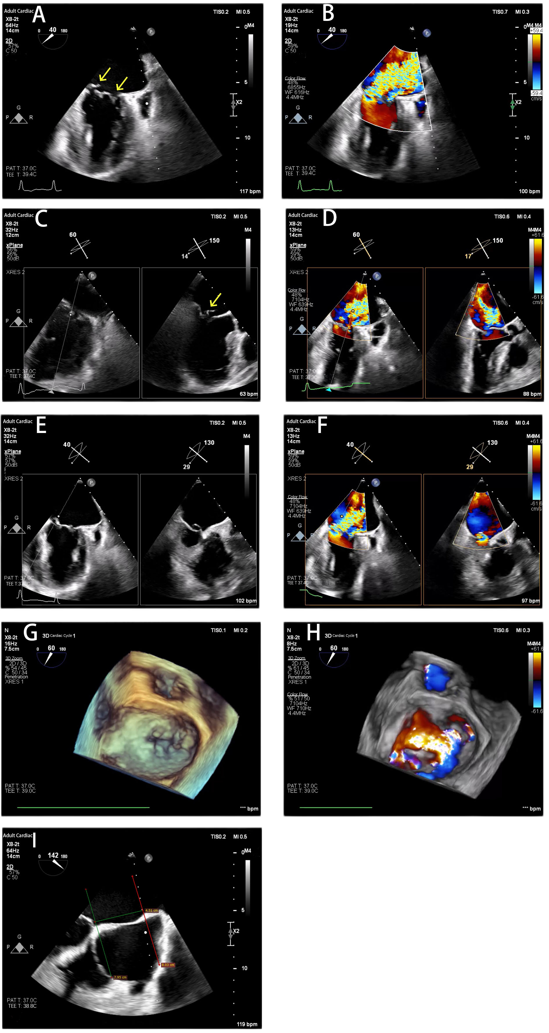
Figure 1. Transesophageal Echocardiography Before the Procedure. (A and B) X-Plane commissural view showed prolapse in zone 1 and zone 3 and severe mitral regurgitation before the procedure; (C and D) X-Plane A1/P1 view showed A1 prolapse and severe mitral regurgitation; (E and F) X-Plane A3/P3 view showed prolapse from P3-C2 and severe mitral regurgitation; (G and H) Three-dimensional TEE shows severe mitral valve regurgitation; (I) The length of the atrial septum was 4.51 cm. The yellow arrow indicated the origin of the Mitral regurgitation. TEE: Transesophageal echocardiography.



