


Transcatheter edge-to-edge repair for bileaflet commissural prolapse with reduced mitral valve orifice area: a case report
 0
0 Abstract
Transcatheter edge-to-edge repair (TEER) is a standard therapy for high-risk patients with severe mitral regurgitation (MR); however, its application in complex anatomies - such as multi-scallop commissural prolapse involving both commissures combined with a reduced mitral valve orifice area (MVOA) - poses significant challenges, including high risks of iatrogenic stenosis and leaflet grasping failure. This case report describes a 76-year-old female who presented with symptomatic severe degenerative MR. Transesophageal echocardiography confirmed multi-scallop commissural prolapse involving both commissures with a reduced MVOA of 3.9 cm2. The patient’s Society of Thoracic Surgeons score was 4.31%. A TEER procedure was performed using the DragonFlyTM system with a staged dual-clip strategy: a short-wide clip was first deployed in the C2 zone, followed by a short-narrow clip in the A1/P1 region. The inherent difficulty of addressing oblique coaptation lines at both commissures was mitigated by the system’s adjustable grasping angle, which allowed precise perpendicular orientation. The risk of iatrogenic stenosis from multiple clips within a small native orifice was managed through a strategic clip-size sequence - prioritizing a short-wide clip for stability followed by a short-narrow clip to minimize added obstruction - along with continuous hemodynamic monitoring. The device’s independent leaflet capture capability was crucial for securing the short, tethered posterior leaflet. This case demonstrates that a meticulously planned, imaging-guided TEER procedure using a staged, size-tailored dual-clip strategy can be a safe and effective treatment for multi-scallop commissural prolapse involving both commissures, even in the presence of a reduced MVOA, achieving durable hemodynamic and clinical improvement.
Keywords
INTRODUCTION
Degenerative mitral regurgitation (DMR) resulting from leaflet prolapse is a common valvular pathology. While surgical repair remains the reference standard, a substantial propo lves, transcatheter edge-to-edge repair (TEER) is being applied to increasingly complex anatomical scenarios beyond simple central A2/P2 pathology.
Among these complex anatomies, bileaflet prolapse - particularly when it involves the commissural areas (A1/P1 and A3/P3) - represents a significant challenge[3]. The technical difficulties are multifaceted: achieving stable clip grasp on multiple, often asymmetrical, prolapsing segments; orienting the device perpendicular to often oblique coaptation lines at the commissures; and avoiding entanglement with the subvalvular apparatus in a potentially crowded ventricular space. Furthermore, when such pathology coexists with a reduced native mitral valve orifice area (MVOA), the risk of creating iatrogenic functional mitral stenosis with the placement of multiple clips becomes a paramount concern[4]. Strategic pre-procedural planning, including precise clip size selection and deployment sequencing, is critical to balance regurgitation reduction against gradient generation.
CASE PRESENTATION
Clinical presentation
A 76-year-old female (body weight 54 kg, height 156 cm) was admitted with a chief complaint of intermittent palpitations for over five years, which had acutely worsened over the preceding week, accompanied by new-onset bilateral lower limb edema. Her past medical history was significant for hypertension, coronary heart disease (with mild non-obstructive disease on imaging), atrial fibrillation, type 2 diabetes mellitus, and chronic kidney disease (stage 3, eGFR 55.23 mL/min). Her surgical history included cholecystectomy and hysteromyomectomy. Physical examination revealed a blood pressure of 111/60 mmHg, an irregularly irregular pulse consistent with atrial fibrillation, and a grade 4/6 holosystolic blowing murmur at the cardiac apex. Mild pitting edema was noted in both lower limbs.
Diagnostic assessment
Biomarker analysis revealed elevated NT-proBNP (1,725 pg/mL) and high-sensitivity troponin T (21.1 ng/L). Transthoracic echocardiography (TTE) and subsequent comprehensive transesophageal echocardiography (TEE) were performed for anatomical and functional assessment [Figure 1]. Key echocardiographic findings were as follows: Left ventricular ejection fraction (LVEF) was 60% with left ventricular end-diastolic dimension (LVEDD) of 47 mm and a left atrial diameter (LAD) of 52 mm. Carpentier Type II DMR with distinct prolapse of the A1 scallop (involving the anterolateral commissure) and the P3 scallop extending into the C2 region (involving the posteromedial commissure). Quantitative assessment confirmed severe mitral regurgitation (MR): effective regurgitant orifice area (EROA) 0.49 cm2, regurgitant volume (RV)
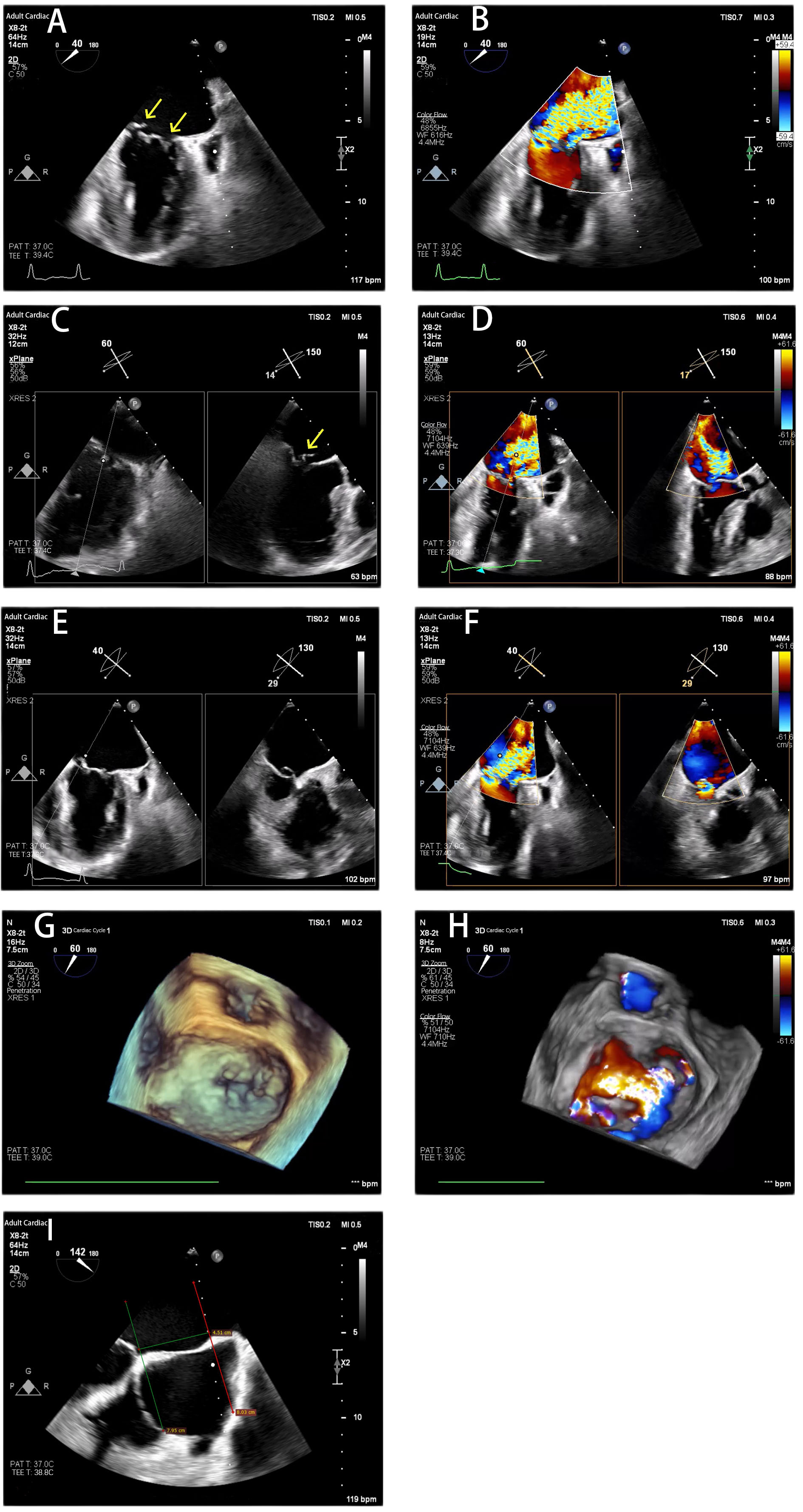
Figure 1. Transesophageal Echocardiography Before the Procedure. (A and B) X-Plane commissural view showed prolapse in zone 1 and zone 3 and severe mitral regurgitation before the procedure; (C and D) X-Plane A1/P1 view showed A1 prolapse and severe mitral regurgitation; (E and F) X-Plane A3/P3 view showed prolapse from P3-C2 and severe mitral regurgitation; (G and H) Three-dimensional TEE shows severe mitral valve regurgitation; (I) The length of the atrial septum was 4.51 cm. The yellow arrow indicated the origin of the Mitral regurgitation. TEE: Transesophageal echocardiography.
The patient was classified as New York Heart Association (NYHA) functional Class III. Her calculated Society of Thoracic Surgeons (STS) score for operative mortality was 4.31%. After a multidisciplinary heart team discussion and consideration of the patient’s preference to avoid open-heart surgery, percutaneous TEER was recommended, and the patient consented to the procedure.
TEER procedure
The procedure was performed under general anesthesia with fluoroscopic and 2D/3D TEE guidance. A key pre-procedural decision was the atrial septal puncture site. To achieve an optimal trajectory for addressing both commissures, a posterior-superior puncture was performed at a height of 4.21 cm from the mitral annular plane [Figure 2A].
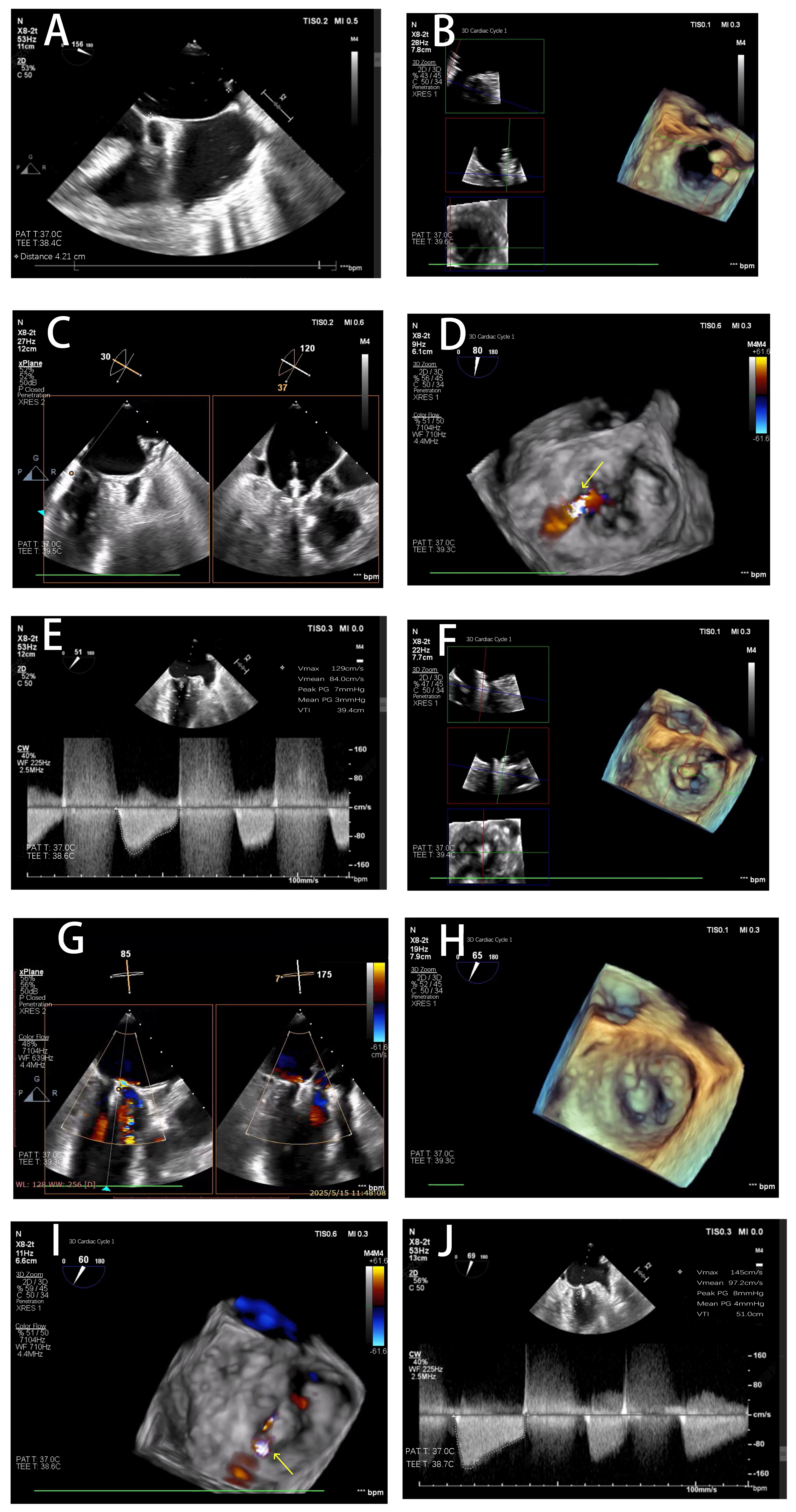
Figure 2. The Procedure of Mitral Transcatheter Edge-to-Edge Repair. (A) A posterior-superior atrial septal puncture was performed at a height of 4.21 cm from the mitral annular plane; (B) The SW0609 clip was oriented perpendicular to the line of coaptation (approximately 11 o’clock to 5 o’clock position) under 3D TEE guidance; (C) Both leaflets were on the clip arms; (D) Complete resolution of the P3-C2 prolapse with no residual regurgitation in that region after the first clip and significant residual MR from the untreated A1 prolapse persisted; (E) The mean trans-mitral pressure gradient (TMPG) measured 3 mmHg after the first clip; (F) The SN0409 clip was re-oriented (1 o’clock to 7 o’clock) to align with the anterolateral commissure; (G) Both leaflets were captured; (H) Stable tissue bridges on 3D imaging after the second clip deployment; (I) Only trace residual lateral MR were existed after the second clip deployment; (J) The final TMPG was 4 mmHg after the procedure. The yellow arrow indicated the residual regurgitation flow after clip deployment. TEE: Transesophageal echocardiography; MR: mitral regurgitation.
A dual-clip strategy was planned, prioritizing the more severe P3-C2 prolapse. Transseptal access was obtained, and the DragonFlyTM delivery system was advanced into the left atrium. A short-wide clip (SW0609) was chosen for the C2 zone to provide a broad grasping area for the prolapsing P3 segment and the adjacent commissural tissue. The clip was oriented perpendicular to the line of coaptation (approximately 11 o’clock to 5 o’clock position) under 3D TEE guidance, carefully advanced into the ventricle, and retracted to capture both leaflets [Figure 2B and C]. After confirming stable grasp and leaflet insertion, the clip was closed and deployed. Post-deployment TEE showed complete resolution of the P3-C2 prolapse with no residual regurgitation in that region [Figure 2D]. The mean trans-mitral pressure gradient (TMPG) measured 3 mmHg [Figure 2E].
Significant residual MR from the untreated A1 prolapse persisted. A second short-narrow clip (SN0409) was selected for the A1/P1 region to minimize the additive effect on the valve gradient. The clip was re-oriented (1 o’clock to 7 o’clock) to align with the anterolateral commissure [Figure 2F]. Precise maneuvering was required to grasp the short and tethered P1 leaflet effectively [Figure 2G]. Following deployment, final assessment showed only trace residual lateral MR, stable tissue bridges on 3D imaging [Figure 2H and I], and a final TMPG of 4 mmHg [Figure 2J]. Both clips were well-positioned without impingement on the left ventricular outflow tract.
Post-procedural course and follow-up
The patient recovered without procedural complications. Post-procedure TTE prior to discharge confirmed the result: MR was reduced to mild, MVOA was 2.3 cm2, and the mean TMPG was 3 mmHg. Rivaroxaban, atorvastatin, sacubitril/valsartan, hydrochlorothiazide, spironolactone, metformin, dapagliflozin, and acarbose were prescribed. No other rhythm-control intervention was performed. The patient’s symptoms improved markedly. At the six-month clinical follow-up, she reported no heart failure symptoms (NYHA Class I), had not been rehospitalized, and her NT-proBNP level had decreased to 306 pg/mL. The patient was in normal sinus rhythm. Repeat echocardiography demonstrated stable clip positions, mild MR, trace TR, preserved LVEF (60%), and positive reverse remodeling of the left-sided cardiac chambers, with LAD of
Echocardiographic parameters before and after transcatheter edge-to-edge repair
| Parameters | Before operation | Discharge | Six-month follow-up |
| LAD, mm | 52 | 45 | 37 |
| LVEDD, mm | 47 | 42 | 43 |
| RAD, mm | 44 | 33 | 40 |
| RVD, mm | 24 | 20 | 20 |
| MR grade | Severe | Mild | Mild |
| TR grade | Moderate | Trace | Trace |
| Mean TMPG, mmHg | 4 | 3 | 3 |
| LVEF, % | 60 | 60 | 60 |
DISCUSSION
We report a case of successful TEER in a patient with multi-scallop commissural prolapse involving both commissures, combined with a reduced MVOA, using the DragonFlyTM transcatheter mitral valve repair system. The percutaneous treatment of commissural pathology remains notably challenging[5]. Unlike central A2/P2 lesions, the coaptation line at the commissures is oblique, making perpendicular clip alignment more technically demanding. Furthermore, the ventricular space is often narrower, increasing the risk of chordal entanglement or interaction with the aortic valve in the case of A1 prolapse. In bileaflet prolapse, the pathology is not isolated; the opposing leaflet segments may have different lengths and tethering characteristics, as seen in this case with a very short P1. This asymmetry complicates simultaneous leaflet grasping and increases the risk of single leaflet device attachment (SLDA) if the grippers cannot achieve equal purchase[6].
Our strategy of addressing the posteromedial (P3-C2) prolapse first was deliberate. This region often has greater prolapse height and width, and securing it first can stabilize the valve architecture, potentially making subsequent clip placement in the opposite commissure more predictable. The use of a short-wide clip (SW0609) in this zone provided a larger surface area for grasping the prolapsing tissue, enhancing stability.
A pre-procedural MVOA of < 4.0 cm2 is a recognized risk factor for post-TEER significant stenosis (often defined as a mean gradient ≥ 5 mmHg)[7]. Placing two clips in such a scenario compounds this risk. The management of this trade-off - between adequate MR reduction and acceptable gradient - is central to planning. Several factors in this case contributed to the favorable hemodynamic outcome (TMPG of
Although the MitraClip and PASCAL are the most frequently used devices in most of the countries, the DragonFlyTM clip system, a novel Chinese device, incorporates a compressible central filler with an adjustable grasping angle (0-45°), independent leaflet capture, and four clip sizes (width 4-6 mm, length 9-12 mm)[8]. MitraClip G4 offers four sizes and mechanical locking, whereas PASCAL features a 10-mm central spacer and passive self-locking. The DragonFlyTM clip system’s specific features facilitated this complex anatomy. Its adjustable grasping angle of up to 45° allowed for fine-tuning of the arm orientation to achieve a perpendicular approach to both the oblique C2 and A1 coaptation lines from a single septal puncture site. This adaptability is crucial in commissural work where “one orientation does not fit all.” Furthermore, the independent leaflet capture capability and the ability to assess leaflet insertion prior to final closure were invaluable, especially for the short, tethered P1 segment. These features reduce the risk of incomplete leaflet capture and subsequent SLDA. The central sealing mesh of the DragonFlyTM may also contribute to a more favorable hemodynamic profile by promoting tissue ingrowth and creating a smoother flow interface, potentially mitigating turbulence that can lead to gradient overestimation.
CONCLUSION
This case demonstrates that transcatheter edge-to-edge repair is a viable and effective therapeutic option for high-surgical-risk patients presenting with complex degenerative mitral regurgitation due to bileaflet commissural prolapse, even in the presence of a reduced MVOA. Success hinges on comprehensive multi-modality imaging, meticulous procedural planning with a staged multi-clip strategy, prudent clip size selection, and the utilization of next-generation device features that enhance grasping control and adaptability. This approach allows for effective regurgitation reduction while maintaining satisfactory transvalvular hemodynamics, leading to significant clinical and echocardiographic improvement.
DECLARATIONS
Authors’ contributions
Concept and design: Chen J
Procedure performance and intraoperative management: Xu C, Yang S, Chen J
Imaging acquisition and analysis: Song H, Zhou Q
Data collection and analysis: Xu C, Yang S
Manuscript drafting: Xu C
Critical revision and final approval of the manuscript: Xu C, Yang S, Song H, Zhou Q, Chen J
Availability of data and materials
Not applicable.
AI and AI-assisted tools statement
Not applicable.
Financial support and sponsorship
None.
Conflicts of interest
All authors declared that there are no conflicts of interest.
Ethical approval and consent to participate
The Ethics Committee of Renmin Hospital of Wuhan University approved the study (No. WDRY2026-K083). The Ethics Committee also granted a waiver of written informed consent.
Consent for publication
Written informed consent was obtained from the patient for the publication of clinical data and relevant imaging materials.
Copyright
© The Author(s) 2026.
REFERENCES
1. Praz F, Borger MA, Lanz J, et al. 2025 ESC/EACTS Guidelines for the management of valvular heart disease. Eur Heart J. 2025;46:4635-736.
2. Amicone S, Zannoni J, Barletta M, et al. Transcatheter mitral edge-to-edge repair: a selection of the mitral regurgitation subtype and definition of the optimal time for intervention. Rev Cardiovasc Med. 2025;26:44073.
3. Li Y, Guo XN, Wu Y, et al. Safety and efficacy of novel morphology classification-guided mitral valve transcatheter edge-to-edge repair for patients with commissural degenerative mitral regurgitation: design and rationale of the TEER-CD trial. Rev Cardiovasc Med. 2025;26:39373.
4. Kharsa C, Sammour YM, Bazari R, et al. Outcomes of mitral transcatheter edge-to-edge repair in patients with small mitral valve area. Struct Heart. 2026;10:100772.
5. Nita N, Paukovitsch M, Felbel D, et al. Transcatheter edge-to-edge repair in anatomically complex degenerative mitral regurgitation: 3-year outcomes from a real-world registry. Clin Res Cardiol. 2025;114:904-14.
6. Bhatia K, Gupta S, Carter K, et al. Single-leaflet device attachment after mitral transcatheter edge-to-edge repair: systematic review and meta-analysis. JACC Cardiovasc Interv. 2024;17:2571-4.
7. Neuss M, Schau T, Isotani A, Pilz M, Schöpp M, Butter C. Elevated mitral valve pressure gradient after mitraclip implantation deteriorates long-term outcome in patients with severe mitral regurgitation and severe heart failure. JACC Cardiovasc Interv. 2017;10:931-9.
Cite This Article
 Open Access
How to Cite
Download Citation
Export Citation File:
Type of Import
Tips on Downloading Citation
Citation Manager File Format
Type of Import
Direct Import: When the Direct Import option is selected (the default state), a dialogue box will give you the option to Save or Open the downloaded citation data. Choosing Open will either launch your citation manager or give you a choice of applications with which to use the metadata. The Save option saves the file locally for later use.
Indirect Import: When the Indirect Import option is selected, the metadata is displayed and may be copied and pasted as needed.
About This Article
Special Topic
Copyright

Data & Comments
Data
0









Comments
Comments must be written in English. Spam, offensive content, impersonation, and private information will not be permitted. If any comment is reported and identified as inappropriate content by OAE staff, the comment will be removed without notice. If you have any queries or need any help, please contact us at [email protected].