


fig5
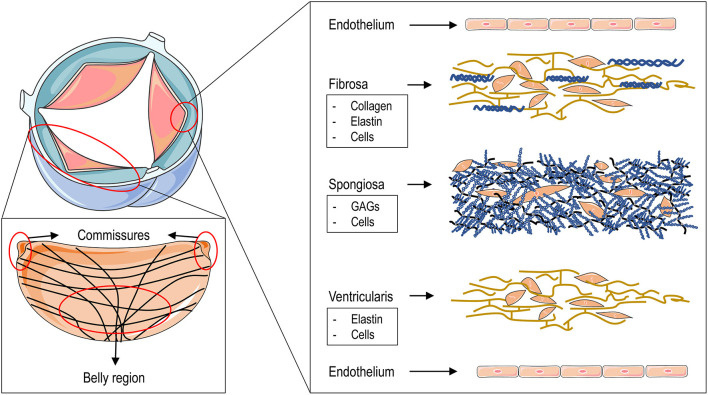
Figure 5. Aortic valve structure and leaflet layers. The red area illustrates the circumferential arrangement of collagen fibers within an aortic leaflet, with crossovers emerging centrally from the commissures. The microstructure of the three layers - fibrous, spongiosa, and ventricular - extends from the aortic to the ventricular surface, each characterized by distinct cellular and extracellular components. Both surfaces are lined by valvular endothelial cells adherent to the basement membrane. Reproduced from Ref.[14] with permission from Rizzi, Ragazzini and Pesce © 2022. GAGs: Glycosaminoglycans.


