


fig18
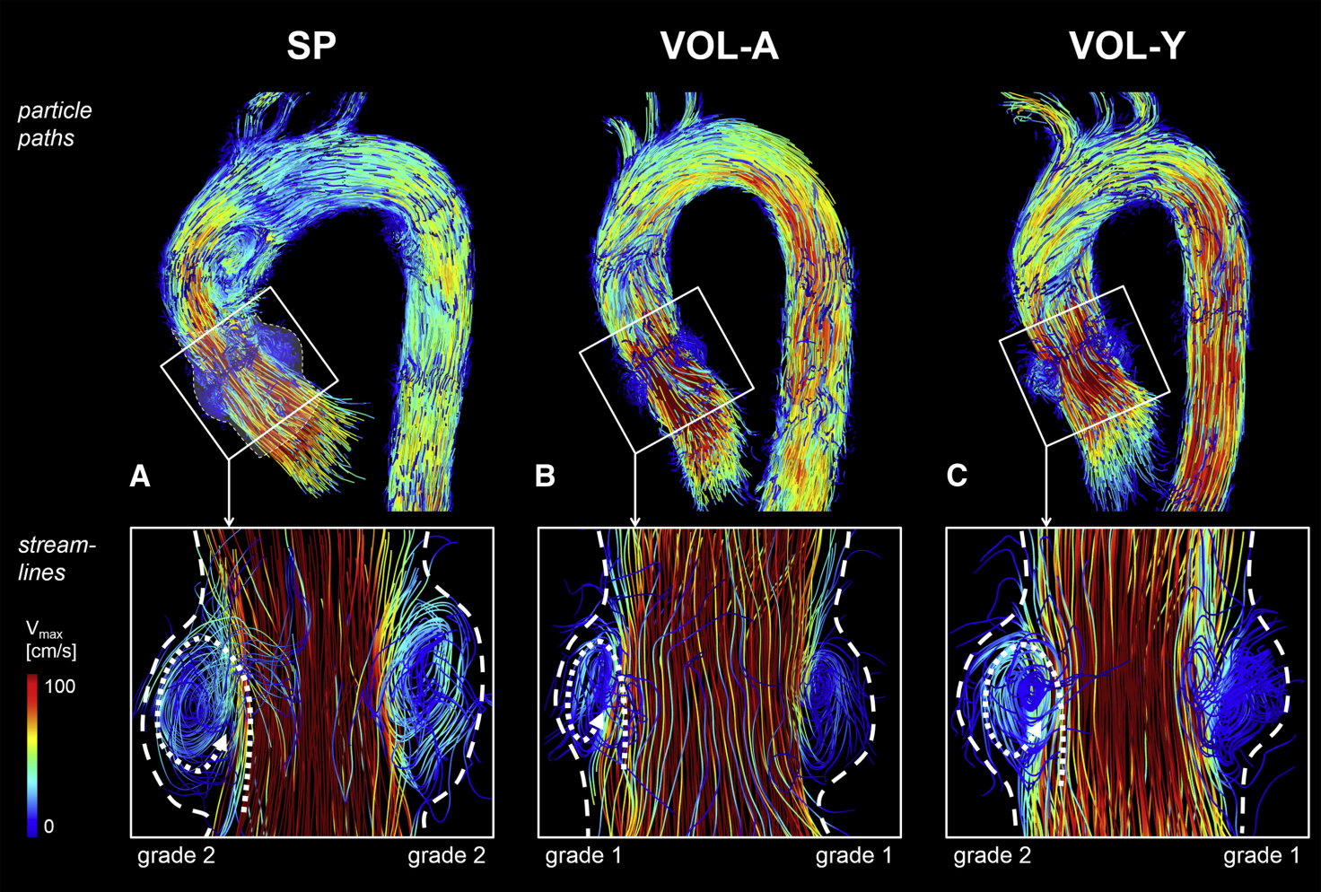
Figure 18. Sinus vortices visualized by 4D flow MRI. Top row: particle pathlines at peak systole in a 60-year-old patient with a sinus prosthesis (SP, A), a 53-year-old age-matched volunteer (VOL-A, B), and a 30-year-old young volunteer (VOL-Y, C). Bottom row: instantaneous streamlines showing right and left coronary sinus vortices, forming behind the open cusps during systole and persisting into early diastole. Vortex configuration in patients closely resembled that of healthy volunteers. Dashed lines indicate sinus borders, and dotted lines indicate vortex direction.. SP: Sinus prosthesis; VOL-A: age-matched volunteer; VOL-Y: young volunteer; Vmax: peak velocity. Reproduced from Ref.[5] with permission.


