


fig2
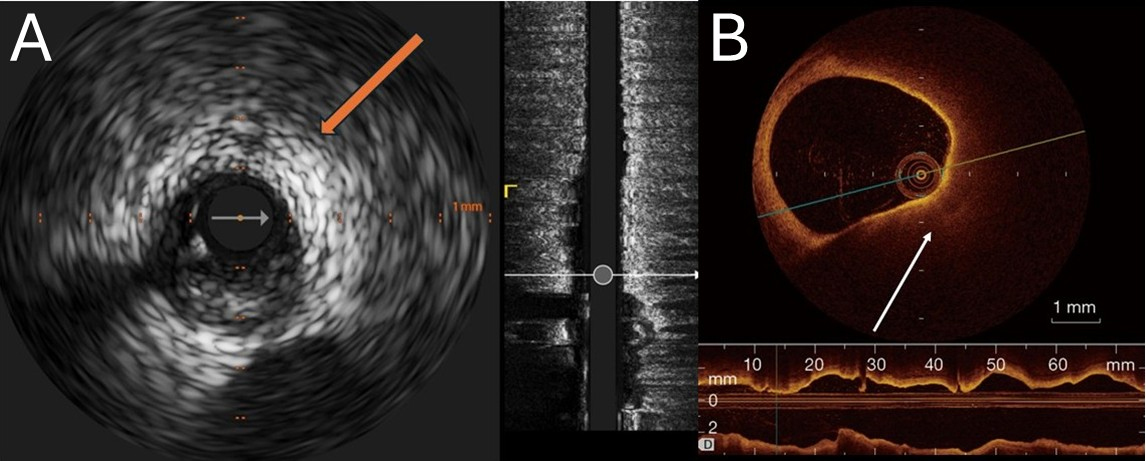
Figure 2. Representative intracoronary imaging of high-burden lipid-rich plaques. (A) Grayscale IVUS of the left circumflex artery showing an eccentric, predominantly hypoechoic plaque with marked deep ultrasound attenuation (arrow) and no bright calcific leading edge, consistent with attenuated plaque; (B) OCT from routine clinical imaging demonstrating a large, signal-poor, diffusely bordered lipid-rich plaque with long contiguous involvement on the longitudinal view. Scale bars =1 mm. IVUS: Intravascular ultrasound; OCT: optical coherence tomography. Images were acquired during routine care at our institution and are fully de-identified.


