


fig2
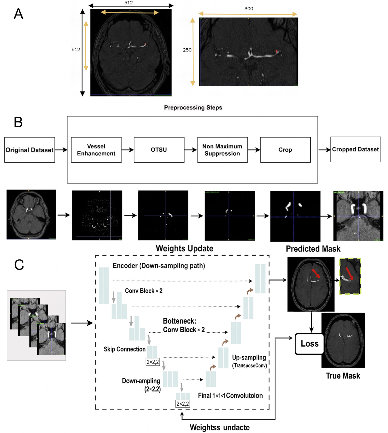
Figure 2. Example of a single 3D TOF-MRA case shown at different stages of our pipeline. (A) Manual cropping; (B) Automatic cropping method with traditional image processing methods: vessel-enhanced image obtained with the multi-scale Hessian filter and final cropped region of interest (ROI) based on the automatic vessel mask, which is then used as input for the aneurysm segmentation network (C).


