fig5

Calcified coronary nodules: pathophysiology, diagnosis, and interventional challenges

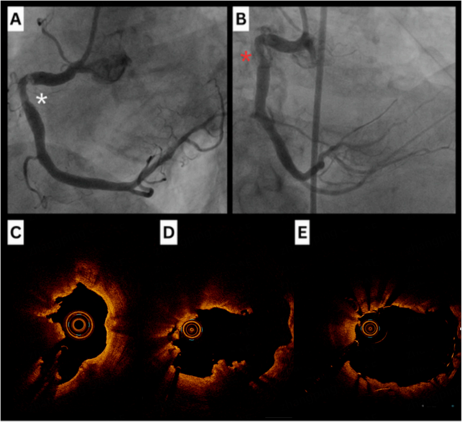
Figure 5. Case of successful rotablation of an eruptive nodule with favorable wire bias. (A and B) Angiographic views of severe mid-RCA disease (asterisk); (C) OCT showing an eruptive calcified nodule. (D) OCT post-RA using a 1.75mm burr and RotaWire Extra-support wire demonstrating embedded bias; (E) OCT showing excellent stent apposition and increased luminal area following PCI with a 4.0 DES and post-dilation using a 5.0 NC balloon. RCA: Right coronary artery; OCT: optical coherence tomography; RA: rotational atherectomy; NC: non-compliant; DES: drug-eluting stent.

Vessel Plus
ISSN 2574-1209 (Online)
Follow Us

Portico

All published articles are preserved here permanently:

https://www.portico.org/publishers/oae/

Portico

All published articles are preserved here permanently:

https://www.portico.org/publishers/oae/