


fig3
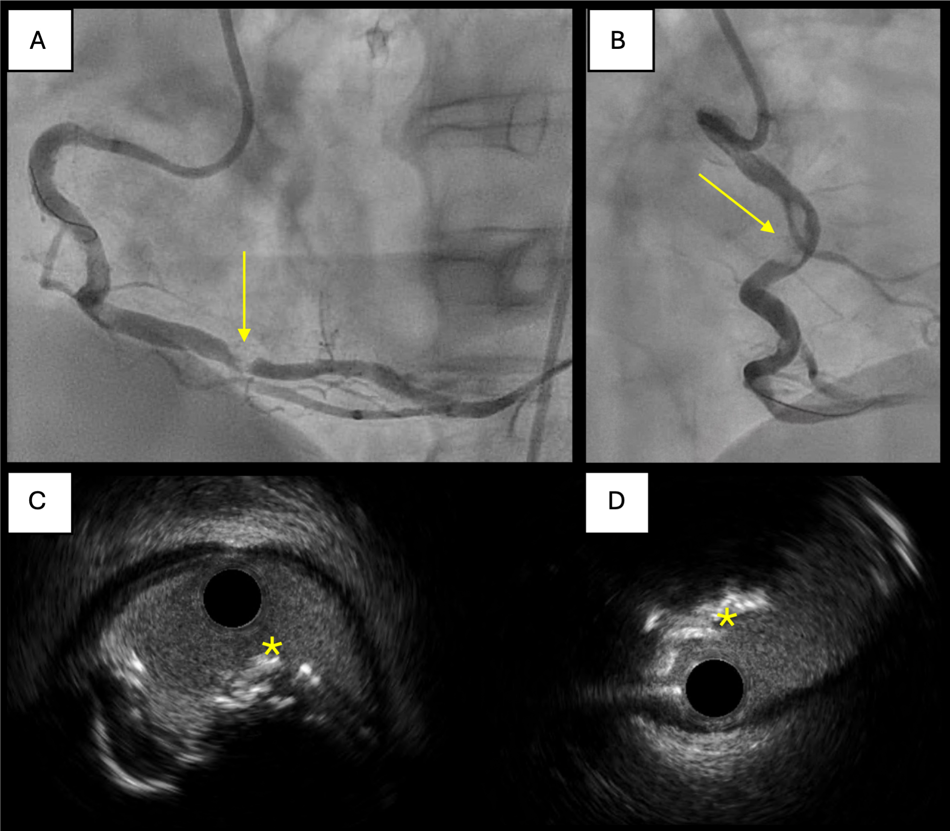
Figure 3. A 69-year-old patient successfully treated for a calcified nodule. The calcified nodule appears as a radiolucent lesion in (A) and (B), indicated by yellow arrows; The calcified lumen is shown in (C) and (D), indicated by yellow asterisks.


