


fig2
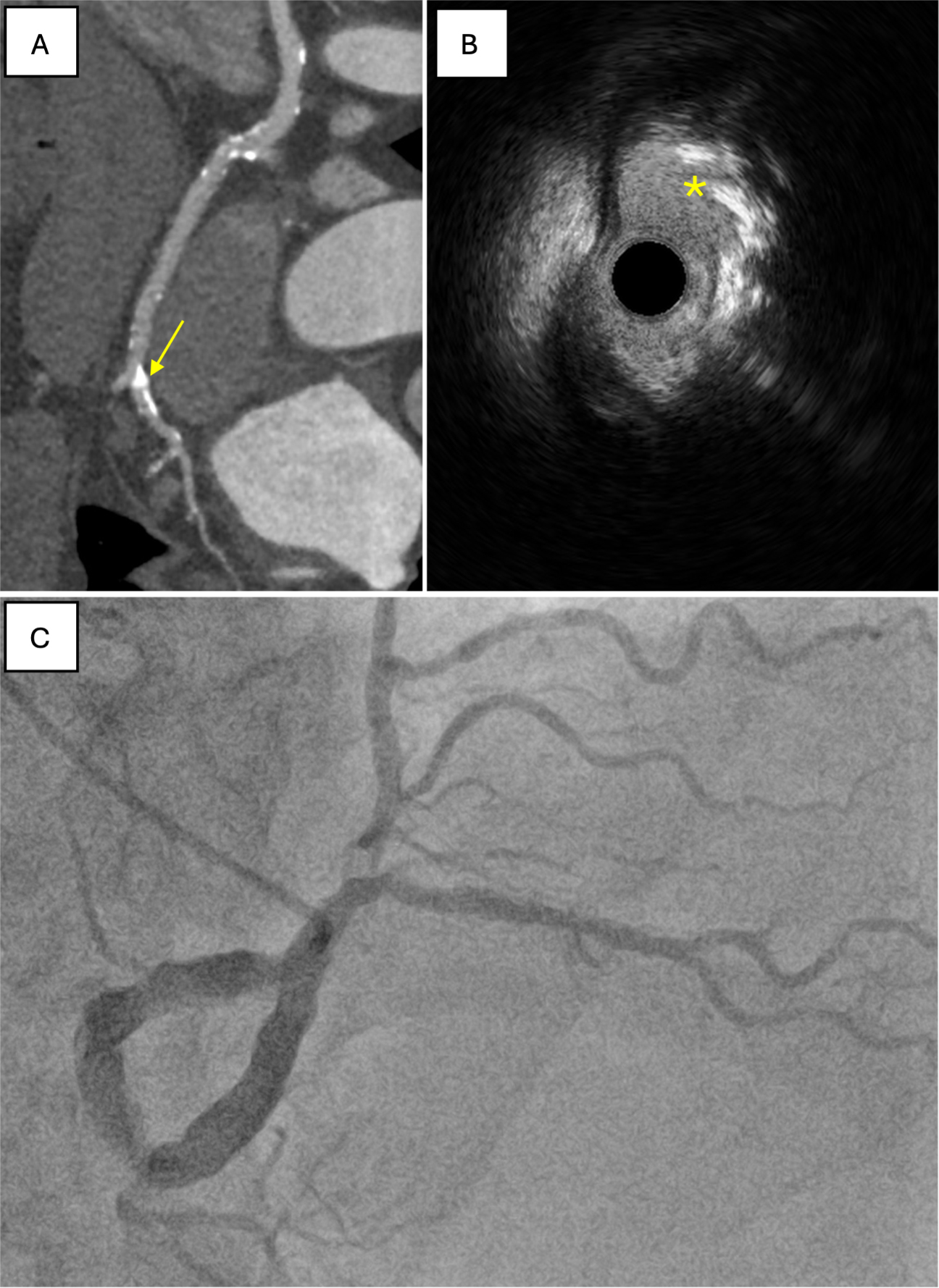
Figure 2. Features of CCNs identifiable on ultra-low dose, 640-slice volumetric CCTA. (A) prospective ECG-gated imaging with intravenous contrast following pre-treatment with sublingual nitrates. The effective dose for the angiographic run was 10.6 mSv/758 DLP. Extensive coronary calcification is visible, indicated by yellow arrows; This is compared with IVUS, where the nodule is marked by a yellow asterisk (B), and with angiography (C). CCNs: Calcified coronary nodules; CCTA: coronary CT angiography; ECG: electrocardiogram; DLP: dose-length product.


