fig4

Functional classification of extrasystoles based on hemodynamic features and risk of arterial vascular complications

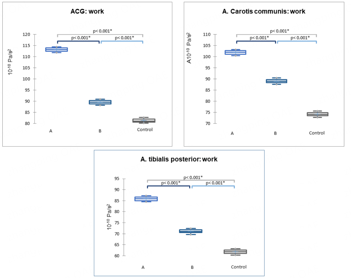
Figure 4. Work parameter based on digital ACG and SG data from the common carotid and posterior tibial arteries (P < 0.001) in subgroups A, B, and the control group. Data are presented as medians (horizontal line), means (cross), 1st and 3rd quartile boundaries (box edges), and minima and maxima (whisker ends). ACG: Apexcardiography; SG: sphygmography.

Vessel Plus
ISSN 2574-1209 (Online)
Follow Us

Portico

All published articles are preserved here permanently:

https://www.portico.org/publishers/oae/

Portico

All published articles are preserved here permanently:

https://www.portico.org/publishers/oae/