


fig4
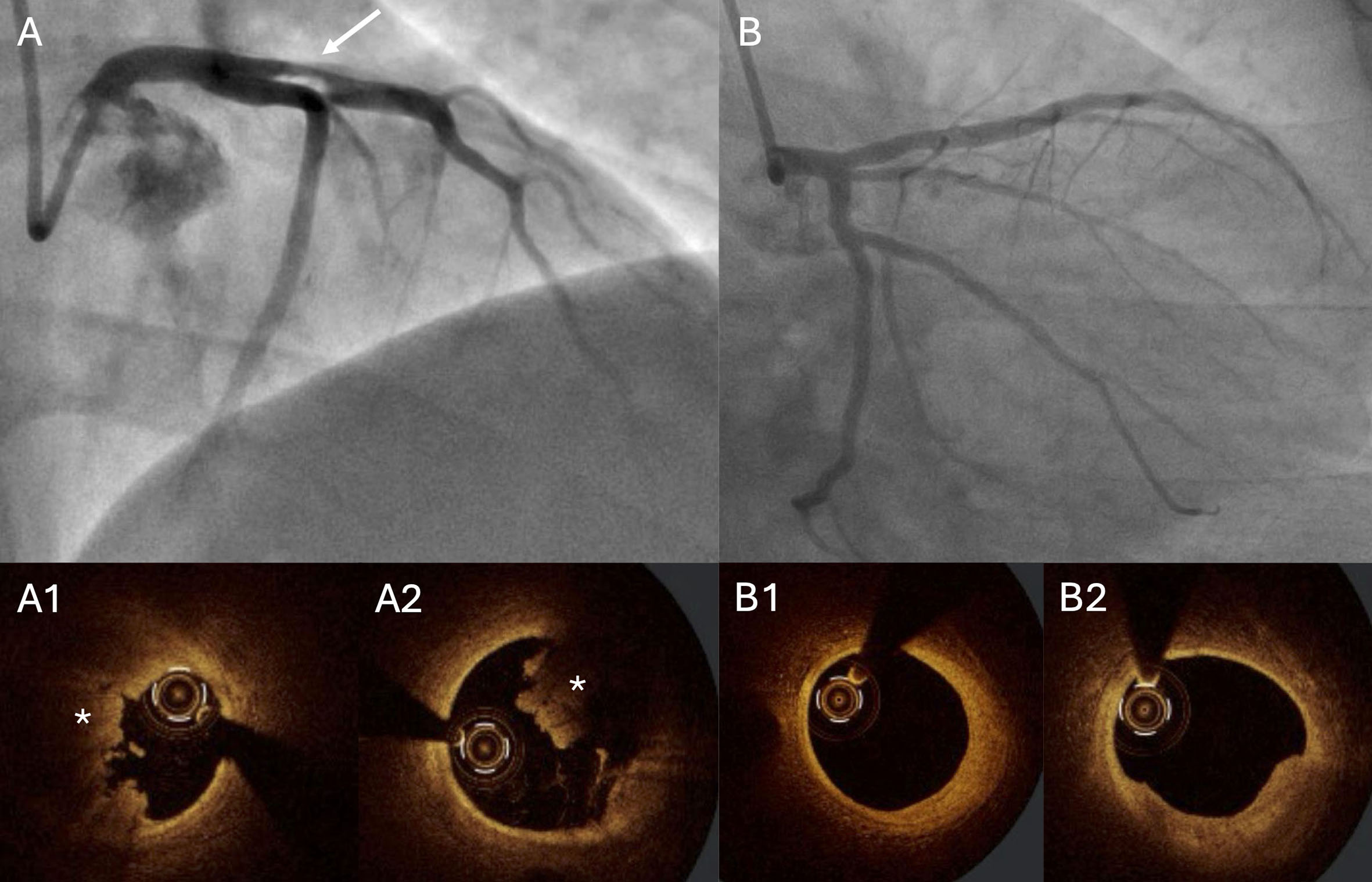
Figure 4. Patient admitted with ST-elevation myocardial infarction. A 34-year-old patient was admitted with anterior wall STEMI. Angiography revealed significant stenosis in the proximal LAD (A; arrow). Thrombectomy was performed, followed by OCT. The OCT image showed a high thrombus burden (A1 and A2; asterisk). PCI was deferred, and the patient received DAPT and anticoagulation (enoxaparin). Angiography with OCT was repeated one week later (B), showing only a small thrombus, fibrous plaque without significant stenosis, and no signs of plaque rupture (B1 and B2). The operators decided not to stent the proximal LAD and recommended OMT. DAPT: Dual antiplatelet therapy; LAD: left anterior descending artery; OCT: optical coherence tomography; OMT: optimal medical therapy; PCI: percutaneous coronary intervention; STEMI: ST-elevation myocardial infarction


