


fig1
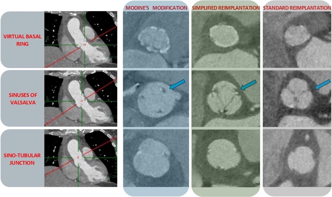
Figure 1. Early postoperative ECG-gated contrast-enhanced computed tomography (CT) scans showing short-axis views at various levels of the aortic root: the virtual basal ring, the sinuses of Valsalva, and the commissures. Note the absence of contrast agent behind the commissures in both the standard and simplified reimplantation techniques, whereas contrast is clearly visible behind the commissures in the Modine modification (indicated by blue arrows).


