


Metabolic memory and immune evasion in MASLD-HCC progression
 0
0 Abstract
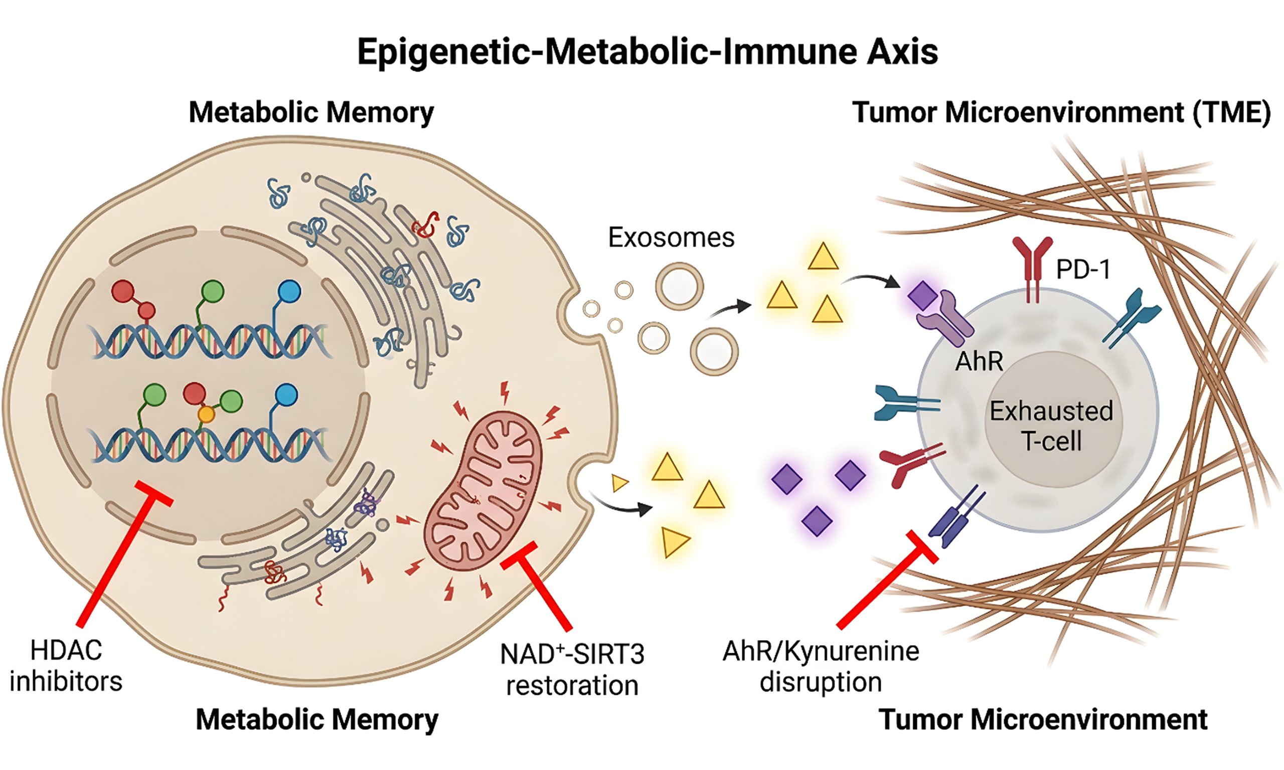
Metabolic dysfunction-associated steatotic liver disease (MASLD) is a leading cause of hepatocellular carcinoma (HCC). Central to its pathogenesis is “metabolic memory” - the persistence of pro-tumorigenic alterations after resolution of the initial metabolic insult. This review examines how metabolic memory, encoded across multiple layers of the epigenome - including DNA methylation, repressive histone modifications, and non-coding RNA networks - and propagated through mitochondrial dysfunction and lipotoxic metabolites, sustains a locked immunosuppressive state that drives immune evasion in MASLD-HCC. We delineate convergent pathways - including the unfolded protein response, dysregulated autophagy, tryptophan-kynurenine-aryl hydrocarbon receptor (AHR) signaling - that couple metabolic memory to immune checkpoint upregulation and T-cell exhaustion. We further discuss how metabolic byproducts such as lactate drive immunosuppression through histone lactylation, and how extracellular vesicles transmit metabolic stress signatures to remotely reprogram immune cells within the tumor microenvironment. From a therapeutic perspective, we evaluate strategies targeting the reversibility of this memory, including NAD+-SIRT3 axis restoration and epigenetic modulators such as HDAC inhibitors, and discuss their potential to sensitize tumors to immunotherapy. We also explore emerging approaches aimed at disrupting the kynurenine-AHR axis and overcoming fibrotic barriers to immune infiltration. Disrupting this “epigenetic-metabolic-immune axis” may be essential for halting disease progression.
Keywords
INTRODUCTION
Metabolic dysfunction-associated steatotic liver disease (MASLD) has emerged as the leading cause of chronic liver disease worldwide, affecting approximately 30% of the global adult population[1,2]. Driven by the escalating epidemics of obesity, type 2 diabetes mellitus, and metabolic syndrome, the disease burden of MASLD continues to rise[3]. Within the natural history of MASLD, approximately 20% of patients progress to metabolic dysfunction-associated steatohepatitis (MASH), and a subset of these cases further advance to cirrhosis and ultimately hepatocellular carcinoma (HCC)[4]. Notably, a substantial proportion of patients with MASLD-associated HCC (MASLD-HCC) do not traverse a clinically evident cirrhotic stage, suggesting the existence of non-canonical carcinogenic pathways[5]. This distinctive feature renders traditional cirrhosis-based surveillance strategies inadequate for the early detection of such HCC cases, which are often associated with a poorer clinical prognosis.
Despite the efficacy of metabolic interventions - including lifestyle modifications, insulin sensitizers, and lipid-lowering agents - in ameliorating hepatic steatosis and inflammation, epidemiological evidence indicates that prior or resolved MASLD continues to confer a persistently elevated risk of HCC development[6,7]. In other words, even when the metabolic derangements are seemingly “corrected”, the liver appears to retain a form of “traumatic memory” that sustains malignant transformation[8,9]. This phenomenon bears a striking resemblance to the well-established concept of “metabolic memory” in diabetes research, wherein early exposure to hyperglycemia instigates epigenetic modifications that drive the relentless progression of vascular and neurological complications despite subsequent glycemic control[10,11]. In the context of hepatic oncology, two critical questions warrant systematic investigation: Does a parallel “metabolic memory” operate in the liver? And if so, what are its molecular vectors?
Concurrently, immune evasion constitutes a cardinal hallmark of tumorigenesis[12]. In the setting of MASLD, the hepatic immune microenvironment undergoes a profound remodeling from “metainflammation” to a state of “immune paralysis”[13,14]. Lipotoxic metabolites, oxidative stress, and chronic endoplasmic reticulum (ER) stress not only inflict hepatocellular injury but also reprogram infiltrating and resident immune cells toward suppressive phenotypes. More critically, these aberrant alterations in the immune microenvironment may persist long after the resolution of the initial metabolic insult, thereby establishing a form of “mnemonic immune evasion”[15]. However, a systematic mechanistic integration of how metabolic memory - through epigenetic regulation, metabolite signaling, and intercellular crosstalk - perpetuates this locked state of immunosuppression remains conspicuously absent.
Before dissecting the mechanisms, it is essential to clarify the terminology that underpins this review. In the context of chronic metabolic diseases, three related but mechanistically distinct concepts are frequently conflated: metabolic reprogramming, metabolic-immune coupling, and metabolic memory. Metabolic reprogramming refers to the adaptive alterations in cellular metabolic pathways - such as the shift from oxidative phosphorylation (OXPHOS) to glycolysis (the Warburg effect) - that enable cells to survive and function under altered environmental conditions. This is a dynamic, reversible, and often homeostatic response to ongoing metabolic stress. Metabolic-immune coupling describes the real-time regulation of immune cell phenotype and function by metabolites; for instance, succinate can acutely stabilize hypoxia-inducible factor 1 alpha (HIF-1α) in pro-inflammatory macrophages, and lactate can immediately suppress T-cell effector function. This coupling is stimulus-dependent and typically resolves once the metabolic stimulus is removed. In contrast, metabolic memory, as operationally defined in this review, refers specifically to the persistence of pro-tumorigenic cellular alterations - encoded through stable epigenetic modifications, durable mitochondrial dysfunction, and self-sustaining metabolic byproduct signaling - that remain long after the initial metabolic insult has been corrected or resolved. Therefore, the aims of this review are fourfold: (1) To systematically delineate the mechanisms underpinning metabolic memory in MASLD-HCC progression, with a particular emphasis on epigenetic modifications, mitochondrial dysfunction, and the enduring effects of metabolic byproducts; (2) To dissect the immune evasion pathways driven by metabolic memory, including sustained upregulation of immune checkpoints, metabolic competition, and lactate-mediated immunosuppression; (3) To explore the critical nodes of convergence between these two pathogenic axes [e.g., ER stress-unfolded protein response (UPR), autophagy, AHR signaling]; and (4) To summarize potential therapeutic strategies targeting the metabolic memory-immune evasion axis and to highlight current knowledge gaps and future research directions. By integrating these advances, we aim to provide novel insights into the distinctive biological behavior of MASLD-related HCC and to establish a theoretical foundation for the development of combined metabolic-immunotherapeutic strategies.
METABOLIC REPROGRAMMING AND METABOLIC MEMORY IN MASLD-HCC
Accumulation of lipotoxic metabolites
The aberrant accumulation of lipotoxic metabolites represents a central mechanism driving metabolic reprogramming and disease progression during the transition from MASH to HCC. These metabolites, which include free fatty acids, cholesterol, and ceramides, not only inflict direct hepatocellular injury but also foster tumorigenesis by remodeling the hepatic microenvironment[16]. Leng et al. demonstrated that the LIX1L protein is markedly upregulated in liver tissues from patients with MASH, in murine models of the disease, and in hepatocytes subjected to palmitate stimulation. Mechanistically, LIX1L promotes CD36 expression by stabilizing CD36 mRNA, thereby exacerbating hepatic lipid accumulation, inflammation, and fibrosis[17]. As a key fatty acid translocase, elevated CD36 expression facilitates enhanced free fatty acid uptake, thereby establishing a lipotoxic positive feedback loop[18]. Furthermore, multi-omics analyses have revealed that lipotoxic species, such as ceramides and diacylglycerols, accumulate significantly in cardiac tissue under conditions of aging and metabolic stress, suggesting that analogous mechanisms of lipotoxic metabolite accrual may play a pivotal role in the establishment of hepatic metabolic memory[19,20]. The persistent presence of these lipid species underpins a pro-inflammatory and pro-fibrotic microenvironment that primes the liver for HCC development.
Epigenetically mediated metabolic memory
Metabolic stress-induced epigenetic alterations constitute a fundamental mechanism for the establishment of “metabolic memory” - a phenomenon whereby gene expression patterns remain persistently altered long after the resolution of the initial insult, thereby propelling the progression from MASLD to HCC[8]. This memory is encoded across multiple layers of the epigenome. Gradual DNA methylation changes have been documented across the histological spectrum of MASLD, correlating with accelerated epigenetic aging[21]. Repressive histone modifications, such as H3K9me3 and H3K27me3, silence tumor suppressors and immunomodulatory genes, linking nutrient excess to durable chromatin remodeling[22]. In addition to these transcriptional controls, this form of cellular memory extends beyond transcriptional regulation to encompass non-coding RNAs and extensive reprogramming at the post-transcriptional level. Studies have demonstrated that chronic ER stress, triggered by prolonged exposure to a high-fat diet (HFD), elicits a distinct post-transcriptional regulatory landscape. Remarkably, approximately 70% of HFD-induced changes in gene expression are governed at the translational level. MicroRNAs and long non-coding RNAs have emerged as critical carriers of metabolic memory, regulating gene expression both intracellularly and via extracellular vesicles (EVs)[23,24]. A specific subset of mRNAs, characterized by suboptimal codon usage and short poly(A) tails under normal dietary conditions, becomes selectively activated via HFD-induced poly(A) tail lengthening[25]. These transcripts are enriched for genes involved in cell cycle regulation, immune response, fibrogenesis, and tissue remodeling, and their expression levels correlate positively with the histologic severity of MASH in both murine models and human biopsy specimens. This mode of regulation - orchestrated by the coordination of polyadenylation and deadenylation via cis-acting elements within the 3’ untranslated region (UTR) - represents a crucial “memory” mechanism at the epitranscriptomic level. Together with the DNA methylation and histone modifications described above, these non-coding RNAs and post-transcriptional mechanisms form a multilayered epigenetic network that enables hepatocytes to adapt to sustained metabolic pressure while potentially locking in pro-carcinogenic gene expression programs[26].
The vicious cycle of mitochondrial dysfunction and oxidative stress
The self-perpetuating vicious cycle established between mitochondrial dysfunction and excessive production of reactive oxygen species (ROS) serves as another critical vector of metabolic memory in MASLD-HCC progression. Persistent metabolic insults, including lipotoxicity and ER stress, compromise mitochondrial integrity, leading to elevated ROS generation. In MASLD, mitochondrial dysfunction manifests through multiple interconnected defects - including β-oxidation imbalance, impaired mitophagy, and mitochondrial DNA (mtDNA) mutations - that collectively drive disease progression from steatosis to HCC[27]. In turn, excessive ROS inflicts further damage upon mtDNA and proteins, thereby establishing a self-amplifying cycle of destruction that can be conceptualized as a form of enduring cellular memory[28]. Notably, mtDNA released from damaged mitochondria can activate the cyclic GMP-AMP synthase-stimulator of interferon genes (cGAS-STING) signaling pathway, upregulating programmed death-ligand 1 (PD-L1) expression and directly linking mitochondrial dysfunction to immune evasion during MASLD malignant transformation[29]. These hepatic findings are consistent with multi-omics studies conducted in aging cardiac tissue, where electron microscopy has revealed structurally impaired and enlarged mitochondria, while proteomic analyses have documented widespread mitochondrial dysregulation and impaired ATP production[30,31], suggesting that mitochondrial dysfunction represents a conserved feature of metabolic memory across organ systems. Although these findings are derived from cardiac research, analogous mechanisms are pervasive in the metabolically stressed liver. Hepatic lipid overload impairs mitochondrial OXPHOS and compromises quality control mechanisms, while mitochondrial oxidative stress in turn promotes genomic instability and creates a pro-tumorigenic microenvironment - establishing a self-reinforcing loop that links metabolic dysregulation to hepatocarcinogenesis[28]. ROS-mediated mtDNA damage is cumulative and largely irreversible, potentially leading to permanent mutations or altered expression of genes encoding critical OXPHOS proteins, thereby chronically compromising cellular energy metabolism and redox homeostasis. This mitochondrial “memory” not only attenuates normal hepatocellular function but also sustains a microenvironment conducive to genomic instability and malignant transformation through the continuous production of ROS and pro-inflammatory signals, thereby accelerating the trajectory toward HCC[32,33]. The core mechanisms and pathological consequences of metabolic memory in MASLD-HCC progression are summarized in Table 1.
Core mechanisms and pathological consequences of metabolic memory in MASLD-HCC progression
| Mechanism category | Key mediators/effectors | Functional impact on hepatocytes | Contribution to immune evasion | Ref. |
| Lipotoxic metabolite accumulation | Free fatty acids, ceramides, cholesterol, DAGs | Induces hepatocellular injury, ER stress, and steatosis; Upregulates CD36 via LIX1L stabilization | Establishes a pro-inflammatory and pro-fibrotic niche that impairs immune surveillance | [16-18] |
| Epigenetic & epitranscriptomic reprogramming | DNA methylation, repressive histone modifications (H3K9me3, H3K27me3), poly(A) tail lengthening (HFD-induced), HDAC activation, ncRNAs | Locks pro-carcinogenic gene expression programs; Selectively activates translational machinery for cell cycle and fibrosis genes | Sustains high expression of PD-L1 and immune checkpoint molecules; Represses MHC-I transcription via HDAC-mediated chromatin compaction | [21,22,25,26,54,58,59] |
| Mitochondrial dysfunction & ROS | mtDNA damage, impaired mitophagy and OXPHOS, excess ROS generation, mtDNA leakage (cGAS-STING activation) | Compromises ATP production and redox homeostasis; Creates a self-amplifying cycle of organelle destruction | Sustains release of DAMPs and ROS, promoting genomic instability; Upregulates PD-L1 via cGASSTING, driving M2-like macrophage polarization | [28-30,33,34] |
THE HEPATIC IMMUNE MICROENVIRONMENT IN THE CONTEXT OF MASLD
The state of “immune paralysis”
During the progression of MASLD, the hepatic immune microenvironment undergoes a complex transition from chronic metainflammation to a state of profound immunosuppression. This shift, frequently termed “immune paralysis”, constitutes a critical foundation for both the development of MASLD-HCC and resistance to immunotherapy[34]. Central to the pathophysiology of MASLD is dysregulated lipid metabolism, which not only directly induces hepatocellular steatosis and injury but also profoundly remodels the tissue microenvironment to favor tumor growth and metastasis[35]. Persistent lipid overload and metabolic stress engender a chronic, low-grade inflammatory state (i.e., metainflammation), characterized by the sustained release of pro-inflammatory cytokines and chemokines. However, in contrast to classical acute inflammation, this protracted inflammatory stimulus fails to effectively mobilize antitumor immunity. Instead, it progressively establishes an immunosuppressive niche by driving metabolic reprogramming and functional exhaustion of immune cells[36]. This transition is reinforced by an age-dependent immunometabolic vicious cycle, in which hepatocyte-derived ROS promotes immune cell senescence while senescent immune cells in turn release pro-inflammatory cytokines that further impair hepatocyte mitochondrial function[37]. For instance, hypoxia-inducible factors (HIFs), which are activated under the hypoxic and oxidative stress conditions prevalent in MASLD, not only orchestrate cellular metabolic adaptation but also profoundly modulate immune cell function to promote the acquisition of immunosuppressive phenotypes[38]. The resulting immunosuppressive microenvironment is characterized by dysfunctional CD8+ T cells, altered myeloid compartments, and heterogeneous immune cell populations that collectively facilitate hepatocarcinogenesis even in the absence of advanced fibrosis[34,39]. This transition from inflammation to immunosuppression renders the liver incapable of mounting effective immune surveillance and clearance in the face of preneoplastic lesions or nascent tumor cells, thereby facilitating the initiation and progression of HCC.
Dysregulation of key immune cell populations
The immunosuppressive characteristics of the hepatic immune microenvironment in MASLD stem from the functional dysregulation of multiple key immune cell subsets. First, the function of cytotoxic CD8+ T cells is severely compromised. The lipotoxic milieu can directly induce CD8+ T cell exhaustion and apoptosis. Moreover, the accumulation of lactate, a byproduct of metabolic reprogramming in tumor cells and stromal cells, depletes metabolic resources available to CD8+ T cells, competitively inhibits their proliferation and effector functions, and thereby cripples their antitumor capacity[39]. Second, regulatory T cells (Tregs) exhibit a relative increase in abundance and altered functionality within the MASLD liver. Although Treg numbers are elevated, their immunosuppressive function may be impaired within the MASLD microenvironment, rendering them incapable of effectively restraining excessive inflammation while potentially facilitating tumor immune evasion through the suppression of effector T cell activity[40]. Furthermore, Tregs play a dichotomous, “double-edged sword” role in the transition from MASLD to HCC, as they may both suppress pro-tumorigenic inflammation and promote cancer cell escape[41]. Third, the functions of innate lymphoid cells, including natural killer T (NKT) cells and mucosal-associated invariant T (MAIT) cells, are also compromised, further attenuating innate immune surveillance in the liver[42]. Finally, phenotypic polarization of macrophages constitutes a cornerstone of the immunosuppressive microenvironment[43]. Driven by lipid loading, hepatic macrophages - comprising both resident Kupffer cells and recruited monocyte-derived macrophages - polarize toward an immunosuppressive and pro-fibrotic M2-like phenotype, frequently designated as tumor-associated macrophages (TAMs)[44,45]. This polarization is regulated by molecular axes such as the TRIM38-HSPA5 pathway, and M2-polarized macrophages create a permissive environment for tumor growth and metastasis by secreting immunosuppressive factors and facilitating tissue remodeling[45]. Collectively, the dysregulation of these immune cell populations constructs an immunosuppressive niche conducive to tumor initiation and progression.
Hepatic stellate cells and extracellular matrix remodeling constitute a physical barrier
In addition to immune cell dysregulation, the activation of hepatic structural cells - specifically hepatic stellate cells (HSCs) - and the subsequent remodeling of the extracellular matrix (ECM) establish critical physical and biochemical barriers within the immune microenvironment of MASLD-HCC. During the progression of MASLD to MASH and fibrosis, persistent inflammatory and injurious signals trigger the activation of quiescent HSCs[46]. Upon activation, HSCs transdifferentiate into myofibroblasts, which engage in robust synthesis and deposition of ECM components such as collagen, culminating in hepatic fibrosis[47]. This excessive ECM deposition not only alters the physical architecture of the liver and increases tissue stiffness but also directly impedes the infiltration and contact of immune cells - particularly cytotoxic T lymphocytes - with neoplastic foci, thereby creating a form of physical sequestration[48]. Moreover, activated HSCs themselves serve as immunomodulatory hubs. They secrete a repertoire of cytokines and chemokines, including transforming growth factor-beta (TGF-β), which further promotes the polarization of M2-like macrophages and the recruitment and functional potentiation of Tregs. Consequently, this superimposes biochemical immunosuppressive signals upon the pre-existing physical barrier[49,50]. In the context of MASLD, HSC activation and the immunosuppressive microenvironment engage in a positive feedback loop. For instance, in models of colorectal cancer liver metastasis, activated HSCs and cancer-associated fibroblasts (CAFs) produce hyaluronan via hyaluronan synthase 2, facilitating ECM remodeling and modulating immune cell behavior to collectively impair antitumor immunity[51]. Thus, HSC activation and ECM remodeling are not merely hallmarks of hepatic fibrosis; they are also pivotal processes that sculpt the immunosuppressive tumor microenvironment (TME) in MASLD-HCC, thereby promoting immune evasion and tumor progression.
MECHANISMS OF IMMUNE EVASION DRIVEN BY METABOLIC MEMORY
Epigenetically locked immunosuppressive phenotypes
Metabolic memory perpetuates immunosuppressive phenotypes by “locking” them into cellular identity through epigenetic mechanisms, ensuring that this state of inhibition persists long after the resolution of the initial metabolic insult (e.g., high-fat or high-glucose environments). DNA methylation and histone modifications orchestrate this process in HCC by silencing immune-related genes - such as CXCL10 and MHC-I - while maintaining PD-L1 expression, thereby shaping a TME characterized by T-cell exhaustion and myeloid-derived suppressor cell (MDSC) enrichment[52]. A principal manifestation of this process is the sustained upregulation of immune checkpoint molecules. For instance, within the TME, elevated expression of PD-L1 on the surface of cancer cells engages programmed cell death protein 1 (PD-1) on T cells, thereby suppressing T cell activation and effector function to facilitate immune evasion[53]. Such upregulation may be instigated by antecedent metabolic perturbations - including hypoxia and lactate accumulation - and subsequently entrenched through epigenetic modifications such as histone alterations or DNA methylation, thus constituting a form of long-term cellular memory[54]. Notably, recent work has identified that RNA-binding proteins can recruit histone methyltransferases to the PD-L1 locus to maintain its sustained expression in HCC, exemplifying the direct coupling of metabolic stress to epigenetic immune checkpoint regulation[55]. Furthermore, alterations in the activity of histone deacetylases (HDACs) represent another critical mechanism. HDACs catalyze the removal of acetyl groups from histones, leading to chromatin compaction and the protracted transcriptional repression of genes essential for antigen presentation, including major histocompatibility complex class I (MHC-I) molecules[56]. The consequent downregulation of MHC-I expression renders tumor cells incapable of effectively presenting tumor antigens to CD8+ T cells, culminating in a failure of immune surveillance[57]. This HDAC-mediated epigenetic silencing thus erects a durable barrier to antitumor immunity that is underpinned by metabolic memory.
Metabolic byproducts as immune evasion signals
Metabolic byproducts generated by dysregulated metabolism are not merely terminal waste products of energy exchange; they function as potent immunomodulatory signals. In the context of MASLD, sustained metabolic stress induces hepatocellular senescence, which is characterized by the secretion of a panoply of inflammatory cytokines and chemokines collectively termed the senescence-associated secretory phenotype (SASP)[58]. The persistent secretion of SASP components, such as interleukin-6 (IL-6) and tumor necrosis factor-alpha (TNF-α), may continue even after the inciting stressor has subsided, creating a state of chronic inflammation within the hepatic microenvironment via paracrine signaling[59]. This chronic inflammation not only inflicts direct hepatocellular injury and promotes fibrosis but also actively suppresses antitumor immunity, thereby sculpting a microenvironment permissive for HCC initiation and progression. This exemplifies the non-cell-autonomous, pro-carcinogenic effects of metabolic memory. On a parallel front, lactate - produced in abundance due to enhanced glycolysis - not only engenders an acidic milieu but also directly modulates gene expression through a process known as lactylation. Histone lactylation is an emerging epigenetic modification wherein elevated lactate levels promote the lactylation of histones, thereby upregulating the transcription of genes associated with immunosuppression[60]. In gastrointestinal cancers, aberrant histone lactylation has been shown to drive M2 macrophage polarization, increase PD-L1 expression, and diminish cytotoxic immune cell infiltration[61]. In HCC specifically, lactylation-driven upregulation of the major vault protein (MVP) inhibits PD-L1 degradation, thereby stabilizing PD-L1 expression and contributing to immunotherapy resistance[62]. This can potentiate the function of Tregs and increase the expression of arginase 1 (Arg1). Arg1 subsequently depletes arginine within the microenvironment, a process that stifles the metabolism and function of effector T cells, thereby reinforcing the immunosuppressive network and transducing the memory of metabolic dysregulation into a sustained immune evasion signal[63].
Metabolic competition and nutrient deprivation in the TME
Metabolic competition within the TME constitutes a fundamental physical mechanism through which metabolic memory drives immune evasion. HCC cells, which typically exhibit the Warburg effect, possess an extraordinarily high avidity for key nutrients such as glucose and glutamine, leading to nutrient scarcity within the TME. Infiltrating immune cells, particularly effector T cells and natural killer (NK) cells that demand substantial bioenergetic resources to execute their cytotoxic functions, are consequently subjected to “metabolic starvation.” This nutrient deprivation not only constrains energy production but also impairs immune cell proliferation, activation, and cytokine elaboration, ultimately driving functional exhaustion or anergy. For example, CD200+ acute myeloid leukemia stem cells have been shown to suppress T cell function by impairing their OXPHOS metabolic activity[64]. Moreover, aberrations in methionine metabolism constitute another critical facet of this competition. The methionine cycle provides S-adenosylmethionine (SAM), the universal methyl donor required for cellular methylation reactions, including DNA and histone methylation. Competitive uptake of methionine by tumor cells can lead to SAM insufficiency within the TME, thereby perturbing the methylation landscape essential for maintaining the proper epigenetic state and functional integrity of T cells[65]. It is important to recognize that SAM pools are highly compartmentalized in MASLD-HCC: tumor cells often maintain high intracellular SAM levels to sustain their own proliferative epigenetic programs, whereas infiltrating T cells within the TME experience SAM deprivation due to methionine competition; circulating SAM concentrations, which are influenced by hepatic metabolic status and dietary intake, reflect the net balance of these compartmentalized pools rather than the local TME concentration experienced by immune cells. In MASLD-HCC specifically, elevated circulating SAM levels and SAM/SAH ratios have been independently associated with HCC development, providing clinical evidence that methionine cycle dysregulation is not merely a consequence of tumor metabolism but may actively contribute to hepatocarcinogenesis[66]. Dysregulation of such epigenetic control may render T cells more susceptible to differentiating into an exhausted phenotype or losing effector functionality, thereby consolidating the immunosuppressive microenvironment. More broadly, aberrant methionine metabolism induces T-cell exhaustion, promotes M2 macrophage polarization, and inhibits NK cell activity through epigenetic reprogramming of both tumor and immune cells[67].
EVs as vectors of memory
EVs serve as critical mediators of intercellular communication and play a prominent role in transmitting metabolic memory and sculpting an immunosuppressive microenvironment. During the progression from MASLD to HCC, metabolically stressed adipocytes, hepatocytes, and incipient preneoplastic cells release substantial quantities of EVs[68]. These EVs carry specific “cargo” - including metabolic enzymes, non-coding RNAs (e.g., miRNAs), and even metabolites - which they deliver to immune cells within the TME, thereby remotely reprogramming the functional state of recipient cells[69]. For instance, EVs derived from adipose tissue may transport enzymes that promote fatty acid oxidation or associated miRNAs, inducing the polarization of macrophages toward an anti-inflammatory, M2-like phenotype[70,71]. Similarly, hepatocyte-derived EVs may transfer signaling molecules that suppress antigen presentation or foster Treg differentiation[72]. Emerging evidence highlights that EVs in MASLD carry bioactive microRNAs that influence lipotoxicity, inflammation, and fibrosis progression, functioning as key intercellular messengers that propagate metabolic stress signals throughout the hepatic microenvironment[73]. In HCC, EVs actively contribute to immune evasion by transmitting PD-L1, pro-angiogenic factors, and chemoresistance signals, thereby directly linking metabolic dysregulation in donor cells to immunosuppressive reprogramming in recipient immune cells[74]. This EV-mediated transmission of information possesses inherent memory-like characteristics, as it converts the specific signatures of metabolic stress experienced by donor cells (e.g., insulin resistance, lipotoxicity) into transmissible signals that persistently modulate the metabolic pathways and gene expression profiles of recipient immune cells. Through this mechanism, EVs propagate and entrench the memory of localized metabolic dysfunction systemically within the immune compartment, thereby orchestrating a systemic downregulation of antitumor immune responses and providing an extracellular conduit for immune evasion in HCC.
CONVERGENT PATHWAYS LINKING METABOLIC MEMORY TO IMMUNE EVASION
ER stress and the UPR
Dysregulation of ER function, precipitated by chronic metabolic stress, represents a central nexus in the progression from MASLD to HCC. Persistent lipid overload and inflammatory signaling trigger ER stress, thereby activating the UPR[75]. Within this framework, the inositol-requiring enzyme 1α (IRE1α)-X-box binding protein 1 (XBP1) signaling axis plays a dual role, serving as a critical molecular bridge that couples metabolic memory to immune evasion. In HCC, phosphorylated PERK and spliced XBP1 are markedly overexpressed, and their dysregulation is associated with aggressive tumor phenotypes, therapeutic resistance, and immune evasion[76]. On one hand, activated IRE1α catalyzes the non-canonical splicing of XBP1 mRNA to generate the spliced isoform XBP1s. As a potent transcription factor, XBP1s directly upregulates a suite of genes integral to de novo lipogenesis (e.g., stearoyl-CoA desaturase 1, SCD1) and lipid droplet biogenesis, thereby exacerbating intrahepatocellular lipid accumulation and reinforcing the pro-carcinogenic metabolic memory[77]. On the other hand, this identical signaling axis has been demonstrated to directly regulate the transcription of the immune checkpoint molecule PD-L1. The IRE1α–XBP1 axis has been implicated in the regulation of PD-L1 expression in several tumor and immune-cell contexts. However, direct transcriptional regulation of the PD-L1/CD274 promoter by XBP1s has not yet been firmly established in HCC cells and requires further validation[78]. This elevated PD-L1 engages the PD-1 receptor on T cells, transducing inhibitory signals that culminate in the exhaustion and functional inactivation of tumor-specific T cells, thereby facilitating immune evasion by HCC cells[79]. Thus, the IRE1α-XBP1 axis functions as a double-edged sword: it simultaneously perpetuates the pro-tumorigenic reprogramming of lipid metabolism and directly establishes a localized immunosuppressive microenvironment. More broadly, the bidirectional crosstalk between ER stress and lipid metabolism - mediated through the UPR, the ubiquitin-proteasome system, and autophagy - constitutes an integrated proteostasis network that drives tumor metabolic reprogramming and adaptation to microenvironmental stress[80]. It stands as a paradigmatic pathway that integrates metabolic dysregulation with immune dysfunction.
Autophagy dysregulation
Autophagy, particularly selective forms such as mitophagy and lipophagy, is indispensable for maintaining hepatocellular metabolic homeostasis and immune surveillance. During the progression of MASLD-HCC, chronic metabolic stress engenders pervasive dysregulation of autophagic flux, a perturbation that constitutes another facet of metabolic memory and directly compromises antitumor immunity. Impaired selective autophagy results in the inadequate clearance of dysfunctional mitochondria - characterized by elevated mitochondrial ROS (mtROS) production - and excessive lipid droplets. The accumulation of damaged mitochondria perpetuates the activation of pathways such as the NOD-like receptor family pyrin domain containing 3 (NLRP3) inflammasome through the sustained release of ROS and damage-associated molecular patterns (DAMPs), thereby cultivating a chronic pro-inflammatory milieu rife with genomic instability that fosters malignant transformation[81]. A comprehensive analysis of selective autophagy pathways in HCC has revealed that mitophagy, lipophagy, ferritinophagy, and reticulophagy each play context-dependent roles - supporting either tumor suppression or tumor progression depending on disease stage - while their dysregulation directly impacts the immune microenvironment and therapeutic responsiveness[82]. Concomitantly, the aberrant accrual of lipid droplets is not merely a hallmark of metabolic imbalance; these organelles can actively promote tumor growth by serving as reservoirs of signaling lipid precursors. In the aging liver, autophagy efficiency progressively declines, contributing to lipid accumulation, oxidative stress, and mitochondrial dysfunction that create a permissive environment for HCC development, while in established HCC, persistent autophagy paradoxically supports tumor cell survival and immune evasion[83]. More critically, autophagy plays a pivotal role in the process of cross-presentation by antigen-presenting cells, notably dendritic cells. Impairment of autophagic function directly compromises the processing and presentation of tumor antigens, rendering CD8+ T cells inadequately primed and activated, thereby blunting the host’s capacity for immune recognition and elimination of malignant cells[84]. Consequently, autophagic dysfunction propels HCC development through two distinct mechanisms: firstly, by permitting the accumulation of metabolic detritus, it directly compromises cellular homeostasis and promotes malignant transformation; secondly, by subverting cross-presentation - a critical step in the initiation of adaptive immunity - it enables tumor cells to evade immune surveillance.
The AHR pathway
The aryl hydrocarbon receptor (AHR), a ligand-activated transcription factor, has recently emerged as a central orchestrator of metabolic-immune crosstalk in oncology. In the context of MASLD-HCC, metabolic memory manifests as a chronic reprogramming of tryptophan catabolism. Tumor cells and tumor-associated immune cells - including MDSCs and Tregs - exhibit elevated expression of indoleamine 2,3-dioxygenase 1 (IDO1) or tryptophan 2,3-dioxygenase (TDO2), enzymes that catalyze the degradation of the essential amino acid tryptophan into metabolites such as kynurenine[85]. These tryptophan-derived catabolites serve as high-affinity endogenous ligands for AHR[86]. The sustained activation of AHR constitutes a pivotal immunosuppressive signaling node. Upon ligand engagement, AHR translocates to the nucleus, heterodimerizes with the aryl hydrocarbon receptor nuclear translocator (ARNT), and directly binds to promoter regions of target genes[87]. Its transcriptional targets encompass a spectrum of key immunosuppressive effectors, including Foxp3 - the master transcription factor requisite for Treg differentiation - and *PD-1*, a critical negative regulator of cytotoxic T cell function[87,88]. Recent pharmacological evidence demonstrates that AHR activation in immune cells promotes Treg and M2 macrophage differentiation, suppresses CD8+ T-cell activity, and increases PD-1 expression; conversely, pharmacological AHR inhibition reprograms the TME by downregulating suppressive cell populations and enhancing stimulatory populations such as M1 macrophages and dendritic cells, thereby reactivating antitumor T-cell function[89]. Hence, the persistent activation of the AHR pathway, fueled by metabolic perturbations (tryptophan depletion and kynurenine accumulation), directly programs the phenotype of immune cells within the TME. It promotes the expansion of Tregs and suppresses effector T cell function, thereby establishing a robust state of immune tolerance[88]. In myelocytomatosis oncogene (MYC)-driven cancer cells, MYC-driven metabolic reprogramming elevates tryptophan uptake and kynurenine production, demonstrating that oncogenic signals converge with metabolic stress to sustain the IDO-Kyn-AHR immunosuppressive axis[90]. This pathway elegantly illustrates how specific metabolites, by engaging cognate receptors, transduce chronic metabolic alterations (i.e., tryptophan metabolic memory) into stable immunosuppressive gene expression programs. It serves as a quintessential example of metabolic memory driving immune evasion.
The convergent pathways linking metabolic memory to immune evasion, along with their therapeutic implications, are summarized in Table 2.
Convergent pathways linking metabolic memory to immune evasion: mechanisms and therapeutic interventions
| Signaling axis | Metabolic trigger/memory vector | Effect on immune microenvironment | Potential therapeutic intervention | Ref. |
| ER Stress-IRE1α/XBP1 Axis | Chronic lipid overload; Sustained UPR activation | Regulation of PD-L1 expression and immunosuppressive signaling; direct transcriptional regulation in HCC remains to be established; Promotes de novo lipogenesis reinforcing the memory loop | IRE1α RNase inhibitors; XBP1 splicing modulators | [77,79,80] |
| Autophagy Dysregulation | Impaired mitophagy and lipophagy; Accumulation of damaged mitochondria and lipid droplets | Impaired cross-presentation of tumor antigens by dendritic cells; Chronic NLRP3 inflammasome activation by DAMPs; Context-dependent dual role (tumor suppression vs. progression) | Autophagy inducers (e.g., TFEB activators); Mitophagy-specific agonists | [83,84,86] |
| Tryptophan-Kynurenine-AHR Axis | IDO1/TDO2-mediated tryptophan depletion; Kynurenine accumulation; MYC-driven metabolic reprogramming | Expansion and stabilization of Tregs via Foxp3 induction; Upregulation of PD-1 on effector CD8+ T cells; M2 macrophage polarization | AHR antagonists (e.g., SB5794); IDO1/TDO2 inhibitors; Dietary tryptophan modulation | [87,89-92] |
| NAD+-SIRT3 Axis | NAD+ depletion secondary to metabolic stress; Mitochondrial dysfunction | Modulation of mitochondrial fitness and memory formation capacity in T cells; Attenuation of oxidative stress and fibrosis in the TME | NAD+ precursors (NMN); SGLT2 inhibitors; Metformin; GLP-1 receptor agonists | [94,95,97,108] |
CLINICAL TRANSLATION AND THERAPEUTIC IMPLICATIONS
Targeting the reversibility of metabolic memory
Metabolic memory constitutes a core pathogenic mechanism driving the progression of numerous chronic diseases, characterized by the persistence of deleterious effects even after the inciting metabolic derangements have been corrected[91]. In diabetic kidney disease, metabolic memory propels progressive renal injury through sustained oxidative stress, inflammation, and epigenetic reprogramming, with dysregulation of the NAD+-SIRT3 axis representing a central mechanistic hub[92]. In the liver, mitochondrial SIRT3 functions as a critical metabolic sensor - its expression is downregulated in MASLD, and its NAD+-dependent deacetylase activity governs lipid metabolism, oxidative stress defense, and fibrogenesis. Restoration of NAD+ levels or SIRT3 activity has been shown to attenuate hepatic steatosis, inflammation, and fibrosis in preclinical MASLD models. Notably, HINT2, a mitochondrial histidine triad nucleotide-binding protein, enhances the NAD+-dependent activation of SIRT3 by promoting mitochondrial NAD+ influx through SLC25A51, and its overexpression ameliorates diet-induced steatosis, fibrosis, and mitochondrial damage in mice[93]. This paradigm provides a theoretical foundation for therapeutically targeting the reversibility of metabolic memory. Preclinical investigations have demonstrated that restoration of NAD+ levels or activation of SIRT3 - achievable through agents such as nicotinamide mononucleotide, metformin, or sodium-glucose cotransporter-2 (SGLT2) inhibitors - can ameliorate mitochondrial dysfunction, mitigate oxidative stress, and attenuate fibrosis, thereby underscoring the tractability of these epigenetic and metabolic pathways[92,94]. Moreover, in the context of MASLD-related HCC chemoprevention, emerging clinical evidence indicates that glucagon-like peptide-1 (GLP-1) receptor agonists and SGLT2 inhibitors are associated with significantly reduced risks of cirrhosis and HCC in patients with MASLD and type 2 diabetes, providing proof-of-principle that metabolic intervention can modulate long-term hepatic outcomes[95,96]. However, robust clinical evidence, particularly pertaining to hepatic endpoints and validated biomarkers of SIRT3 activity, remains conspicuously limited. The enduring nature of metabolic memory underscores the paramount importance of early intervention. Studies conducted in the context of metabolic bariatric surgery have revealed that molecular imprints - driven by epigenetic, inflammatory, and mitochondrial mechanisms - may persist despite postoperative weight reduction and predispose to metabolic relapse. This observation highlights the necessity for intensive “metabolic normalization” therapy during the early stages of disease or in the immediate aftermath of intervention to reset the metabolic set-point and forestall the establishment and consolidation of adverse cellular memory[97]. Consequently, the development of selective liver-targeted SIRT3 activators, the integration of metabolic pharmacotherapies such as GLP-1 receptor agonists and SGLT2 inhibitors, and the validation of SIRT3 activity biomarkers represent prioritized directions for future research aimed at evaluating whether therapeutic modulation of the NAD+-SIRT3 axis can alleviate metabolic memory and forestall disease progression.
Immunotherapy: opportunities and challenges
MASLD-HCC exhibits a characteristically poor response to immune checkpoint inhibitors (ICIs), a phenomenon largely attributable to the distinctive immune microenvironment sculpted by the disease. The state of chronic, low-grade inflammation and profound immunosuppression imposed by metabolic memory constitutes a pivotal contributing factor. Recent work has dissected the multifaceted mechanisms underlying this resistance, including dysregulated immune cell crosstalk networks, metabolic adaptations that reshape T-cell function, and premature engagement of inhibitory checkpoints - all of which are exacerbated by the MASLD-specific metabolic milieu[98,99]. In MASLD-HCC, neutrophils are polarized into a protumorigenic N2-like phenotype that establishes an immunosuppressive niche by directly suppressing cytotoxic T-cell function and promoting angiogenesis through vascular endothelial growth factor (VEGF) and matrix metalloproteinase (MMP) secretion, thereby contributing to immunotherapy resistance. In HCC specifically, T-cell exhaustion represents a substantial barrier to effective immunotherapy. This exhaustion is driven and stabilized by epigenetic reprogramming - including DNA methylation, repressive histone modifications, and chromatin remodeling - that locks CD8+ T cells into a dysfunctional state. Targeting these epigenetic barriers, for instance through HDAC8 inhibition to enhance H3K27 acetylation, has been shown to promote memory T-cell phenotypes and improve tumor control when combined with anti-PD-L1 therapy in HCC models[100]. In ulcerative colitis, specific metabolic regulators have been identified as diagnostic biomarkers, and their upregulation exhibits a robust positive correlation with the infiltration abundance of immune cell subsets such as memory B cells and activated memory CD4+ T cells, thereby illuminating the intimate nexus between metabolic status and the functional programming of distinct immune cell populations[101]. These insights offer valuable clues for deciphering the mechanistic basis of immunotherapeutic resistance in MASLD-HCC.
Combination therapeutic strategies are being actively explored to reverse this entrenched immunosuppressive microenvironment. HDAC inhibitors, by virtue of their capacity for epigenetic modulation, harbor the theoretical potential to reverse the functional exhaustion of immune cells instigated by metabolic memory and may therefore synergize with ICIs. A landmark study demonstrated that selective class I HDAC inhibition with CXD101 resensitized immunecheckpointblockade (ICB)-resistant HCC tumors to anti-PD-1 therapy by restoring STAT1-driven antitumor immunity via enhanced chromatin accessibility and H3K27 hyperacetylation of interferon-gamma (IFNγ)-responsive genes. This pharmacological reprogramming induced a self-reinforcing STAT1-GSDME pyroptotic circuitry and CD8+ T cell-dependent memory responses, providing the mechanistic basis for an ongoing phase II clinical trial (NCT05873244)[102]. Beyond HDACs, combined enhancer of zeste homolog 2 (EZH2) and DNA methyltransferase 1 (DNMT1) inhibition has been shown to upregulate Th1 chemokines (CXCL9/CXCL10) and cancer-testis antigens in HCC, thereby enhancing T-cell trafficking into the TME and augmenting anti-PD-L1 efficacy[103]. Furthermore, metabolic modulators such as metformin and SGLT2 inhibitors, beyond their direct salutary effects on metabolic parameters, have been shown in models of diabetic kidney disease to exert anti-inflammatory and mitochondria-protective actions through modulation of the NAD+-SIRT3 axis[92]. These agents may potentiate the efficacy of immunotherapy by “metabolically reprogramming” immune cells within the TME - for instance, by enhancing the metabolic fitness and memory formation capacity of T cells. The concept of targeting the kynurenine pathway - which links tryptophan metabolism to AHR-mediated immunosuppression - has been revisited, with renewed emphasis on dual IDO1/TDO2 inhibition and AHR antagonism as strategies to overcome ICI resistance in appropriate patient populations[104]. Illustratively, in the context of chimeric antigen receptor (CAR)-T cell therapy, the composition of memory T cell subsets and their intrinsic metabolic adaptability constitute critical determinants predictive of therapeutic efficacy[105]. Furthermore, overcoming the physical and biochemical barriers imposed by the fibrotic TME is essential for effective immunotherapy. A nanoparticle-based co-targeting strategy that simultaneously reprograms CAFs via TGF-β/Smad signaling inhibition and degrades PD-L1 has been shown to remodel the stromal and immune microenvironments, inducing robust immune activation in humanized HCC models[106]. This principle suggests that systemic and local modulation of metabolism may serve as a viable strategy to augment antitumor immune responses. Ultimately, a stage-adapted, multi-targeted combinatorial approach that integrates epigenetic remodeling, metabolic reprogramming, and immune checkpoint blockade may be required to disrupt the metabolic memory-driven immunosuppressive axis and achieve durable therapeutic responses in MASLD-HCC.
CONCLUSION
The progression of MASLD-HCC represents a complex, multi-step process driven by metabolic dysregulation, orchestrated by epigenetic modifications, and culminating in the profound remodeling of the immune microenvironment. The central thesis of this review posits that metabolic memory - operating through an “epigenetic-metabolic-immune axis” - establishes a recalcitrant pathological “scar” within the liver, which constitutes the primary driving force underlying sustained immune evasion and relentless disease progression in MASLD-HCC. This axial mechanism integrates seemingly disparate phenomena - namely, metabolic abnormalities, dysregulated gene expression, and immunosuppression - into a cohesive and coherent pathological network. This review has delineated how this memory is encoded across multiple layers of the epigenome - from DNA methylation and repressive histone modifications (H3K9me3, H3K27me3) to non-coding RNA networks and epitranscriptomic regulation - and how it is propagated through mitochondrial dysfunction, metabolic byproducts such as lactate-driven histone lactylation, and EV-mediated intercellular transmission of metabolic stress signatures.
From an expert perspective, research in this domain is undergoing a paradigmatic shift from phenomenological description to mechanistic dissection. While early investigations predominantly focused on the causal relationships between lipid accumulation and inflammation, contemporary frontier work is delving into the intricate mechanisms by which metabolites function as signaling moieties. These molecules persistently alter the phenotype and functional attributes of immune cells - for instance, by polarizing macrophages toward pro-fibrotic/pro-tumorigenic phenotypes or by inducing T cell exhaustion - through the covalent modification of histones, DNA, or the regulation of non-coding RNAs. Reconciling the diverse viewpoints within the literature necessitates the critical recognition that the effects of metabolic memory are both stage-specific and cell-type-specific. In the early stages of disease, epigenetic alterations induced by metabolic stress may retain a degree of reversibility. However, as the disease trajectory advances, these modifications become progressively accumulated and entrenched, driving a transition of the hepatic microenvironment toward a state of heightened immunosuppression and fibrogenesis. This is exemplified by the convergent pathways dissected herein - the IRE1α-XBP1 axis that simultaneously drives lipogenesis and PD-L1 expression, the dysregulation of autophagy that compromises both mitochondrial quality control and antigen cross-presentation, and the sustained activation of the AHR pathway by kynurenine that programs Treg expansion and effector T-cell suppression. At this juncture, isolated metabolic interventions often yield negligible therapeutic benefit. This paradigm accounts for the clinical observation that merely optimizing glycemic control or lipid profiles frequently fails to restrain tumor progression in advanced MASLD or established HCC.
The mechanistic insights gleaned from this axis carry profound implications for clinical translation. The recognition that metabolic dysfunction is not merely a risk factor but a causal driver of hepatocarcinogenesis has become a defining theme in contemporary liver oncology[107-109]. They compellingly suggest that future effective therapeutic regimens must comprise multi-targeted, stage-adapted combinatorial interventions. In the early phase of disease, aggressive management of metabolic perturbations - including insulin resistance and lipotoxicity - is paramount for preventing the initial formation of “metabolic scars” and therefore constitutes a form of primary prevention. Emerging evidence suggests that GLP-1 receptor agonists and SGLT2 inhibitors may contribute to this goal, with recent clinical data demonstrating reduced risks of cirrhosis and HCC in patients with MASLD and type 2 diabetes receiving these agents. For patients presenting with significant hepatic fibrosis or early-stage HCC, therapeutic strategies must transcend the metabolic plane. Such approaches necessitate the adjunctive deployment of agents targeting key epigenetic-modifying enzymes - such as HDACs or methyltransferases - to erase or neutralize deleterious metabolic memory, in conjunction with ICIs to dismantle the immunosuppressive barrier erected by this axis. As highlighted in this review, pharmacological activation of STAT1-GSDME pyroptotic circuitry via selective class I HDAC inhibition has demonstrated the capacity to resensitize ICB-resistant HCC to anti-PD-1 therapy, providing a mechanistic foundation for ongoing clinical trials. Furthermore, strategies aimed at breaking the physical barrier imposed by fibrotic ECM remodeling - including nanoparticle-based co-targeting of CAFs and PD-L1 - and at disrupting the kynurenine-AhR immunosuppressive axis through IDO1/TDO2 inhibition or AHR antagonism, represent emerging avenues that target distinct nodes of the epigenetic-metabolic-immune axis. This tripartite strategy of “metabolic regulation-epigenetic remodeling-immune activation” holds considerable promise for disrupting the vicious cycle of MASLD-HCC progression and may provide novel solutions to this burgeoning global health challenge. Future investigations are warranted to further delineate the precise metabolite-epigenetic target-immune effector cascades within this axis, to map the “memory landscape” at single-cell resolution across the diverse cellular compartments of the liver, thereby guiding the rational development of precision pharmacotherapies.
DECLARATIONS
Authors’ contributions
Design of the review: Song Q, Tian C, Sui Y
Literature review and manuscript writing: Song Q
Manuscript revision: Sui Y
Availability of data and materials
Not applicable.
AI and AI-assisted tools statement
During the preparation of this manuscript, the AI tool DeepSeek (version V3, released 2024-12-26) was used solely for language editing. The tool did not influence the study design, data collection, analysis, interpretation, or the scientific content of the work. All authors take full responsibility for the accuracy, integrity, and final content of the manuscript.
Financial support and sponsorship
This work was supported by the Shenzhen Science and Technology Program (JCYJ20240813145222029 to Sui Y).
Conflicts of interest
All authors declared that there are no conflicts of interest.
Ethical approval and consent to participate
Not applicable.
Consent for publication
Not applicable.
Copyright
© The Author(s) 2026.
REFERENCES
1. Huang DQ, Wong VWS, Rinella ME, et al. Metabolic dysfunction-associated steatotic liver disease in adults. Nat Rev Dis Primers. 2025;11:14.
2. Israelsen M, Francque S, Tsochatzis EA, Krag A. Steatotic liver disease. Lancet. 2024;404:1761-78.
3. Byrne CD, Armandi A, Pellegrinelli V, Vidal-Puig A, Bugianesi E. Μetabolic dysfunction-associated steatotic liver disease: a condition of heterogeneous metabolic risk factors, mechanisms and comorbidities requiring holistic treatment. Nat Rev Gastroenterol Hepatol. 2025;22:314-28.
4. European Association for the Study of the Liver (EASL), European Association for the Study of Diabetes (EASD), European Association for the Study of Obesity (EASO). EASL-EASD-EASO Clinical Practice Guidelines on the management of metabolic dysfunction-associated steatotic liver disease (MASLD). Obes Facts. 2024;17:374-444.
5. Rivera-Esteban J, Muñoz-Martínez S, Higuera M, et al. Phenotypes of metabolic dysfunction-associated steatotic liver disease-associated hepatocellular carcinoma. Clin Gastroenterol Hepatol. 2024;22:1774-89.e8.
6. Cho SH, Kim G, Lee KN, et al. Metabolic dysfunction-associated steatotic liver disease and risk of hepatocellular carcinoma in type 2 diabetes mellitus. Diabetes Metab J. 2025;49:1298-307.
7. Jeong S, Oh YH, Ahn JC, et al. Evolutionary changes in metabolic dysfunction-associated steatotic liver disease and risk of hepatocellular carcinoma: a nationwide cohort study. Clin Mol Hepatol. 2024;30:487-99.
8. Njei B, Al-Ajlouni YA. Epigenetic reprogramming in metabolic dysfunction-associated steatotic liver disease: from metabolic memory to precision medicine. Front Physiol. 2026;17:1791638.
9. Hlady RA, Robertson KD. Epigenetic memory of environmental exposures as a mediator of liver disease. Hepatology. 2024;80:451-64.
10. Chen Z, Malek V, Natarajan R. Update: the role of epigenetics in the metabolic memory of diabetic complications. Am J Physiol Renal Physiol. 2024;327:F327-39.
11. Chen Z, Natarajan R. Epigenetic modifications in metabolic memory: what are the memories, and can we erase them? Am J Physiol Cell Physiol 2022;323:C570-82.
12. Puleo J, Polyak K. A Darwinian perspective on tumor immune evasion. Biochim Biophys Acta Rev Cancer. 2022;1877:188671.
13. Volponi C, Gazzillo A, Bonavita E. The tumor microenvironment of hepatocellular carcinoma: untying an intricate immunological network. Cancers. 2022;14:6151.
14. Seyhan D, Allaire M, Fu Y, et al. Immune microenvironment in hepatocellular carcinoma: from pathogenesis to immunotherapy. Cell Mol Immunol. 2025;22:1132-58.
15. Ju Y, Xu K, Chen X, Wu T, Yuan Y. Metabolic-immune microenvironment crosstalk mediating ICI resistance in MASH-HCC. Trends Endocrinol Metab. 2026;37:262-76.
16. Iturbe-Rey S, Maccali C, Arrese M, et al. Lipotoxicity-driven metabolic dysfunction-associated steatotic liver disease (MASLD). Atherosclerosis. 2025;400:119053.
17. Leng Y, Zhang Y, Cheng Y, et al. LIX1L aggravates MASH-HCC progression by reprogramming of hepatic metabolism and microenvironment via CD36. Pharmacol Res. 2025;211:107567.
18. Li J, Li P, Wu X, et al. Protocatechuic acid reduces liver fatty acid uptake in HFD-fed mice associated with the inhibition of DHHC5-mediated CD36 palmitoylation. Mol Nutr Food Res. 2025;69:e70012.
19. Fonseka O, Raja R, Ross C, et al. XBP1s-EDEM2 prevents the onset and development of HFpEF by ameliorating cardiac lipotoxicity. Circulation. 2025;151:1583-605.
20. Robles-Martinez L, Morin KH, Nikolova-Karakashian M. Ceramide homeostasis in hepatic lipid droplets. Biochem Soc Trans. 2025;53:509-18.
21. Van Dijck E, Van Laere S, Logie E, et al. Gradual DNA methylation changes reveal transcription factors implicated in metabolic dysfunction-associated steatotic liver disease progression and epigenetic age acceleration. Clin Epigenetics. 2025;17:138.
22. Zhelezniakova N, Aleksandrova T. The influence of epigenetic mechanisms on the development of metabolic dysfunction associated steatotic liver disease: a review. J Gastrointestin Liver Dis. 2026;35:109-18.
23. Asero C, Franzè MS, Cacciola I, Gangemi S. MASLD under the microscope: how microRNAs and microbiota shape hepatic metabolic disease progression. Int J Mol Sci. 2025;26:8633.
24. Wade H, Pan K, Zhang B, Zheng W, Su Q. Mechanistic role of long non-coding RNAs in the pathogenesis of metabolic dysfunction-associated steatotic liver disease and fibrosis. eGastroenterology. 2024;2:e100115.
25. Belloc E, Calderone V, Naranjo-Suarez S, et al. Posttranscriptional reprogramming controls MASLD progression through chronic ER stress adaptation. Sci Adv. 2026;12:eaea4125.
26. Lui KH, Geisberg JV, Moqtaderi Z, Struhl K. 3’ untranslated regions are modular entities that determine polyadenylation profiles. Mol Cell Biol. 2022;42:e0024422.
27. Mu C, Wang S, Wang Z, et al. Mechanisms and therapeutic targets of mitochondria in the progression of metabolic dysfunction-associated steatotic liver disease. Ann Hepatol. 2025;30:101774.
28. Sharafi F, Rismani E, Rhmanian M, Khosravi A, Zarrabi A, Vosough M. Mitochondrial dysfunction in hepatocellular carcinoma: from metabolism to targeted therapies. Mol Cell Biochem. 2025;480:6015-38.
29. Xu M, Ruan TT, Tang H, et al. Up-regulated programmed cell death protein-1/its ligand 1 expression promotes metabolic dysfunction-associated steatotic liver disease malignant progression. World J Gastrointest Oncol. 2025;17:104842.
30. Nakao K, Maeda S, Iemitsu M. Exercise training improves mitochondrial oxidative energy metabolism through PGC-1α-dependent transcriptional pathway in the aged rat heart. Exp Gerontol. 2026;217:113100.
31. Szekeres R, Priksz D, Bombicz M, et al. Exercise types: physical activity mitigates cardiac aging and enhances mitochondrial function via PKG-STAT3-opa1 axis. Aging Dis. 2024;16:3040-54.
32. Wang Q, Yu P, Liu C, He X, Wang G. Mitochondrial fragmentation in liver cancer: emerging player and promising therapeutic opportunities. Cancer Lett. 2022;549:215912.
33. Wang D, Tian J, Yan Z, et al. Mitochondrial fragmentation is crucial for c-Myc-driven hepatoblastoma-like liver tumors. Mol Ther. 2022;30:1645-60.
34. Jin J, Cheng K, Chen M, Liang H, Zhang W. Immunotherapy resistance in MASLD-related hepatocellular carcinoma: special immune microenvironment and gut microbiota. Int J Biol Sci. 2025;21:5666-90.
35. Kim SJ, Hyun J. Altered lipid metabolism as a predisposing factor for liver metastasis in MASLD. Mol Cells. 2024;47:100010.
36. Yang J, He J, Feng Y, Xiang M. Obesity contributes to hepatocellular carcinoma development via immunosuppressive microenvironment remodeling. Front Immunol. 2023;14:1166440.
37. Xu Y, Li Q, Jiao X. Immunosenescence and metabolic reprogramming in MASLD: an age-dependent immunometabolic vicious cycle and therapeutic opportunities. Front Cell Dev Biol. 2025;13:1650677.
38. Zhao Y, Wang Y, Li F, Yu G. Hypoxia-inducible factors in the immunometabolism of metabolic dysfunction-associated steatotic liver disease (MASLD): molecular mechanisms and therapeutic implications. Front Physiol. 2026;17:1779019.
39. Liu Y. The emerging challenge of non-cirrhotic MASLD-related hepatocellular carcinoma: etiology, immunopathology, and precision oncology. Liver Cancer. 2025;Epub ahead of print.
40. Chaudhary S, Rai R, Pal PB, et al. Western diet dampens t regulatory cell function to fuel hepatic inflammation in metabolic dysfunction-associated steatotic liver disease. Cells. 2026;15:165.
41. Wang H, Tsung A, Mishra L, Huang H. Regulatory T cell: a double-edged sword from metabolic-dysfunction-associated steatohepatitis to hepatocellular carcinoma. EBioMedicine. 2024;101:105031.
42. Papanastasatou M, Verykokakis M. Innate-like T lymphocytes in chronic liver disease. Front Immunol. 2023;14:1114605.
43. Qiu JG, Sang P, Zhou FM, et al. Novel role of MKRN2 in regulating tumor growth through host microenvironment and macrophage M1 to M2 switch. Cancer Lett. 2025;633:218035.
44. Chan MM, Daemen S, Beals JW, et al. Steatosis drives monocyte-derived macrophage accumulation in human metabolic dysfunction-associated fatty liver disease. JHEP Rep. 2023;5:100877.
45. Yang H, Jung S, Choi EY. E3 ubiquitin ligase TRIM38 regulates macrophage polarization to reduce hepatic inflammation by interacting with HSPA5. Int Immunopharmacol. 2025;157:114662.
46. Subramanian P, Hampe J, Tacke F, Chavakis T. Fibrogenic pathways in metabolic dysfunction associated fatty liver disease (MAFLD). Int J Mol Sci. 2022;23:6996.
47. Akkız H, Gieseler RK, Canbay A. Liver fibrosis: from basic science towards clinical progress, focusing on the central role of hepatic stellate cells. Int J Mol Sci. 2024;25:7873.
48. Anton A, Friedman SL, Cogliati B. Hepatic fibrosis and liver cancer. Semin Liver Dis. 2026;46:31-50.
49. Ezhilarasan D, Najimi M. Deciphering the possible reciprocal loop between hepatic stellate cells and cancer cells in the tumor microenvironment of the liver. Crit Rev Oncol Hematol. 2023;182:103902.
50. Zhao H, Wang W, Zhu P, Shi Z. Macrophage-tregs crosstalk: the “hub” of the immune network in MASLD. Front Immunol. 2025;16:1680878.
51. Cho GJ, Kim SM, Yang YM, Seki E. From steatosis to metastasis: microenvironmental reprogramming of the liver in metabolic dysfunction-associated steatotic liver disease. Clin Mol Hepatol. 2026;32:1135-57.
52. Fernandez-Barrena MG, Uriarte I, Sarobe P, Avila MA. Epigenetic mechanisms in HCC immune landscape: therapeutic implications. Semin Immunol. 2025;79:101980.
53. Yi M, Niu M, Xu L, Luo S, Wu K. Regulation of PD-L1 expression in the tumor microenvironment. J Hematol Oncol. 2021;14:10.
54. Li C, Li J, Hu L, Zeng D. Fusobacterium nucleatum enhances immune escape in colorectal cancer by inducing H3K18 lactylation of CD274 (PD-L1) protein through lactate accumulation. APMIS. 2026;134:e70134.
55. Han H, Gong C, Zhang Y, et al. RBM30 recruits DOT1L to activate STAT1 transcription and drive immune evasion in hepatocellular carcinoma. Oncogene. 2025;44:3955-73.
56. Yang S, Xie S, Shi X, et al. Characterizing HDAC pathway copy number variation in pan-cancer. Pathol Oncol Res. 2022;28:1610288.
57. Zhou Y, Bastian IN, Long MD, et al. Activation of NF-κB and p300/CBP potentiates cancer chemoimmunotherapy through induction of MHC-I antigen presentation. Proc Natl Acad Sci U S A. 2021;118:e2025840118.
58. Huang Y, Yang X, Meng Y, et al. The hepatic senescence-associated secretory phenotype promotes hepatocarcinogenesis through Bcl3-dependent activation of macrophages. Cell Biosci. 2021;11:173.
59. Herbstein F, Sapochnik M, Attorresi A, et al. The SASP factor IL-6 sustains cell-autonomous senescent cells via a cGAS-STING-NFκB intracrine senescent noncanonical pathway. Aging Cell. 2024;23:e14258.
60. Jian X, Cheng C, Lu W, Peng H, Yang D. Histone lactylation: unveiling a novel pathway for the impact of lactate on physiological and pathological processes (Review). Int J Mol Med. 2026;57:1-12.
61. Huang M. Histone lactylation in gastrointestinal cancers: developing immunotherapeutic drugs targeting epigenetics. Arch Pharm Res. 2025;48:831-42.
62. Liu S, Pan Y, Liu W, et al. Lactylation-driven MVP upregulation boosts immunotherapy resistance by inhibiting PD-L1 degradation in hepatocellular carcinoma. J Immunother Cancer. 2025;13:e012230.
63. Zhang H, Zhu X, Friesen TJ, et al. Annexin A2/TLR2/MYD88 pathway induces arginase 1 expression in tumor-associated neutrophils. J Clin Invest. 2022;132:e153643.
64. Herbrich S, Baran N, Cai T, et al. Overexpression of CD200 is a stem cell-specific mechanism of immune evasion in AML. J Immunother Cancer. 2021;9:e002968.
65. Pandit M, Kil YS, Ahn JH, et al. Methionine consumption by cancer cells drives a progressive upregulation of PD-1 expression in CD4 T cells. Nat Commun. 2023;14:2593.
66. Yalley NN, Armasu SM, Fan WZ, et al. Associations of S-adenosylmethionine and S-adenosylhomocysteine with hepatocellular carcinoma. Metabolites. 2025;15:740.
67. Huang Y, Chen Z, Wu H. Advances in methionine metabolism for the remodeling of the tumor metabolic-immune microenvironment. J China Pharm Univ. 2025;56:280-6.
68. Guan Y, Kim Y, Wang Y, et al. Adipocyte death promotes hepatic infiltration of S100A8+ macrophages and steatotic liver disease progression in mice. J Clin Invest. 2025;135:e190635.
69. Guo WW, Dongzhi C, Ma T, et al. CCT2 orchestrates glycolysis and exosome-mediated M2 macrophage polarization in HCC tumorigenesis. Int J Biol Sci. 2026;22:3389-410.
70. Wang B, Pi Q, Mohsin A, Gao WQ, Guo M, Xu H. Exosomal miR-1246 of adipose stem cells attenuates obesity by polarizing M2 macrophages, reducing fat mass, and beiging of white adipose tissue. J Adv Res. 2026;81:195-209.
71. Zhao H, Shang Q, Pan Z, et al. Exosomes from adipose-derived stem cells attenuate adipose inflammation and obesity through polarizing M2 macrophages and beiging in white adipose tissue. Diabetes. 2018;67:235-47.
72. Jennings H, McMorrow S, Chlebeck P, et al. Normothermic liver perfusion derived extracellular vesicles have concentration-dependent immunoregulatory properties. J Extracell Vesicles. 2024;13:e12485.
73. Boonkaew B, Charoenthanakitkul D, Suntornnont N, Ariyachet C, Tangkijvanich P. Extracellular vesicles in metabolic dysfunction-associated steatotic liver disease: From intercellular signaling to clinical translation. World J Hepatol. 2025;17:108259.
74. Xu K, Wu Q, Lingyun Z, et al. Extracellular vesicles as a promising platform of precision medicine in liver cancer. Pharmacol Res. 2025;217:107800.
75. Song MJ, Malhi H. The unfolded protein response and hepatic lipid metabolism in non alcoholic fatty liver disease. Pharmacol Ther. 2019;203:107401.
76. Karin M, Kim JY. Endoplasmic reticulum stress at the forefront of fatty liver diseases and cancer. Pharmacol Rev. 2025;77:100096.
77. Du H, Li J, Wei X, et al. Methylparaben induces hepatic glycolipid metabolism disorder by activating the IRE1α-XBP1 signaling pathway in male mice. Environ Int. 2024;184:108445.
78. Batista A, Rodvold JJ, Xian S, et al. IRE1α regulates macrophage polarization, PD-L1 expression, and tumor survival. PLoS Biol. 2020;18:e3000687.
79. Yi X, Wang H, Yang Y, et al. SIRT7 orchestrates melanoma progression by simultaneously promoting cell survival and immune evasion via UPR activation. Signal Transduct Target Ther. 2023;8:107.
80. Wu Y, Luo H, Pan Z, Chen W, Bi L. Bidirectional crosstalk between ER stress and lipid metabolism: from proteostasis to tumor adaptation. Cell Death Discov. 2025;12:37.
81. Myint M, Oppedisano F, De Giorgi V, et al. Inflammatory signaling in NASH driven by hepatocyte mitochondrial dysfunctions. J Transl Med. 2023;21:757.
82. Hsieh CH. Selective autophagy in hepatocellular carcinoma: mechanisms, roles and therapeutic implications (Review). Exp Ther Med. 2025;30:245.
83. Zapatería B, Arias E. Aging, cancer, and autophagy: connections and therapeutic perspectives. Front Mol Biosci. 2024;11:1516789.
84. Niture S, Lin M, Rios-Colon L, Qi Q, Moore JT, Kumar D. Emerging roles of impaired autophagy in fatty liver disease and hepatocellular carcinoma. Int J Hepatol. 2021;2021:6675762.
85. Zhou Q, Shi Y, Chen C, Wu F, Chen Z. A narrative review of the roles of indoleamine 2,3-dioxygenase and tryptophan-2,3-dioxygenase in liver diseases. Ann Transl Med. 2021;9:174.
86. Dong F, Annalora AJ, Murray IA, et al. Endogenous tryptophan-derived ah receptor ligands are dissociated from CYP1A1/1B1-dependent negative-feedback. Int J Tryptophan Res. 2023;16:11786469231182508.
87. Diao X, Shang Q, Guo M, et al. Structural basis for the ligand-dependent activation of heterodimeric AHR-ARNT complex. Nat Commun. 2025;16:1282.
88. Dong Z, Dai H, Zhang X, et al. The IDO1/AhR-HIF-1α metabolic axis: ARNT competition as a central antagonistic switch in autoimmune pathogenesis. Front Immunol. 2026;17:1772536.
89. Cha D, Choi SJ, Park H, et al. Preclinical pharmacological and toxicological evaluation of SB5794, a novel aryl hydrocarbon receptor modulator on the kynurenine-AhR axis. Biomol Ther. 2026;34:146-53.
90. Venkateswaran N, Lafita-Navarro MC, Hao YH, et al. MYC promotes tryptophan uptake and metabolism by the kynurenine pathway in colon cancer. Genes Dev. 2019;33:1236-51.
91. Lachin JM, Nathan DM; DCCT/EDIC Research Group. Understanding metabolic memory: the prolonged influence of glycemia during the diabetes control and complications trial (DCCT) on future risks of complications during the study of the epidemiology of diabetes interventions and complications (EDIC). Diabetes Care. 2021;44:2216-24.
92. Zhang Y, Wang Y, Qiao Y, et al. Targeting the NAD+-SIRT3 axis to mitigate metabolic memory in diabetic kidney disease. Ren Fail. 2026;48:2648912.
93. Wang Q, Guo Y, Chen S, et al. Histidine triad nucleotide-binding protein 2 attenuates metabolic dysfunction-associated steatotic liver disease through NAD+-dependent sirtuin-3 activation. Exp Mol Med. 2025;57:990-1004.
94. Wang H, Sun Y, Pi C, et al. Nicotinamide mononucleotide supplementation improves mitochondrial dysfunction and rescues cellular senescence by NAD+/Sirt3 pathway in mesenchymal stem cells. Int J Mol Sci. 2022;23:14739.
95. Kuo CC, Chuang MH, Li CH, et al. Glucagon-like peptide-1 receptor agonists and liver outcomes in patients with MASLD and type 2 diabetes. Aliment Pharmacol Ther. 2025;61:1163-74.
96. Cheraghpour M, Hatami B, Singal AG. Lifestyle and pharmacologic approaches to prevention of metabolic dysfunction-associated steatotic liver disease-related hepatocellular carcinoma. Clin Gastroenterol Hepatol. 2025;23:685-94.e6.
97. Fazeli SA, Soleimani Samarkhazan H. Metabolic memory following metabolic bariatric surgery: mechanisms, clinical implications, and strategies for long-term success. Obes Surg. 2025;35:5606-24.
98. Zhang Y, Xiong S, Zheng W, et al. Reshaping the immunosuppressive niche in hepatocellular carcinoma: crosstalk networks, metabolic reprogramming, and therapeutic strategies. Biochem Pharmacol. 2026;248:117858.
99. Sun J, Yang F, Li Y, Li S, Song J. Neutrophil polarization landscapes in HCC: from immunosuppressive niches to antitumor immunity resuscitation. Cancer Lett. 2026;651:218527.
100. Nouri K, Asadollahei N, Haghir-Sharif-Zamini Y, et al. T cell exhaustion in hepatocellular carcinoma: a substantial barrier in immunotherapy. J Cell Mol Med. 2026;30:e71044.
101. Duan Q, Liu P, Chen H, Ding Y, Xu X. Identification of metabolism regulators as diagnostic markers for ulcerative colitis and their correlation with immune infiltration. Pol J Pathol. 2025;76:110-9.
102. Tu Y, Wu H, Zhong C, et al. Pharmacological activation of STAT1-GSDME pyroptotic circuitry reinforces epigenetic immunotherapy for hepatocellular carcinoma. Gut. 2025;74:613-27.
103. Hong YK, Li Y, Pandit H, et al. Epigenetic modulation enhances immunotherapy for hepatocellular carcinoma. Cell Immunol. 2019;336:66-74.
104. Kang I, Theodoropoulos G, Wangpaichitr M. Targeting the kynurenine pathway: another therapeutic opportunity in the metabolic crosstalk between cancer and immune cells. Front Oncol. 2024;14:1524651.
105. Yoo KH, Szymura S, Dong Z, Kandel A, Cha SC, Kwak LW. Predictive markers for the efficacy of CAR T-cell therapy: the interplay between CAR T-cell fitness and systemic immunity. Blood Adv. 2025;9:6432-42.
106. Yin X, Zhao X, Shen Y, et al. Nanoparticle-mediated dual targeting of stromal and immune components to overcome fibrotic and immunosuppressive barriers in hepatocellular carcinoma. J Control Release. 2025;383:113783.
107. Zhang Y, Rui W, Liu X, et al. Lipid metabolism in liver transplantation: from challenge to chance. Metab Target Organ Damage 2025;510.20517/mtod.2025.03.
108. Liu Q, Fan C, Xu B, et al. Thrombospondin 1 is associated with MASH and hepatic macrophage inflammatory responses via CD47. Metab Target Organ Damage 2026;6:25.
Cite This Article
 Open Access
How to Cite
Download Citation
Export Citation File:
Type of Import
Tips on Downloading Citation
Citation Manager File Format
Type of Import
Direct Import: When the Direct Import option is selected (the default state), a dialogue box will give you the option to Save or Open the downloaded citation data. Choosing Open will either launch your citation manager or give you a choice of applications with which to use the metadata. The Save option saves the file locally for later use.
Indirect Import: When the Indirect Import option is selected, the metadata is displayed and may be copied and pasted as needed.
About This Article
Special Topic
Copyright

Data & Comments
Data
0









Comments
Comments must be written in English. Spam, offensive content, impersonation, and private information will not be permitted. If any comment is reported and identified as inappropriate content by OAE staff, the comment will be removed without notice. If you have any queries or need any help, please contact us at [email protected].