

Non-invasive assessment in MASLD: from biomarker discovery to clinical pathway integration
 0
Graphical Abstract
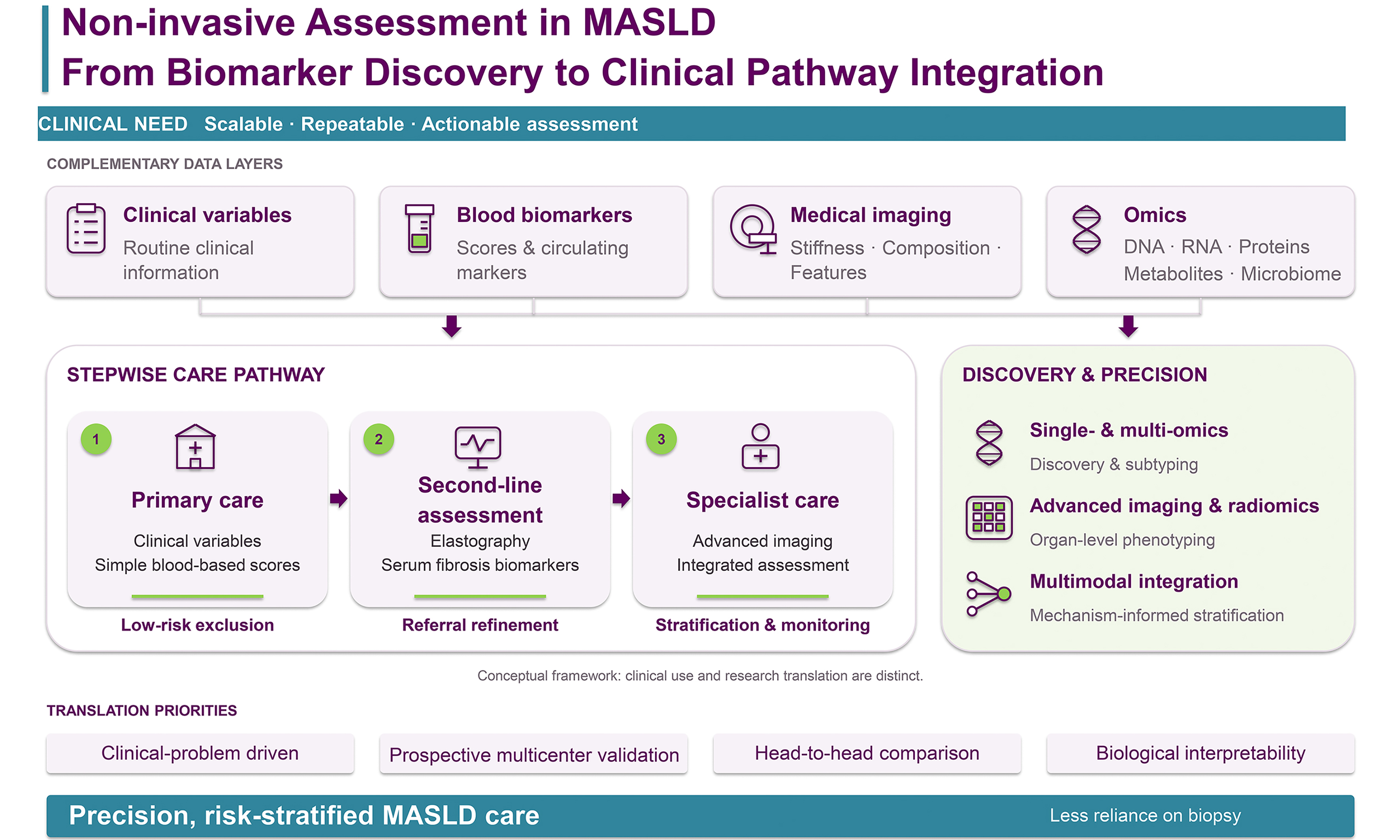
Abstract
Metabolic dysfunction-associated steatotic liver disease (MASLD) is one of the leading causes of chronic liver disease worldwide. Although liver biopsy remains the histological reference standard for assessing steatohepatitis, fibrosis stage, and therapeutic response, its invasiveness, sampling variability, and limited suitability for repeated monitoring have driven the development of non-invasive approaches. Clinical scores based on routine variables, omics-derived biomarkers, quantitative imaging techniques, and multimodal integrative models have substantially expanded the landscape of MASLD assessment. This review summarizes current progress in non-invasive evaluation of MASLD from a clinically oriented perspective, covering traditional blood-based and clinical tools, omics approaches, quantitative imaging, and multimodal integration. Particular attention is given to how these tools may serve different clinical purposes, including targeted screening or case-finding, specialist referral, fibrosis stratification, prognostic assessment, longitudinal monitoring, and precision, risk-stratified care. Major translation barriers remain, including the predominance of technology-driven instead of clinically driven study designs, the imperfect nature of biopsy-based reference standards, insufficient head-to-head comparisons across modalities, limited external validation of high-dimensional models, and uncertainty regarding real-world implementation. We argue that the future of non-invasive assessment in MASLD lies not in identifying a universally superior single modality, but in developing a clinical-context-specific, stepwise, and biologically interpretable framework that aligns non-invasive tools with distinct clinical needs and decision points.
Keywords


 Download PDF
INTRODUCTION
Metabolic dysfunction-associated steatotic liver disease (MASLD) is a leading cause of chronic liver disease worldwide, affecting approximately 32% of adults[1]. Its prevalence continues to increase, imposing a substantial and growing burden on healthcare systems globally[2-5]. A multicountry modeling study projected that the number of MASLD cases across eight countries could increase by up to 30% between 2016 and 2030[6]. Clinical assessment of MASLD focuses primarily on three key disease features: steatosis, inflammatory activity, and fibrosis. Among these features, fibrosis stage is the strongest histological predictor of liver-related outcomes and mortality, with the greatest risks observed in patients with advanced fibrosis or cirrhosis. This prognostic importance is further highlighted by the rapidly increasing contribution of MASLD to the global burden of hepatocellular carcinoma (HCC)[7]. Although MASLD-related HCC most commonly develops in patients with advanced fibrosis or cirrhosis, it can also occur before cirrhosis in a subset of patients[8]. Early identification of high-risk individuals and dynamic risk stratification across the disease spectrum are therefore essential. Accordingly, the primary objective of non-invasive assessment extends beyond detecting steatosis to identifying clinically significant fibrosis and predicting disease progression[9-11]. Evidence demonstrating the prognostic value of fibrosis stage in patients with MASLD, including those with metabolic dysfunction-associated steatohepatitis (MASH), further supports its use as a surrogate endpoint in clinical assessment[9].
Liver biopsy remains the reference standard for histological assessment of MASLD. However, biopsy is associated with invasive risks, including internal bleeding, complications, and even life-threatening events, making it unsuitable as a tool for long-term, real-time monitoring of disease progression[12]. In addition, each biopsy samples only about 1/50,000 of the liver, and such a limited specimen cannot adequately represent the condition of the entire organ, thereby leading to sampling error[13-15]. Finally, liver fibrosis is a dynamic process, whereas fibrosis staging based on biopsy is only semi-quantitative and cannot satisfactorily capture disease progression[16-19]. Because of these limitations, together with the substantial clinical demand, there is an urgent need for non-invasive assessment methods, as biopsy is not suitable for large-scale, repeated, dynamic use in clinical management. Over the past several decades, a wide range of non-invasive approaches has emerged. The analysis of omics and imaging data has led to the development of numerous techniques with promising diagnostic performance to assist clinicians in the evaluation of MASLD.
Rather than seeking a single best-performing tool, the key to non-invasive assessment of MASLD lies in building a stratified, interpretable, and clinically translatable framework that aligns with different clinical scenarios and management goals. In this context, this article reviews non-invasive assessment strategies for MASLD with emphasis not only on diagnostic performance, but also on their utility in screening, risk stratification, prognosis, and long-term monitoring. Because primary care and specialist liver centers differ substantially in clinical goals and available resources, this review focuses on what each of these settings actually needs. It also emphasizes methods that are interpretable and translatable, rather than covering end-to-end deep learning models in detail. The following sections are organized accordingly, beginning with current clinical tools, then progressing to emerging omics and imaging approaches, and finally exploring their future applications in the precision management of MASLD.
Unlike most previous reviews that focus on diagnostic accuracy and comparison of individual non-invasive tests, this review focuses on the clinical positioning of non-invasive tools across different MASLD care pathways. We emphasize how blood-based scores, omics-derived biomarkers, quantitative imaging, and multimodal models may be deployed differently for population screening, referral prioritization, fibrosis stratification, prognostic assessment, longitudinal monitoring, and precision management[7,10,11,20-23]. We further discuss the potential for integrating complementary tools within the same clinical task into multimodal strategies, with the aim of developing a comprehensive, high-performance, and interpretable precision-medicine pathway. We also distinguish tools with current clinical readiness from those that remain largely investigational. Figure 1 summarizes the MASLD disease continuum, the clinical roles of established and emerging non-invasive tools, their positions in a stepwise care pathway, and the main barriers to translation. Table 1 further compares their evidence-supported uses and boundaries of interpretation.
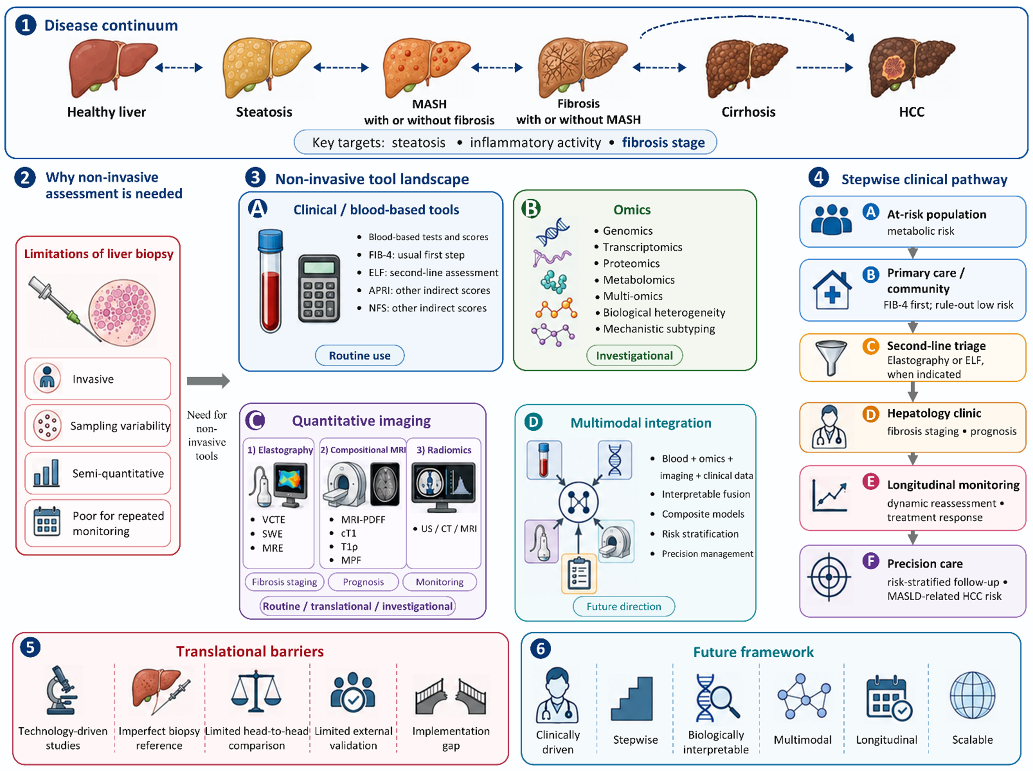
Figure 1. Non-invasive assessment across the MASLD disease continuum. The figure shows the limitations of liver biopsy, the roles of established and emerging tools in a stepwise clinical pathway, and key barriers and future directions for clinical translation. The pathway is conceptual. APRI: Aspartate aminotransferase-to-platelet ratio index; CT: computed tomography; cT1: corrected T1; ELF: enhanced liver fibrosis; FIB-4: fibrosis-4 index; HCC: hepatocellular carcinoma; MASLD: metabolic dysfunction-associated steatotic liver disease; MASH: metabolic dysfunction-associated steatohepatitis; MPF: macromolecular proton fraction; MRE: magnetic resonance elastography; MRI: magnetic resonance imaging; MRI-PDFF: MRI-derived proton density fat fraction; NFS: nonalcoholic fatty liver disease fibrosis score; SWE: shear wave elastography; T1ρ: spin-lattice relaxation time in the rotating frame; US: ultrasound; VCTE: vibration-controlled transient elastography.
Potential application of the MASLD non-invasive assessment tools in different clinical tasks
| Tool category | Representative approaches | Clinical question/setting | What the evidence supports | Boundary of interpretation |
| Blood-based scores | AST/ALT ratio, APRI, BARD, FIB-4, NFS; ELF, PRO-C3 | Primary care and diabetology triage; specialist prognostication | Reliable for ruling out, not confirming; FIB-4 is a widely used first-line test; both prognostic for decompensation and mortality; two-step pathways reduce unnecessary referral[7,10,11,20] | Large indeterminate zone; no discrimination of individual stages; age confounds indirect scores[10,11,21,22] |
| Single-omics | Genomics, transcriptomics, proteomics, metabolomics and metagenomics for biomarker discovery and diagnosis | Research and selected cohorts | Risk variants robustly replicated but of modest effect; credible replicated circulating candidates; potential metabolomic and microbiome markers of disease activity and fibrosis[7,10,11] | Evidence strength differs by layer; genotype adds marginally over routine clinical data; mostly exploratory, cross-sectional, and optimistic in single-cohort pilots[10] |
| Multi-omics integration | Multi-omics models for disease classification, fibrosis stratification | Research and selected cohorts | Adding a molecular layer improves within-cohort discrimination; panels for at-risk steatohepatitis can outperform simple indirect scores; microbiome-derived predicted metabolites support a feasible blood test[7,10] | Within-cohort discrimination gain is not incremental clinical utility; needs external validation; non-routine assays and limited cost-effectiveness; each modality's contribution must be shown[7,11] |
| Elastography | VCTE, 2D-SWE, MRE; stiffness-containing composites | Specialist care after triage: refine fibrosis risk, prognosis | VCTE the most validated and superior to simple blood panels; MRE more accurate; both prognostic, with serial rise carrying independent risk[7,10,11] | Confounded by necroinflammation, cholestasis, congestion, food intake and (for MRE) iron; serial change is not histological regression; MRE limited by cost and access[10,11,20,21] |
| Quantitative imaging | MRI-PDFF; iron-corrected T1, T1ρ, MPF | Trials and specialist care: quantify hepatic fat and assess fibrosis | MRI-PDFF the most accurate and reproducible fat measure; fibrosis-oriented parameters remain exploratory[7,10,11,21] | Require external validation[7,11,22] |
| Radiomics | Radiomics models based on ultrasound, CT or MRI for fibrosis or steatosis assessment | Research: image-derived features add beyond existing clinical, biochemical and imaging measures | Rationale is images as mineable data feeding decision support[24,25] | Limited by reproducibility; segmentation the most contentious step; studies underpowered, single-centre, externally unvalidated; interpretability gap vs. radiographic features[24-26] |
Literature search and study selection
This narrative review was informed by a targeted search of PubMed and Web of Science for relevant studies published through January 2026, without a formal lower publication-date limit. We focused primarily on studies published between 2005 and 2026, while retaining earlier foundational publications where relevant. Search terms included combinations of “MASLD” or “NAFLD”, “fibrosis”, “non-invasive test”, “magnetic resonance elastography”, “PDFF”, “radiomics”, and “omics”. Additional relevant studies were identified by screening the reference lists of relevant articles, review articles, and clinical guidelines. Priority was given to large prospective or multicenter studies, meta-analyses, studies using histological or clinically meaningful outcome reference standards, and studies with direct relevance to clinical risk stratification, prognostic assessment, or longitudinal monitoring in MASLD. Studies included in the tables were selected as representative examples based on study design, sample size, methodological quality, and clinical relevance; they were not intended to constitute an exhaustive systematic evidence synthesis.
Clinical parameter-based non-invasive tools
Clinical application of non-invasive tools differs between primary care and hepatology specialist settings. In primary care, the main goal is to identify low-risk patients who can be managed without much intervention and high-risk patients who need further tests or specialist referral. In contrast, hepatology clinics focus on more refined diagnostic assessment, fibrosis staging, prognostic stratification, treatment selection, and longitudinal monitoring[20]. These tasks demand more from non-invasive tools, requiring them to be reliable, robust, and consistent. Assessment based on routine laboratory parameters and simple clinical variables is the common non-invasive strategy in MASLD. These tools are inexpensive, accessible, and scalable, making them particularly suitable for first-line triage and population-level risk stratification. Existing evidence indicates that non-invasive tools such as Fibrosis-4 index (FIB-4), Nonalcoholic fatty liver disease fibrosis score (NFS) and Aspartate aminotransferase-to-platelet ratio index (APRI) are clinically useful in the assessment of patients with MASLD[27-30]. Because these parameters are derived from routine clinical indicators and the calculations are simple and inexpensive, these methods are highly accessible in large-scale population screening and primary care settings. As a result, they have become the most commonly used first-line screening tools and have been incorporated into clinical guidelines[22,23].
Studies have moved beyond comparing these models to fibrosis stages and now focus more on their ability to predict liver-related events, HCC, and overall mortality[29-31]. For example, Tayob et al. validated the epatocellular carcinoma early detection screening (HES) algorithm, which integrates alpha-fetoprotein (AFP), age, alanine aminotransferase (ALT), platelet count, and longitudinal changes in AFP, and demonstrated that it improved the detection of early-stage HCC within 6 months compared with AFP alone in a community-based cirrhosis cohort[32]. Fujii et al. further showed that, among patients with biopsy-proven MASLD, a baseline platelet count was associated with an increased risk of HCC development[33]. Collectively, these findings suggest that routinely available laboratory parameters may contribute not only to fibrosis assessment, but also to HCC screening and prognostic stratification.
Most conventional scoring systems are based on indirect indicators such as age, aminotransferase levels, platelet count, and metabolism-related parameters. Although they are clinically useful, they remain limited in their ability to capture biological heterogeneity and individualized disease trajectories[27,34,35]. Ioannou et al. developed a prediction model using seven routinely available variables - age, sex, diabetes, body mass index, platelet count, serum albumin, and the aspartate aminotransferase (AST)-to-ALT ratio - in patients with MASLD and alcohol-related liver disease[29]. The model achieved an area under the curve (AUC) of 0.75 in the validation cohort, indicating moderate discriminative performance and suggesting that the integration of conventional clinical parameters may support HCC risk stratification. Particularly in the context of dynamic monitoring, repeated measurement of indices such as FIB-4 has shown some prognostic value in several large population-based studies[34], but its ability to identify true histological progression in individual patients is not stable, and findings have even differed across studies[27,35].
Unlike indirect scores derived from routine clinical variables, the Enhanced Liver Fibrosis (ELF) test reflects extracellular-matrix turnover through hyaluronic acid, amino-terminal propeptide of type III procollagen, and tissue inhibitor of metalloproteinase-1. In MASLD, ELF has shown good diagnostic performance for advanced fibrosis (AUC 0.83), although its predictive value depends substantially on the underlying disease prevalence and selected threshold[36]. ELF also has prognostic relevance: higher ELF scores were independently associated with liver-related outcomes in patients with chronic liver disease, and this association has subsequently been confirmed in a large population-based cohort, particularly among individuals with obesity, diabetes, or elevated ALT[37,38]. Therefore, ELF is best positioned as a clinically validated complementary test for fibrosis risk refinement, rather than a stand-alone universal rule-in tool.
Routine clinical scores are easy to scale and access, but they do not reflect much of the underlying biology. By contrast, omics-driven integrative strategies are better for understanding disease mechanisms and supporting therapeutic development.
Table 2 summarizes the representative studies of blood-based biomarkers, conventional fibrosis scores, and clinically translatable composite models discussed in this section. Overall, routine clinical scores and blood-based markers work best as first-line tools for population-level risk stratification, primary care screening, and referral prioritization. Their main clinical value is in excluding advanced fibrosis among low-risk populations and identifying patients who require second-line assessment with elastography, ELF test, or specialist evaluation. However, because these scores are based largely on indirect variables such as age, aminotransferases, and platelet count, they are less suitable for refined fibrosis staging, precise individual-level monitoring, or mechanistic disease characterization. Therefore, these tools should be used as triage tools at the entry of a stepwise pathway rather than as standalone methods for comprehensive MASLD assessment.
Representative studies of blood-based biomarkers and clinical scoring systems for non-invasive assessment of MASLD
| Author(s) | Year | Study design | Sample size | Clinical task | Biomarker/Model |
| Srivastava et al.[28] | 2019 | Prospective | 3,012 | Screening | FIB-4 |
| Siddiqui et al.[27] | 2019 | Retrospective | 1,904 | Diagnosis | APRI, FIB-4, AST/ALT ratio, NFS |
| Ioannou et al.[29] | 2019 | Retrospective | 7,068 | Prognosis | HCC risk prediction models |
| Hagstrom et al.[34] | 2020 | Retrospective | 40,729 | Prognosis | FIB-4 |
| Balkhed et al.[35] | 2022 | Retrospective | 135 | Prognosis | FIB-4, NFS, APRI, dAAR |
| Labenz et al.[39] | 2022 | Prospective | 11,859 | Screening | SEAL algorithm |
| Younes et al.[31] | 2021 | Retrospective | 1,173 | Prognosis | APRI, BARD, FIB-4, NFS, HFS |
| Parkes et al.[37] | 2010 | Prospective | 457 | Prognosis | ELF |
| Saarinen et al.[38] | 2023 | Prospective | 6,040 | Prognosis | ELF |
| Tayob et al.[32] | 2021 | Retrospective | 38,431 | Screening | Updated HES algorithm, AFP |
| Fujii et al.[33] | 2023 | Retrospective | 1,398 | Prognosis | Platelet count |
OMICS-DRIVEN NON-INVASIVE ASSESSMENT
Single-omics assessment
Early omics research was limited to single-omics approaches, investigating MASLD separately from the perspectives of DNA, RNA, proteins, and metabolites. The research objectives ranged from pathological exploration, diagnosis, and prognostic evaluation to treatment monitoring[40-43]. Some studies have further extended the research to the identification of HCC-related risk, suggesting that single-omics strategies may contribute to disease subtyping and fibrosis assessment, and provide complementary information for identifying high-risk individuals with MASLD who are susceptible to liver cancer[44-46]. The major strength of omics-based research is its ability to interrogate MASLD biology at the molecular level. By characterizing genetic susceptibility, transcriptional regulation, protein expression, metabolic remodeling, and microbial interactions, omics approaches can identify candidate biomarkers, reveal mechanistic pathways, and support biologically informed patient stratification[47].
Genomics can help characterize genetic susceptibility to MASLD by providing relatively stable upstream biological information. Unlike dynamic biomarkers that reflect changes in disease activity, genetic markers are better suited for baseline and long-term risk stratification than real-time disease monitoring. From a clinical perspective, their potential value lies in complementing conventional clinical factors and fibrosis assessment models to identify individuals who may be more susceptible to MASLD progression, advanced fibrosis, or MASLD-related HCC. However, genomics should currently be regarded as an adjunctive risk-enrichment tool instead of a standalone approach for disease monitoring, treatment-response assessment, or selection of populations for HCC surveillance[48,49]. In relation to HCC risk, genetic evidence supports an inherited susceptibility to MASLD-related HCC. The PNPLA3 rs738409 variant has been consistently associated with steatotic liver disease severity, fibrosis progression, and an increased risk of HCC. In addition, polygenic risk scores incorporating multiple liver-disease-associated variants have shown promise for non-invasive risk stratification of individuals at higher risk of severe liver outcomes, although their predictive performance remains under investigation. Collectively, these findings suggest that genetic background influences not only hepatic steatosis and fibrosis progression, but also susceptibility to liver cancer[45,50,51]. Representative studies on variants such as PNPLA3, TM6SF2, GCKR, and MBOAT7 have shown that host genetic background affects inter-individual heterogeneity in disease severity and clinical outcomes[45,48,52-54]. However, because genomic information is largely static and cannot directly reflect short-term changes in inflammation, fibrotic remodeling, or treatment response, its role in MASLD should therefore be more appropriately viewed as complementary to dynamic biomarkers rather than as a replacement for them.
Transcriptomics shows how molecular processes change and drive MASLD progression, beyond just genetic risk. Its principal contribution lies in providing information on steatosis, inflammation, and fibrosis, thereby bridging upstream risk factors and downstream clinical phenotypes. Transcriptomic studies have also identified pathway-level drivers of disease progression, highlighting their value in mechanistic stratification and therapeutic target discovery[55]. However, RNA signals may vary according to disease stage, sample type, and cellular context, and the multicellular architecture of the liver further complicates their interpretation. Transcriptomics is useful for understanding mechanisms and finding biomarkers, but its clinical use will need better standardization, validation, and integration with other non-invasive methods.
Proteomics reveals the functional effector layer of MASLD by detecting proteins involved in hepatocellular injury, inflammatory signaling, and matrix remodeling. Because proteins are more directly linked to biological functions, and many proteins can be detected in blood, proteomics has strong clinical applicability for the development of non-invasive biomarkers. Sanyal et al. developed proteomic models for selected histological components of NAFLD, with training/paired-validation AUCs of 0.95/0.79 for steatosis, 0.83/0.72 for inflammation, 0.87/0.83 for ballooning, and 0.92/0.85 for fibrosis. The combined model for at-risk NASH achieved AUCs of 0.93/0.85, respectively[56]. Nevertheless, issues such as technical variability, lack of standardization, and limited external validation continue to constrain its routine application.
Metabolomics captures the downstream dynamic layer of disease biology by reflecting the persistent metabolic disturbances closely associated with steatosis, inflammation, and fibrosis in MASLD. Compared with genomics, metabolomics is more sensitive to the current physiological state and may therefore be better suited for identifying active disease processes, monitoring disease progression, or assessing treatment-related changes[57-60]. This is also what gives metabolomics its translational appeal for blood- or urine-based biomarkers, particularly for large-scale screening and repeated assessment[57,61,62].
In the field of MASLD-related HCC, serum metabolomics and lipidomics have shown considerable potential for clinical translation. Lewinska et al. reported that patients with MASLD-HCC exhibited serum lipid profiles that clearly differed from those of patients with non-malignant MASLD and healthy controls[63]. Based on these differences, they developed a MASLD-HCC Diagnostic Score, which could distinguish MASLD-HCC from non-malignant MASLD, healthy controls, and HCC of other etiologies, with better diagnostic performance than AFP[63]. Lu et al. further identified a circulating metabolite signature in multicenter MASLD and MASLD-HCC cohorts that could predict the presence of HCC in the setting of MASLD[64]. Model performance was further improved when cirrhosis status was incorporated, and the metabolite signature was also associated with overall survival in patients with MASLD-HCC[64]. Overall, these studies suggest that serum metabolic signatures may not only facilitate HCC detection but also reflect a high-risk state during the progression from MASLD to HCC. Its potential value lies in identifying metabolic reprogramming associated with disease progression and high-risk states[58,60]. However, because metabolic profiles are highly susceptible to environmental influences, reproducibility across populations and the standardization required for clinical use remain important challenges.
Table 3 summarizes representative single-omics studies. Across these studies, genomics appears most relevant to relatively stable susceptibility and long-term risk assessment, whereas transcriptomic, proteomic, and metabolomic approaches provide more dynamic biological information but remain largely investigational because of limited standardization and external validation.
Representative studies of single-omics approaches for non-invasive assessment of MASLD
| Author(s) | Year | Study design | Sample size | Clinical task | Omics |
| Liu et al.[45] | 2014 | Retrospective | 375 | Prognosis | Genomics |
| De Vincentis et al.[65] | 2022 | Retrospective | 266,687 | Prognosis | Genomics |
| Eslam et al.[66] | 2016 | Retrospective | 1,992 | Diagnosis | Genomics |
| Dongiovanni et al.[48] | 2018 | Retrospective | 9,414 | Prognosis | Genomics |
| Verweij et al.[67] | 2022 | Retrospective | 542,904 | Prognosis | Genomics |
| Bianco et al.[46] | 2021 | Retrospective | 367,041 | Prognosis | Genomics |
| Chen et al.[68] | 2023 | Retrospective | 54,773 | Prognosis | Genomics |
| Abul-Husn et al.[51] | 2018 | Retrospective | 46,544 | Prognosis | Genomics |
| Jamialahmadi et al.[49] | 2021 | Retrospective | 425,671 | Screening | Genomics |
| Valenti et al.[53] | 2010 | Retrospective | 824 | Prognosis | Genomics |
| Baselli et al.[50] | 2022 | Retrospective | 301 | Prognosis | Genomics |
| Walker et al.[52] | 2020 | Retrospective | 27,744 | Prognosis | Genomics |
| Emdin et al.[69] | 2021 | Retrospective | 4,829 | Prognosis | Genomics |
| Grimaudo et al.[70] | 2020 | Prospective | 471 | Prognosis | Genomics |
| Govaere et al.[71] | 2020 | Retrospective | 403 (381 NAFLD + 22 controls); discovery n = 206, validation n = 175 | Diagnosis/Prognosis | Transcriptomics |
| Sun et al.[44] | 2024 | Retrospective | 546 | Prognosis | Transcriptomics |
| Conway et al.[72] | 2023 | Retrospective | 1,208 | Prognosis | Transcriptomics |
| Baboota et al.[55] | 2022 | Retrospective | 58 | Diagnosis/Prognosis | Transcriptomics |
| Tobaruela-Resola et al.[73] | 2025 | Retrospective | 100 | Diagnosis | Transcriptomics |
| Starmann et al.[74] | 2012 | Retrospective | 87 | Diagnosis | Transcriptomics |
| Bell et al.[75] | 2010 | Retrospective | 85 | Diagnosis | Proteomics |
| Feng et al.[76] | 2023 | Retrospective | 285 | Diagnosis | Proteomics |
| Yuan et al.[77] | 2020 | Retrospective | 56 | Diagnosis | Proteomics |
| Luo et al.[78] | 2021 | Retrospective | 216 | Diagnosis | Proteomics |
| Govaere et al.[79] | 2023 | Retrospective | 306 | Diagnosis | Proteomics (n = 306); hepatic transcriptomics in paired subset (n = 52) |
| Jiang et al.[80] | 2023 | Retrospective | 2,236 overall; 80 in the proteomic subset | Diagnosis | Proteomics |
| Sanyal et al.[56] | 2023 | Retrospective | 636 | Diagnosis/Treatment | Proteomics |
| Rodríguez-Suárez et al.[81] | 2010 | Retrospective | 18 | Diagnosis | Proteomics |
| McGlinchey et al.[58] | 2022 | Retrospective | 627 | Diagnosis | Metabolomics |
| Zhu et al.[57] | 2022 | Retrospective | 83 | Diagnosis | Metabolomics |
| Perakakis et al.[59] | 2019 | Retrospective | 80 | Diagnosis | Metabolomics |
| Chen et al.[60] | 2024 | Retrospective | 250 | Diagnosis | Metabolomics |
| Khusial et al.[62] | 2019 | Retrospective | 559 | Screening | Metabolomics |
| Moolla et al.[61] | 2020 | Retrospective | 275 | Diagnosis | Metabolomics |
| Kalhan et al.[82] | 2011 | Retrospective | 60 | Diagnosis | Metabolomics |
| Lu et al.[64] | 2023 | Retrospective | 113 | Diagnosis | Metabolomics |
Multi-omics assessment
Each individual omics layer shows one part of MASLD biology and therefore provides an incomplete representation of this multifactorial disease. For example, genomics cannot capture the influence of dietary and environmental factors on MASLD. By integrating multi-level biological information and combining it with artificial intelligence (AI), multi-omics enables the construction of models from large-scale datasets and diverse data modalities to extract and identify disease-related information. This approach improves model performance as well as helps establish a more complete mechanistic chain of disease development[40]. In multifactorial diseases such as MASLD, any single dimension is insufficient to directly characterize the origins and progression of the disease. In contrast, the integration of multidimensional information makes it possible to build more complex and interpretable models that illustrate the interactions and transformations among various biomolecules. The integration may enhance the biological interpretability of multi-omics models, particularly when coupled with experimental validation, and could help move the field beyond descriptive association studies towards research on mechanistic hypotheses. For diseases such as HCC, cross-layer integration is important, as HCC risk is unlikely to be determined by a single molecular layer alone. It reflects the combined effects of multiple factors, including genetic susceptibility, inflammation and fibrotic progression, metabolic dysregulation, and host-microbiome interactions[44,83,84].
Several studies have systematically compared the diagnostic performance of single-omics strategies with that of multi-omics integration, consistently confirming the superiority of integrative approaches within specific study cohorts. Wood et al. and Zhou et al. developed diagnostic models and found that model performance was optimal after integrating all omics data[85,86]. Beyond diagnosis, this integrative strategy is also applicable to the identification of severe clinical outcomes, particularly cirrhosis and HCC, because a multi-omics framework is more likely to simultaneously capture the genetic background preceding tumorigenesis, high-risk inflammatory and fibrosis stage, and their associated molecular information[44,87].
The integration of different omics datasets can significantly improve diagnostic performance. The likely reason is that different omics datasets carry complementary information, and this complementarity represents a major advantage of multi-omics. Compared with the relatively narrow and task-specific applications typically seen in single-omics studies, multi-omics may offer broader clinical and biological utility. Some multi-omics studies have established a workflow of exploring pathological mechanisms, building diagnostic models, and validating clinical performance, moving from descriptive association to mechanistic inference and, in selected cases, experimental causal testing. Both Caussy, Loomba and Oh et al. used microbial features to reveal the influence of the gut microbiota on liver pathology and subsequently validated this relationship in predictive models[87,88]. Quesada-Vázquez et al. further advanced mechanistic research toward functional validation: after integrating metabolomic, transcriptomic, and microbial data and identifying a negative association between plasma histidine and steatosis, they verified the therapeutic effect of histidine supplementation in multiple animal models, thereby completing a closed loop from association to causality[89]. Therefore, the strengths of multi-omics research should not be interpreted solely as improvements in predictive performance. Rather, its added value may lie in generating biologically meaningful hypotheses and identifying candidate pathways that can be tested in longitudinal and experimental studies. This approach may enhance the translational relevance of multi-omics research, but it should not be regarded as a mature tool for clinical prognostication or selection of patients for surveillance at present. Although multi-omics offers many advantages over single-omics approaches, most current studies on multi-omics remain at the research stage. Owing to factors such as testing costs, the complexity of clinical analytical workflows, and insufficient standardization, few studies have been used in real-world clinics. At the same time, most existing studies have been conducted in specific centers or selected populations, while multicenter external validation remains inadequate, resulting in limited reproducibility and generalizability.
Table 4 summarizes representative multi-omics studies. The main potential advantage of multi-omics over single-omics lies in integrating complementary biological layers and generating mechanistic hypotheses; however, most evidence remains retrospective and exploratory, with limited readiness for routine clinical implementation. In summary, omics-based approaches give a useful biological insight into MASLD heterogeneity and may support biomarker discovery, molecular subtyping, prognostic modeling, and treatment-response research. For MASLD-related HCC, multi-omics approaches may be valuable for risk enrichment and mechanistic discovery, as they can identify molecular changes linking metabolic dysfunction, inflammation, fibrosis, and hepatocarcinogenesis. At present, however, most omics tools remain insufficiently standardized and externally validated for routine clinical screening, referral decisions, or longitudinal monitoring. Their most appropriate current role is therefore in mechanistic investigation, discovery of candidate biomarkers, and research-oriented precision stratification, with promise for future translation into clinically deployable panels once analytical reproducibility, cost-effectiveness, and real-world validity are established. In contrast to omics approaches, which characterize molecular changes, quantitative imaging provides organ-level information on steatosis, stiffness, tissue composition, and architectural heterogeneity.
Representative studies of multi-omics integration strategies for MASLD assessment
| Author(s) | Year | Study design | Sample size | Clinical task | Omics |
| Wood et al.[85] | 2017 | Retrospective | 576a (training 443; validation 133) | Diagnosis | Genomics, Proteomics, Phenomics (clinical variables) |
| Oh et al.[87] | 2020 | Retrospective | 163 | Diagnosis | Microbiomics, Metabolomics |
| Younossi et al.[90] | 2005 | Retrospective | 98 | Biomarker discovery | Transcriptomics, Proteomics |
| Atabaki-Pasdar et al.[91] | 2020 | Retrospective | 1,514 | Diagnosis | Genomics, Transcriptomics, Proteomics, Metabolomics |
| Zhou et al.[86] | 2016 | Retrospective | 318 | Diagnosis | Metabolomics, Genomics |
| Wen et al.[92] | 2026 | Retrospective | 166 | Diagnosis | Transcriptomics, Epigenomics |
| Quesada-Vázquez et al.[89] | 2023 | Retrospective | 651 | Treatment | Metabolomics, Transcriptomics, Microbiomics |
| Leung et al.[93] | 2022 | Prospective | 180 | Prognosis | Microbiomics, Metabolomics |
| Sveinbjornsson et al.[84] | 2022 | Retrospective | 36,116 | Diagnosis/Prognosis | Genomics, Transcriptomics, Proteomics |
| Li et al.[83] | 2025 | Retrospective | 61 | Diagnosis | Transcriptomics, Metabolomics, Proteomics |
| Anspach et al.[94] | 2026 | Retrospective | 8 (multi-omics discovery); 424 TCGA-LIHC samples (transcriptomic validation) | Association discovery study | Transcriptomics, Metabolomics, Lipidomics |
NON-INVASIVE IMAGING ASSESSMENT
At the organ and tissue levels, non-invasive assessment of MASLD depends mainly on imaging-based biomarkers. Once liver fibrosis progresses to cirrhosis, patients face an increased risk of portal hypertension, hepatic decompensation, and HCC. Previous studies have demonstrated that patients with steatohepatitis are at substantial risk of developing HCC in the presence of advanced fibrosis or cirrhosis[95]. Among patients with biopsy-confirmed MASLD, fibrosis stage is one of the strongest predictors of liver-related mortality and the risk of HCC[96,97]. Advanced fibrosis and cirrhosis remain the most important risk substrate for MASLD-related HCC; however, HCC risk is not determined solely by fibrosis stage. Therefore, non-invasive imaging-based assessment of fibrosis should be regarded as an important component of HCC risk enrichment, rather than as a tool that directly measures hepatocarcinogenesis.
Numerous imaging-based methods have been reported for the evaluation of liver fibrosis; however, a comprehensive review of all available techniques is beyond the scope of this article. The methods discussed in this review can broadly fall into three groups. The first group uses the mechanical properties of liver tissue, taking liver stiffness itself as the imaging biomarker. This includes Vibration-controlled transient elastography (VCTE), Shear wave elastography (SWE), and Magnetic resonance elastography (MRE). The second group involves leveraging endogenous contrast mechanisms to acquire information on tissue composition through specifically designed composition imaging sequences, followed by post-processing to derive quantitative compositional indices, such as T1, T1ρ, and quantitative magnetization transfer (MT). The third group consists of approaches based on image post-processing and quantitative modeling, in which quantitative features are extracted from routine imaging and liver fibrosis is indirectly assessed through mathematical analysis or diagnostic models; representative methods include radiomics.
Owing to its availability and lower cost, VCTE remains one of the most widely used non-invasive techniques for the clinical assessment of liver fibrosis[7]. Although MRE generally outperforms VCTE in terms of diagnostic performance and reproducibility, its use is limited by higher costs, less accessibility, and the need for specialized equipment. Assessment methods based on elastography or other mechanical parameters generally require dedicated equipment, specialized functional modules, or compatible probes. In contrast, compositional imaging methods based on endogenous contrast mechanisms and radiomics do not require additional standalone examination devices, thereby offering practical advantages in clinical implementation and application flexibility.
Assessment based on stiffness
Elastography has become a key non-invasive tool in the clinical management of MASLD. Its clinical use spans primary screening of high-risk populations, accurate diagnosis of advanced fibrosis, longitudinal monitoring of disease progression, and prognostic evaluation of liver-related outcomes[98]. VCTE, SWE, and MRE are currently the main elastography-based methods used for fibrosis assessment in MASLD; however, their value lies not so much in their technical differences as in their distinct positions within the clinical pathway. In many clinical pathways, FIB-4 is used for initial risk assessment, followed by VCTE when further evaluation is needed. SWE provides an ultrasound-based alternative with good feasibility within routine imaging workflows, whereas MRE is generally reserved for settings requiring higher precision and reproducibility. Rather than viewing these modalities as competing technologies, it may be more useful to regard them as complementary tools with different levels of accessibility, accuracy, and resource needs. It should be emphasized that elastography-based techniques use liver stiffness as a surrogate marker of fibrosis. They do not directly measure extracellular matrix composition or interrogate the molecular pathophysiology underlying MASLD progression.
Among elastography-based techniques, VCTE is still one of the most widely used non-invasive tools in routine clinical practice because of its ease of operation, rapid acquisition, and established role in fibrosis triage[99-107]. A cohort study of 6,295 individuals from Europe and Asia found VCTE-based fibrosis screening in primary care to be cost-effective[108]. SWE can be integrated into conventional ultrasound platforms, offering practical advantages in workflow flexibility, and has demonstrated good reproducibility in several MASLD cohorts[109-116]. A study evaluating the repeatability and reproducibility of SWE and VCTE showed that, in patients with MASLD, SWE had superior interday and interoperator reproducibility to VCTE and achieved the prespecified performance threshold[117]. MRE generally provides higher diagnostic accuracy and reproducibility, particularly in obese patients and specialist care settings, although its broader application is limited by cost, availability, and examination complexity[118,119]. Overall, these techniques have become central to non-invasive fibrosis assessment, although all of them are susceptible to confounding by factors other than fibrosis itself, such as inflammation, congestion, and cholestasis[11].
Table 5 summarizes representative elastography studies. Collectively, VCTE has the broadest evidence base for scalable fibrosis triage and prognostic assessment, SWE offers an ultrasound-integrated alternative, and MRE provides higher diagnostic precision in specialist settings.
Representative studies of elastography approaches for MASLD assessment
| Author(s) | Year | Study design | Sample size | Clinical task | Technique |
| Boursier et al.[107] | 2022 | Retrospective | 1,057 | Prognosis | VCTE |
| Wong et al.[105] | 2012 | Prospective | 193 | Diagnosis | VCTE |
| Petta et al.[106] | 2021 | Retrospective | 1,039 | Prognosis | VCTE |
| Shili-Masmoudi et al.[104] | 2020 | Prospective | 2,245 | Prognosis | VCTE |
| Wong et al.[100] | 2010 | Prospective | 246 | Diagnosis | VCTE |
| Braude et al.[103] | 2023 | Retrospective | 7,079 | Prognosis | VCTE |
| Boursier et al.[101] | 2016 | Retrospective | 452 | Diagnosis | VCTE |
| Serra-Burriel et al.[108] | 2019 | Prospective | 6,295 | Screening | VCTE |
| Mendoza et al.[116] | 2022 | Prospective | 104 | Diagnosis | VCTE, SWE |
| Ozturk et al.[114] | 2020 | Retrospective | 116 | Diagnosis | SWE |
| Lee et al.[110] | 2021 | Prospective | 102 | Diagnosis | SWE |
| Pierce et al.[117] | 2024 | Prospective | 40 | Diagnosis | SWE, VCTE |
| Jang et al.[109] | 2022 | Prospective | 132 | Diagnosis | SWE |
| Imajo et al.[120] | 2016 | Retrospective | 142 | Diagnosis | MRE |
| Park et al.[121] | 2017 | Prospective | 104 | Diagnosis | MRE |
| Gidener et al.[122] | 2021 | Retrospective | 829 | Prognosis | MRE |
Assessment based on non-invasive compositional imaging
The pathological hallmark of liver fibrosis is excessive deposition of collagen-rich extracellular matrix[97]. This remodeling expands the extracellular matrix compartment and often shifts tissue water distribution as well. These tissue alterations provide a biological basis for compositional magnetic resonance imaging (MRI) sensitive to relaxation and MT. Quantitative MRI methods, including T1 mapping[123-125] and T1ρ mapping[126-130], have been investigated as potential non-invasive approaches for assessing liver fibrosis. Both T1 and T1ρ values have been reported to increase with fibrosis severity.
However, both T1 mapping and T1ρ mapping are sensitive to hepatic iron content, which can confound fibrosis assessment. To address this limitation, iron-corrected T1 mapping[123-125] has been introduced for liver fibrosis evaluation, and iron correction strategies for hepatic T1ρ mapping[131] have also been reported. In addition to iron deposition, inflammation may also influence T1 and T1ρ measurements. Furthermore, reliable quantification requires close agreement between the assumed signal model and the acquired data. Careful sequence design, acquisition optimization, and post-processing can therefore help minimize possible sources of bias and improve measurement robustness.
Because collagen forms a gel-like macromolecular network, it is expected to exhibit a strong MT effect. Although the liver contains other proteins, these molecules are generally more mobile than collagen and are therefore expected to contribute less to MT. Triglycerides and lipids are hydrophobic and have limited MT effects. These properties suggest that quantitative MT imaging may provide a means to evaluate collagen deposition in the liver and thereby assess liver fibrosis. This concept has been demonstrated in both animal[132] and human studies[133-135]. Importantly, quantitative MT-based methods appear to be relatively less affected by hepatic iron deposition, which is an advantage for compositional imaging of the liver.
Conventional quantitative MT methods typically require T1 information and relatively long acquisition times, posing challenges for robust clinical imaging of the liver. In contrast, spin-lock radiofrequency-based methods[136,137] have shown advantages for quantitative MT imaging because they can be designed to achieve higher saturation efficiency with reduced direct water saturation, enabling rapid mapping of macromolecular proton fraction (MPF) in the liver.
Compared with stiffness-based methods, compositional imaging approaches that more directly interrogate collagen-related tissue properties provide a distinct physical mechanism for fibrosis assessment. These methods do not require exogenous contrast agents or additional hardware and may have potential for detecting early-stage fibrosis. In early fibrosis, liver stiffness may not change substantially, whereas direct imaging of collagen-associated tissue alterations may provide greater sensitivity to early extracellular matrix remodeling. Such capability could be important for the development of antifibrotic therapies, patient stratification, and treatment monitoring. By introducing complementary biophysical mechanisms, compositional imaging may help address current limitations in non-invasive liver fibrosis assessment. Nevertheless, despite their promise, these techniques should be regarded as research tools for tissue characterization rather than as tools for hepatocellular carcinoma risk prediction; further technical development, standardization, and large-scale clinical validation are required before routine clinical implementation.
Assessment based on radiomics
Radiomics is another imaging-based strategy that can often be applied to routinely acquired clinical images without requiring additional scanner hardware, although robust implementation requires standardized acquisition, segmentation, preprocessing, and analysis pipelines. The basic idea is to extract large numbers of quantitative features from medical images automatically, and then use those features to build models for diagnosis, prognosis, treatment-response assessment, or disease monitoring[24-26,138]. To improve reproducibility and comparability in radiomics research, Zwanenburg et al. standardized 169 radiomic features[139]. Because of their widespread clinical use, computed tomography (CT) and ultrasound have provided relatively large datasets for radiomics research, although their roles in MASLD care differ. Models developed by Zhang et al. and Meng et al. have both achieved encouraging results[140,141]. In comparison, although Chen et al. and Xu et al. reported good performance using T2- and T1-weighted MRI sequences, respectively, limitations in sample size may have constrained the generalizability of these models[142,143].
However, most current radiomics studies in MASLD have focused mainly on diagnosis, whereas real-world clinical settings include both primary care clinics that require first-line screening tools and specialist clinics that address diagnosis, prognosis, and monitoring. Some clinical indicators currently used in primary care still play an important role in radiomics research. In a representative study, Tang et al. showed that a combined clinical-radiomics model achieved an AUC of 0.930 for the diagnosis of moderate-to-severe fatty liver, whereas the radiomics-only model reached only 0.807; notably, the clinical model alone had already achieved an AUC of 0.904, meaning that the addition of radiomics was only a modest absolute improvement of 0.026 in AUC[144]. This finding should not be interpreted as definitive evidence that radiomics has limited value in the management of MASLD. Rather, it suggests that radiomics may improve performance, but its incremental benefit over readily available clinical variables may be modest in some diagnostic settings. Many radiomics studies therefore still require large-scale, multicenter validation to establish model robustness, generalizability, and true clinical value. The future of radiomics in MASLD will depend on its ability to provide reliable and useful clinical information beyond existing low-cost tools, rather than just slightly improving diagnostic accuracy. Table 6 summarizes representative radiomics studies across CT, ultrasound, MRI, and positron emission tomography (PET)/CT. Despite encouraging diagnostic results, most studies remain retrospective and single-center, and the incremental clinical value of radiomics beyond conventional clinical and imaging variables remains uncertain.
Representative studies of radiomics-based models for MASLD assessment
| Author(s) | Year | Study design | Sample size | Clinical task | Modality |
| Zhang et al.[141] | 2025 | Retrospective | 840 | Diagnosis | CT |
| Lubner et al.[145] | 2017 | Retrospective | 289 | Diagnosis | CT |
| Wang et al.[146] | 2022 | Retrospective | 443 | Diagnosis | CT |
| Dichtel et al.[147] | 2023 | Retrospective | 16 | Diagnosis | CT |
| Cui et al.[148] | 2021 | Retrospective | 332 | Diagnosis | CT |
| Tang et al.[144] | 2023 | Retrospective | 227 | Diagnosis | CT |
| Li et al.[149] | 2025 | Retrospective | 254 | Diagnosis | CT |
| Chen et al.[150] | 2021 | Retrospective | 22 | Diagnosis | PET/CT |
| Meng et al.[140] | 2023 | Retrospective | 618 | Prognosis | Ultrasound |
| Xia et al.[151] | 2024 | Retrospective | 87 rats | Diagnosis | Ultrasound (Experimental animal study) |
| Chen et al.[143] | 2022 | Retrospective | 203 | Diagnosis | MRI |
| Xu et al.[142] | 2021 | Retrospective | 53 | Diagnosis | MRI |
| Cannella et al.[152] | 2019 | Retrospective | 54 | Diagnosis | MRI |
In summary, imaging-based tools occupy different positions across the MASLD care pathway. VCTE fits best for second-line triage and fibrosis risk stratification after initial blood-based screening, particularly in primary care or referral pathways. SWE can provide similar elastography-based information within conventional ultrasound workflows. MRE offers higher accuracy and reproducibility and is useful in specialist settings, clinical trials, and cases with indeterminate or discordant first-line tests. MRI-derived proton density fat fraction (MRI-PDFF) is well suited for quantifying steatosis and monitoring treatment-related changes in liver fat, whereas emerging compositional MRI methods and radiomics are still mostly at the research stage. Thus, established elastography-based methods, MRE, and MRI-PDFF are clinically useful for staging, prognostic assessment, and longitudinal follow-up, whereas newer quantitative imaging and radiomics approaches require further standardization and external validation before routine implementation.
MULTI-MODALITY INTEGRATION
Over the past decades, non-invasive assessment of MASLD has evolved from reliance on isolated laboratory indices and conventional imaging measurements toward a broader framework incorporating blood-based biomarkers, elastography, advanced MRI techniques, AI, radiomics, and omics profiling. Different diagnostic approaches should not be viewed as being in simple competition with one another; rather, they reflect distinct layers of disease biology. Blood-based biomarkers and composite scores, such as FIB-4 and ELF, provide systemic surrogate indicators of liver injury, inflammation and fibrosis risk. Imaging-based methods, including VCTE, SWE, MRE, and MRI-PDFF, quantify hepatic fat content, liver stiffness, and fibrosis-related structural alterations at the organ level. Omics and radiomics approaches further capture molecular perturbations linking metabolic, genetic, transcriptomic, and proteomic layers, whereas radiomics quantifies image-derived phenotypes and tissue heterogeneity. Integrating these complementary data may help link molecular changes to organ-level manifestations of MASLD[153,154]. From this perspective, a more relevant clinical question is which combination of tests provides actionable information for a specific decision point in the MASLD care pathway.
In primary care and community settings, the priority is not detailed biological analysis, but scalable identification of individuals who are unlikely to have advanced disease and those who require referrals. Therefore, a sequential strategy using FIB-4 as a first-line screening tool, followed by VCTE-derived liver stiffness measurement or ELF testing in patients at intermediate or high risk, remains of substantial practical value[155,156]. In this framework, a low FIB-4 result supports rule-out of advanced fibrosis and continued management with metabolic risk-factor control and periodic reassessment in primary care. Intermediate results should prompt second-line testing because they do not reliably distinguish low- from high-risk disease, whereas persistently high-risk or abnormal second-line results should lead to specialist evaluation. This approach is supported by large multicenter longitudinal studies. In a cohort of 12,950 patients with MASLD, sequential assessment with FIB-4 followed by VCTE-derived liver stiffness measurement (VCTE-LSM) stratified patients into low-, intermediate-, and high-risk groups, with 5-year liver-related event rates of 0.5%, 1.0%, and 10.8%, respectively[155]. In a prospective real-world study of patients with type 2 diabetes, those with elevated FIB-4 underwent transient elastography and specialist assessment; 21.4% were subsequently identified as having previously unrecognized advanced MASLD, supporting the clinical yield of this sequential approach[157].
By contrast, specialist hepatology clinics face a different set of challenges, including refinement of fibrosis stage, identification of patients at risk of progression, selection of candidates for therapeutic intervention, and monitoring of longitudinal change. These tasks may benefit more from higher-resolution phenotyping, including elastography, advanced MRI-based methods, selected omics approaches, and composite non-invasive scores that combine imaging or elastography with laboratory variables, such as FibroScan-AST (FAST)[158], MRE combined with FIB-4 (MEFIB)[159], and MRI-aspartate aminotransferase (MAST)[160]. FAST combines FibroScan-derived parameters with AST to identify active MASH with significant fibrosis[158]. MEFIB integrates MRE with FIB-4 to enrich for patients with significant or advanced fibrosis and to support referral and risk stratification[159]. MAST further combines MRE, MRI-PDFF, and AST, enabling joint assessment of MASH activity and fibrosis burden[160]. Accordingly, the value of a non-invasive test should be judged not only by its aggregate diagnostic accuracy in heterogeneous cohorts, but also by its ability to inform specific clinical decisions.
Importantly, the value of multimodal integration lies both in the complementarity of data and in its potential to connect mechanistic understanding with clinical application[154,161,162]. Clinical biomarkers can help identify high-risk populations, imaging can localize and quantify organ-level injury, and omics can help explain why disease progression differs among individuals[154]. When interpreted together, these data may support a more comprehensive model of MASLD and could facilitate patient stratification, individualized prognosis, treatment selection, and long-term follow-up.
Overall, the future of non-invasive MASLD assessment is unlikely to depend on one modality replacing another. Rather, the field is moving toward a layered, clinically contextualized framework in which blood-based biomarkers support broad triage, imaging refines phenotypic and prognostic assessment, and omics provides deeper biological resolution for mechanism-informed stratification. Such a strategy may ultimately provide a more practical, reproducible, and biologically meaningful framework for reducing reliance on biopsy, while preserving histology for selected cases in which diagnostic uncertainty or trial requirements justify invasive assessment.
CHALLENGES FROM BENCH TO BEDSIDE
Although omics, quantitative imaging, and AI have pushed the development of non-invasive assessment for MASLD, a stable, unified, and broadly generalizable strategy has yet to emerge. One reason is that much of the current literature remains technology-driven rather than clinically driven. Model development is often centered on maximizing AUC in retrospective datasets, whereas the intended use case, target population, clinical pathway, and downstream decision point are insufficiently specified. As a result, high-dimensional or technically sophisticated models may show impressive performance in selected cohorts yet add limited value in practice if they do not outperform simpler tools, cannot be reproduced across centers, or are difficult to implement at scale. For MASLD, where screening and longitudinal follow-up must be scalable and sustainable, parsimony, external validity, calibration, and deployability may matter more than marginal improvements in discrimination.
A second limitation is that the reference standard itself is imperfect. Although liver biopsy remains the conventional histologic reference standard, it samples only a very small fraction of hepatic parenchyma, inter-observer agreement for steatohepatitis activity components is only moderate, and fibrosis staging remains semi-quantitative. These inherent shortcomings of the “gold standard” restrict, to some extent, the optimization and validation of non-invasive assessment methods. Given sampling variability and interobserver disagreement, the apparent performance of non-invasive tests may be constrained by a practical ceiling imposed by biopsy-based classification itself. This suggests that the field may ultimately require more robust reference frameworks, including composite outcome-based endpoints, central pathology review, AI-assisted digital pathology tools for MASH histology assessment, and longitudinal outcomes instead of sole reliance on single-time-point histology.
Another structural problem is the relative lack of direct head-to-head evidence across modalities and populations. The European LITMUS and US NIMBLE consortia have begun to fill this gap by evaluating non-invasive tests in more standardized and collaborative frameworks. However, robust direct comparisons across biomarkers, imaging modalities, and composite models in unselected real-world populations remain limited. Evidence from collaborative and external-validation studies suggests that the apparent performance of many tools is lower in less-selected populations than in the enriched tertiary or case-control cohorts in which they were often developed. In addition, the relative ranking of tests may change when disease prevalence, spectrum effects, and cohort enrichment are considered. Therefore, studies or guidelines that rely mainly on derivation-cohort AUCs without adequately considering enrichment effects may overstate real-world performance.
Evidence also remains relatively limited for the precise identification and dynamic monitoring of early fibrosis, especially at potentially reversible stages of disease. This gap is particularly important because future disease-burden reduction will depend not only on detecting advanced fibrosis and cirrhosis, but also on identifying high-risk individuals earlier in the disease course, thereby providing a critical window for intervention to delay fibrosis progression, reduce liver-related events, and lower the risk of HCC. Current evidence indicates that MASLD is emerging as a major cause of HCC, and that a proportion of cases can arise in the absence of cirrhosis, highlighting the clear limitations of the traditional strategy of defining surveillance candidates solely on the basis of cirrhosis status[163,164]. However, this observation does not imply that universal HCC surveillance should be implemented for all non-cirrhotic patients with MASLD. A more rational future direction is to develop validated non-invasive risk-enrichment frameworks that integrate fibrosis burden, metabolic risk factors, liver functional status, genetic susceptibility, longitudinal biomarker trajectories, and imaging phenotypes, thereby more precisely identifying patients who may require prioritization for HCC surveillance. At the quantitative imaging level, important practical limitations also remain. Liver stiffness-based methods are valuable for fibrosis assessment, but their implementation and interpretation are constrained by modality-specific limitations. Ultrasound-based elastography can be affected by operator dependence, body habitus, fasting status, and technical failure, whereas MRE is less widely available and more costly. Across stiffness-based approaches, liver stiffness may also be influenced by non-fibrotic processes such as inflammation, cholestasis, venous congestion, and edema. Elastography remains a cornerstone of fibrosis assessment because of its maturity and prognostic relevance, yet stiffness is still an indirect surrogate rather than a direct measurement of fibrotic architecture. MRI-based techniques may provide complementary phenotypic information. Although established methods such as MRI-PDFF and MRE are relatively well validated, many emerging MRI-based approaches still require broader standardization, cross-platform reproducibility assessment, and external validation. For advanced imaging to become clinically consequential, standardization, external validation, and workflow feasibility must be treated as primary endpoints.
Similarly, major translational challenges remain for high-dimensional approaches such as radiomics and multi-omics, as well as for AI models built upon them. Radiomics and other quantitative imaging signatures may capture tissue heterogeneity beyond conventional stiffness-based assessment, but their clinical utility remains constrained by variability in image acquisition, segmentation strategies, feature extraction, and cross-platform reproducibility. Multi-omics offers powerful opportunities for biological discovery, yet its translational gap remains substantial because molecular signatures are often affected by batch effects, platform heterogeneity, and differences in analytical pipelines. In both settings, high-dimensional signals may appear highly predictive in selected datasets while remaining difficult to reproduce, interpret biologically, or implement clinically. Moreover, AI can facilitate pattern recognition and multimodal integration, but it does not eliminate the fundamental risks of overfitting, shortcut learning, and hidden cohort-specific bias. Prospective validation, transparent reporting, calibration assessment, and evaluation across demographic and metabolic subgroups are therefore essential before such models can be adopted in clinical pathways. Therefore, the challenge is not simply to generate more features, but to identify which image- and molecular-derived signatures are robust, biologically meaningful, and actionable in real clinical pathways.
Future directions: toward precision and risk-stratified MASLD care
Future development of non-invasive assessment in MASLD should shift from a model-performance-driven paradigm to a clinical problem-driven paradigm. The goal is not to focus on incremental improvements in diagnostic accuracy or predictive performance; future studies should be designed around specific clinical tasks and clearly defined use cases. Study design should move from retrospective case-control comparisons toward prospective, multicenter, pathway-based evaluation. In parallel, endpoint selection should focus on clinical needs, encompassing histological stage and progression to advanced fibrosis, liver-related events such as hepatic decompensation and HCC, and treatment response. Compared with costly efforts to achieve increasingly fine-grained histopathological classification, clinical practice is often more concerned with disease progression, long-term prognosis, and the consequences of management decisions.
With the burden of MASLD-related end-stage liver disease continuing to increase, future research priorities should also shift further upstream toward early risk identification, targeted case-finding, and prevention. Rather than relying primarily on intervention at advanced stages, establishing robust, accurate, accessible, and scalable risk-identification tools in primary care and community healthcare settings has clear practical value for reducing disease burden and allocating resources sensibly. Therefore, the development of initial risk-identification strategies suitable for primary care, community settings, and high-risk populations has clear practical value for future non-invasive assessment systems. In this setting, simple and scalable tools are likely to remain the entry point of care pathways, with more advanced tests reserved for second-line refinement.
Children and adolescents represent an important special population in MASLD assessment because disease phenotype, histological patterns, growth-related physiology, and clinical decision thresholds may differ from those in adults. Adult-derived scores such as FIB-4 and NFS are strongly influenced by age and routine laboratory variables and therefore should not be directly extrapolated to pediatric MASLD without dedicated validation. In pediatric settings, the primary clinical tasks are usually identification of clinically significant steatosis, exclusion of advanced fibrosis, monitoring of cardiometabolic risk, and selection of patients who require specialist referral or biopsy. Imaging-based tools may be particularly attractive because they avoid invasive sampling and can support longitudinal follow-up. MRI-PDFF provides quantitative assessment of hepatic steatosis and is useful in clinical studies and treatment monitoring, whereas VCTE and other elastography methods may help stratify fibrosis risk when pediatric-specific protocols, probes, and cutoffs are available. However, evidence for longitudinal change thresholds, prognostic prediction, and treatment-response monitoring in children remains more limited than in adults. Therefore, pediatric MASLD requires age-appropriate validation of non-invasive tools rather than direct adoption of adult thresholds, and future studies should define pediatric-specific cutoffs, monitoring intervals, and clinically meaningful endpoints.
Multimodal integration will remain central to moving MASLD assessment toward precision medicine, but it should be pursued in a clinically disciplined manner. The goal is not to aggregate every available data type, but to determine which combinations of blood biomarkers, imaging, omics, pathology, and clinical variables provide actionable information at each stage of care. Early studies of composite models suggest that integrating clinical, laboratory, and imaging information can improve risk stratification. In the future, if digital pathology, electronic health records, and multi-omics data can be further incorporated, and if AI-based methods can be rigorously validated for integrating high-dimensional information, it may become possible to construct more precise risk-stratification systems, capture the heterogeneity of MASLD more fully, and support treatment decisions on a multimodal basis.
Longitudinal monitoring should receive much greater emphasis in future research. Repeated non-invasive assessment may be better suited than static single-time-point staging to capture dynamic changes in fibrosis, inflammatory activity, and treatment response. For longitudinal monitoring, available guidance and cohort data support a risk-adapted approach. In adults with MASLD, FIB-4 is commonly used as the first-line reassessment tool; it may be repeated every 2-3 years in lower-risk individuals and every 1-2 years in patients with prediabetes, type 2 diabetes, or multiple metabolic risk factors. A FIB-4 value below 1.3 generally supports continued follow-up in primary care, whereas values of 1.3 or higher should prompt second-line assessment with VCTE, ELF, or specialist evaluation; values above 2.67 indicate higher risk and usually warrant referral[22,23]. For VCTE, liver stiffness values below 8 kPa generally indicate low risk, values of 8 to
This is particularly relevant for identifying early fibrosis and potentially reversible disease states, as well as for evaluating therapeutic efficacy over time. A more dynamic assessment paradigm may ultimately prove more clinically meaningful than attempting to reproduce biopsy categories at a single time point.
Finally, in the long term, the MASLD field needs a more comprehensive and robust evaluation framework than liver biopsy alone. With deeper investigation into disease mechanisms and continued progress in multimodal data integration methods, it may become possible to develop a non-invasive assessment system that combines biological interpretability, clinical feasibility, reproducibility, and longitudinal relevance, thereby reducing, and in selected contexts partially replacing, the central role of liver biopsy in disease staging, risk stratification, and treatment-response assessment. This would not only help overcome the inherent sampling error and semi-quantitative limitations of biopsy but also facilitate drug development and therapeutic monitoring in MASLD. At the same time, implementation science should become an integral part of the research agenda, because a non-invasive tool is clinically meaningful only if it can be adopted, interpreted, acted upon, and sustained in real healthcare systems.
Limitations
This review has several limitations. First, because liver fibrosis is among the most prognostically important histological features in MASLD, we focused primarily on fibrosis, with more limited discussion of steatosis and inflammatory activity. Second, given the emphasis on interpretability and translational potential, this review does not systematically evaluate emerging technologies, such as deep learning approaches, or provide a comprehensive methodological comparison across techniques. Third, although we have strengthened the discussion of the increasing risk of MASLD-related HCC, this Review is not intended to provide a systematic overview of HCC surveillance, diagnosis, or treatment. Accordingly, the discussion of HCC-related evidence is focused primarily on the positioning of non-invasive tools for MASLD assessment. Finally, most of the evidence discussed derives from adult populations, with limited consideration of pediatric and other special populations.
Conclusion
The future of non-invasive assessment in MASLD lies not in identifying a universally superior single test, but in developing clinically driven, stepwise, and biologically interpretable frameworks that align different modalities with specific needs across the care pathway. Such an approach may provide a more practical and scalable alternative to biopsy-centered assessment for risk stratification, longitudinal monitoring, and precision, risk-stratified MASLD care.
DECLARATIONS
Acknowledgments
We would like to thank Dr. Wong V from the Chinese University of Hong Kong for his valuable comments on this review.
Authors’ contributions
Conceptualised the study: Shen Q
Performed the literature review: Shen Q
Led the drafting and revision of the manuscript: Shen Q
Contributed to the literature collection, organization of specific thematic sections, and critical revision of the manuscript: Kang H, Shan Q, Yu Z, Yang D, Yang Z
Supervised the overall study design and manuscript preparation: Chen W
All authors contributed to the article, reviewed the manuscript, and approved the final version for publication.
Availability of data and materials
Not applicable.
AI and AI-assisted tools statement
During the preparation of this manuscript, the authors used ChatGPT (OpenAI; GPT-5.4, released 2026-03-05) solely for language editing, including language polishing and improvements in clarity and readability. In addition, the image-generation feature within ChatGPT (OpenAI; GPT-6 Astra, released 2026-09-03) was used solely to generate illustrative elements for Figure 1, including the liver icon, based on the authors’ instructions. These AI-generated elements were reviewed by the authors for scientific accuracy and incorporated into the final figure. These tools did not influence the study design, data collection, analysis, interpretation, or the scientific content of the work. All authors take full responsibility for the accuracy, integrity, and final content of the manuscript.
Financial support and sponsorship
This study was supported by a grant from the Innovation and Technology Commission of the Hong Kong SAR (Project MRP/046/20X) and a grant from the Research Grants Council of the Hong Kong SAR (Project GRF 14213322).
Conflicts of interest
All authors declared that there are no conflicts of interest.
Ethical approval and consent to participate
Not applicable.
Consent for publication
Not applicable.
Copyright
© The Author(s) 2026.
REFERENCES
1. Teng ML, Ng CH, Huang DQ, et al. Global incidence and prevalence of nonalcoholic fatty liver disease. Clin Mol Hepatol. 2023;29:S32-42.
2. Le MH, Le DM, Baez TC, et al. Global incidence of non-alcoholic fatty liver disease: A systematic review and meta-analysis of 63 studies and 1,201,807 persons. J Hepatol. 2023;79:287-95.
3. Quek J, Chan KE, Wong ZY, et al. Global prevalence of non-alcoholic fatty liver disease and non-alcoholic steatohepatitis in the overweight and obese population: a systematic review and meta-analysis. Lancet Gastroenterol Hepatol. 2023;8:20-30.
4. Estes C, Razavi H, Loomba R, Younossi Z, Sanyal AJ. Modeling the epidemic of nonalcoholic fatty liver disease demonstrates an exponential increase in burden of disease. Hepatology. 2018;67:123-33.
5. Younossi ZM, Golabi P, Paik JM, Henry A, Van Dongen C, Henry L. The global epidemiology of nonalcoholic fatty liver disease (NAFLD) and nonalcoholic steatohepatitis (NASH): a systematic review. Hepatology. 2023;77:1335-47.
6. Estes C, Anstee QM, Arias-Loste MT, et al. Modeling NAFLD disease burden in China, France, Germany, Italy, Japan, Spain, United Kingdom, and United States for the period 2016-2030. J Hepatol. 2018;69:896-904.
7. Wong VW, Adams LA, de Lédinghen V, Wong GL, Sookoian S. Noninvasive biomarkers in NAFLD and NASH - current progress and future promise. Nat Rev Gastroenterol Hepatol. 2018;15:461-78.
8. Sanyal AJ, Van Natta ML, Clark J, et al. ; NASH Clinical Research Network (CRN). Prospective study of outcomes in adults with nonalcoholic fatty liver disease. N Engl J Med. 2021;385:1559-69.
9. Taylor RS, Taylor RJ, Bayliss S, et al. Association between fibrosis stage and outcomes of patients with nonalcoholic fatty liver disease: a systematic review and meta-analysis. Gastroenterology. 2020;158:1611-25.e12.
10. Anstee QM, Castera L, Loomba R. Impact of non-invasive biomarkers on hepatology practice: Past, present and future. J Hepatol. 2022;76:1362-78.
11. Sanyal AJ, Castera L, Wong VW. Noninvasive assessment of liver fibrosis in NAFLD. Clin Gastroenterol Hepatol. 2023;21:2026-39.
12. Castéra L, Nègre I, Samii K, Buffet C. Patient-administered nitrous oxide/oxygen inhalation provides safe and effective analgesia for percutaneous liver biopsy: a randomized placebo-controlled trial. Am J Gastroenterol. 2001;96:1553-7.
14. Davison BA, Harrison SA, Cotter G, et al. Suboptimal reliability of liver biopsy evaluation has implications for randomized clinical trials. J Hepatol. 2020;73:1322-32.
15. Ratziu V, Charlotte F, Heurtier A, et al. ; LIDO Study Group. Sampling variability of liver biopsy in nonalcoholic fatty liver disease. Gastroenterology. 2005;128:1898-906.
16. Ishak K, Baptista A, Bianchi L, et al. Histological grading and staging of chronic hepatitis. J Hepatol. 1995;22:696-9.
17. Bedossa P. An algorithm for the grading of activity in chronic hepatitis C. Hepatology. 1996;24:289-93.
18. Kleiner DE, Brunt EM, Van Natta M, et al. ; Nonalcoholic Steatohepatitis Clinical Research Network. Design and validation of a histological scoring system for nonalcoholic fatty liver disease. Hepatology. 2005;41:1313-21.
19. Bedossa P; FLIP Pathology Consortium. Utility and appropriateness of the fatty liver inhibition of progression (FLIP) algorithm and steatosis, activity, and fibrosis (SAF) score in the evaluation of biopsies of nonalcoholic fatty liver disease. Hepatology. 2014;60:565-75.
20. Wong VWS, Zelber-Sagi S, Cusi K, et al. Management of NAFLD in primary care settings. Liver Int. 2022;42:2377-89.
21. Machado MV, Cortez-Pinto H. Non-invasive diagnosis of non-alcoholic fatty liver disease. A critical appraisal. J Hepatol. 2013;58:1007-19.
22. Kanwal F, Shubrook JH, Adams LA, et al. Clinical care pathway for the risk stratification and management of patients with nonalcoholic fatty liver disease. Gastroenterology. 2021;161:1657-69.
23. European Association for the Study of the Liver (EASL), European Association for the Study of Diabetes (EASD), European Association for the Study of Obesity (EASO). EASL-EASD-EASO Clinical Practice Guidelines on the management of metabolic dysfunction-associated steatotic liver disease (MASLD). J Hepatol. 2024;81:492-542.
24. Gillies RJ, Kinahan PE, Hricak H. Radiomics: images are more than pictures, they are data. Radiology. 2016;278:563-77.
25. Lambin P, Leijenaar RTH, Deist TM, et al. Radiomics: the bridge between medical imaging and personalized medicine. Nat Rev Clin Oncol. 2017;14:749-62.
26. Morin O, Vallières M, Jochems A, et al. A deep look into the future of quantitative imaging in oncology: a statement of working principles and proposal for change. Int J Radiat Oncol Biol Phys. 2018;102:1074-82.
27. Siddiqui MS, Yamada G, Vuppalanchi R, et al. ; NASH Clinical Research Network. Diagnostic accuracy of noninvasive fibrosis models to detect change in fibrosis stage. Clin Gastroenterol Hepatol. 2019;17:1877-85.e5.
28. Srivastava A, Gailer R, Tanwar S, et al. Prospective evaluation of a primary care referral pathway for patients with non-alcoholic fatty liver disease. J Hepatol. 2019;71:371-8.
29. Ioannou GN, Green P, Kerr KF, Berry K. Models estimating risk of hepatocellular carcinoma in patients with alcohol or NAFLD-related cirrhosis for risk stratification. J Hepatol. 2019;71:523-33.
30. Lee J, Vali Y, Boursier J, et al. Prognostic accuracy of FIB-4, NAFLD fibrosis score and APRI for NAFLD-related events: a systematic review. Liver Int. 2021;41:261-70.
31. Younes R, Caviglia GP, Govaere O, et al. Long-term outcomes and predictive ability of non-invasive scoring systems in patients with non-alcoholic fatty liver disease. J Hepatol. 2021;75:786-94.
32. Tayob N, Corley DA, Christie I, et al. Validation of the updated hepatocellular carcinoma early detection screening algorithm in a community-based cohort of patients with cirrhosis of multiple etiologies. Clin Gastroenterol Hepatol. 2021;19:1443-50.e6.
33. Fujii H, Fujii M, Iwaki M, et al. ; Japan Study Group of Nonalcoholic Fatty Liver Disease (JSG-NAFLD). Multicenter, retrospective, cohort study shows platelet counts predict hepatocellular carcinoma development in patients with nonalcoholic fatty liver disease. Hepatol Res. 2023;53:391-400.
34. Hagström H, Talbäck M, Andreasson A, Walldius G, Hammar N. Repeated FIB-4 measurements can help identify individuals at risk of severe liver disease. J Hepatol. 2020;73:1023-9.
35. Balkhed W, Åberg FO, Nasr P, Ekstedt M, Kechagias S. Repeated measurements of non-invasive fibrosis tests to monitor the progression of non-alcoholic fatty liver disease: a long-term follow-up study. Liver Int. 2022;42:1545-56.
36. Vali Y, Lee J, Boursier J, et al. ; LITMUS systematic review team†. Enhanced liver fibrosis test for the non-invasive diagnosis of fibrosis in patients with NAFLD: a systematic review and meta-analysis. J Hepatol. 2020;73:252-62.
37. Parkes J, Roderick P, Harris S, et al. Enhanced liver fibrosis test can predict clinical outcomes in patients with chronic liver disease. Gut. 2010;59:1245-51.
38. Saarinen K, Färkkilä M, Jula A, et al. Enhanced liver Fibrosis® test predicts liver-related outcomes in the general population. JHEP Rep. 2023;5:100765.
39. Labenz C, Arslanow A, Nguyen-Tat M, et al. Structured early detection of asymptomatic liver cirrhosis: results of the population-based liver screening program SEAL. J Hepatol. 2022;77:695-701.
40. Perakakis N, Stefanakis K, Mantzoros CS. The role of omics in the pathophysiology, diagnosis and treatment of non-alcoholic fatty liver disease. Metabolism. 2020;111S:154320.
41. Bourganou MV, Chondrogianni ME, Kyrou I, et al. Unraveling metabolic dysfunction-associated steatotic liver disease through the use of omics technologies. Int J Mol Sci. 2025;26:1589.
42. Vitorino R. Transforming clinical research: the power of high-throughput omics integration. Proteomes. 2024;12:25.
43. Ghosh S, Zhao X, Alim M, Brudno M, Bhat M. Artificial intelligence applied to 'omics data in liver disease: towards a personalised approach for diagnosis, prognosis and treatment. Gut. 2025;74:295-311.
44. Sun J, Shi R, Wu Y, et al. Integration of transcriptomic analysis and multiple machine learning approaches identifies NAFLD progression-specific hub genes to reveal distinct genomic patterns and actionable targets. J Big Data. 2024;11:40.
45. Liu YL, Patman GL, Leathart JB, et al. Carriage of the PNPLA3 rs738409 C >G polymorphism confers an increased risk of non-alcoholic fatty liver disease associated hepatocellular carcinoma. J Hepatol. 2014;61:75-81.
46. Bianco C, Jamialahmadi O, Pelusi S, et al. Non-invasive stratification of hepatocellular carcinoma risk in non-alcoholic fatty liver using polygenic risk scores. J Hepatol. 2021;74:775-82.
47. Lan T, Tacke F. Diagnostics and omics technologies for the detection and prediction of metabolic dysfunction-associated steatotic liver disease-related malignancies. Metabolism. 2024;161:156015.
48. Dongiovanni P, Stender S, Pietrelli A, et al. Causal relationship of hepatic fat with liver damage and insulin resistance in nonalcoholic fatty liver. J Intern Med. 2018;283:356-70.
49. Jamialahmadi O, Mancina RM, Ciociola E, et al. Exome-wide association study on alanine aminotransferase identifies sequence variants in the GPAM and APOE associated with fatty liver disease. Gastroenterology. 2021;160:1634-46.e7.
50. Baselli GA, Jamialahmadi O, Pelusi S, et al. ; EPIDEMIC Study Investigators. Rare ATG7 genetic variants predispose patients to severe fatty liver disease. J Hepatol. 2022;77:596-606.
51. Abul-Husn NS, Cheng X, Li AH, et al. A protein-truncating HSD17B13 variant and protection from chronic liver disease. N Engl J Med. 2018;378:1096-106.
52. Walker RW, Belbin GM, Sorokin EP, et al. A common variant in PNPLA3 is associated with age at diagnosis of NAFLD in patients from a multi-ethnic biobank. J Hepatol. 2020;72:1070-81.
53. Valenti L, Al-Serri A, Daly AK, et al. Homozygosity for the patatin-like phospholipase-3/adiponutrin I148M polymorphism influences liver fibrosis in patients with nonalcoholic fatty liver disease. Hepatology. 2010;51:1209-17.
54. Sookoian S, Pirola CJ. Meta-analysis of the influence of I148M variant of patatin-like phospholipase domain containing 3 gene (PNPLA3) on the susceptibility and histological severity of nonalcoholic fatty liver disease. Hepatology. 2011;53:1883-94.
55. Baboota RK, Rawshani A, Bonnet L, et al. BMP4 and Gremlin 1 regulate hepatic cell senescence during clinical progression of NAFLD/NASH. Nat Metab. 2022;4:1007-21.
56. Sanyal AJ, Williams SA, Lavine JE, et al. Defining the serum proteomic signature of hepatic steatosis, inflammation, ballooning and fibrosis in non-alcoholic fatty liver disease. J Hepatol. 2023;78:693-703.
57. Zhu Q, Li H, Ao Z, et al. Lipidomic identification of urinary extracellular vesicles for non-alcoholic steatohepatitis diagnosis. J Nanobiotechnology. 2022;20:349.
58. McGlinchey AJ, Govaere O, Geng D, et al. Metabolic signatures across the full spectrum of non-alcoholic fatty liver disease. JHEP Rep. 2022;4:100477.
59. Perakakis N, Polyzos SA, Yazdani A, et al. Non-invasive diagnosis of non-alcoholic steatohepatitis and fibrosis with the use of omics and supervised learning: a proof of concept study. Metabolism. 2019;101:154005.
60. Chen J, Lu RS, Diaz-Canestro C, et al. Distinct changes in serum metabolites and lipid species in the onset and progression of NAFLD in Obese Chinese. Comput Struct Biotechnol J. 2024;23:791-800.
61. Moolla A, de Boer J, Pavlov D, et al. Accurate non-invasive diagnosis and staging of non-alcoholic fatty liver disease using the urinary steroid metabolome. Aliment Pharmacol Ther. 2020;51:1188-97.
62. Khusial RD, Cioffi CE, Caltharp SA, et al. Development of a plasma screening panel for pediatric nonalcoholic fatty liver disease using metabolomics. Hepatol Commun. 2019;3:1311-21.
63. Lewinska M, Santos-Laso A, Arretxe E, et al. The altered serum lipidome and its diagnostic potential for Non-Alcoholic Fatty Liver (NAFL)-associated hepatocellular carcinoma. EBioMedicine. 2021;73:103661.
64. Lu H, George J, Eslam M, et al. Discriminatory changes in circulating metabolites as a predictor of hepatocellular cancer in patients with metabolic (Dysfunction) associated fatty liver disease. Liver Cancer. 2023;12:19-31.
65. De Vincentis A, Tavaglione F, Jamialahmadi O, et al. A polygenic risk score to refine risk stratification and prediction for severe liver disease by clinical fibrosis scores. Clin Gastroenterol Hepatol. 2022;20:658-73.
66. Eslam M, Hashem AM, Romero-Gomez M, et al. ; International Liver Disease Genetics Consortium (ILDGC). FibroGENE: a gene-based model for staging liver fibrosis. J Hepatol. 2016;64:390-8.
67. Verweij N, Haas ME, Nielsen JB, et al. Germline mutations in CIDEB and protection against liver disease. N Engl J Med. 2022;387:332-44.
68. Chen VL, Oliveri A, Miller MJ, et al. PNPLA3 genotype and diabetes identify patients with nonalcoholic fatty liver disease at high risk of incident cirrhosis. Gastroenterology. 2023;164:966-77.e17.
69. Emdin CA, Haas M, Ajmera V, et al. Association of genetic variation with cirrhosis: a multi-trait genome-wide association and gene-environment interaction study. Gastroenterology. 2021;160:1620-33.e13.
70. Grimaudo S, Pipitone RM, Pennisi G, et al. Association between PNPLA3 rs738409 C>G variant and liver-related outcomes in patients with nonalcoholic fatty liver disease. Clin Gastroenterol Hepatol. 2020;18:935-44.e3.
71. Govaere O, Cockell S, Tiniakos D, et al. Transcriptomic profiling across the nonalcoholic fatty liver disease spectrum reveals gene signatures for steatohepatitis and fibrosis. Sci Transl Med. 2020;12:eaba4448.
72. Conway J, Pouryahya M, Gindin Y, et al. Integration of deep learning-based histopathology and transcriptomics reveals key genes associated with fibrogenesis in patients with advanced NASH. Cell Rep Med. 2023;4:101016.
73. Tobaruela-Resola AL, Milagro FI, Elorz M, et al. Circulating miR-122-5p, miR-151a-3p, miR-126-5p and miR-21-5p as potential predictive biomarkers for Metabolic Dysfunction-Associated Steatotic Liver Disease assessment. J Physiol Biochem. 2025;81:1275-88.
74. Starmann J, Fälth M, Spindelböck W, et al. Gene expression profiling unravels cancer-related hepatic molecular signatures in steatohepatitis but not in steatosis. PLoS One. 2012;7:e46584.
75. Bell LN, Theodorakis JL, Vuppalanchi R, et al. Serum proteomics and biomarker discovery across the spectrum of nonalcoholic fatty liver disease. Hepatology. 2010;51:111-20.
76. Feng G, Zhang X, Zhang L, et al. ; CHESS-MAFLD Consortium. Novel urinary protein panels for the non-invasive diagnosis of non-alcoholic fatty liver disease and fibrosis stages. Liver Int. 2023;43:1234-46.
77. Yuan X, Sun Y, Cheng Q, et al. Proteomic analysis to identify differentially expressed proteins between subjects with metabolic healthy obesity and non-alcoholic fatty liver disease. J Proteomics. 2020;221:103683.
78. Luo Y, Wadhawan S, Greenfield A, et al. SOMAscan proteomics identifies serum biomarkers associated with liver fibrosis in patients with NASH. Hepatol Commun. 2021;5:760-73.
79. Govaere O, Hasoon M, Alexander L, et al. ; LITMUS Investigators. A proteo-transcriptomic map of non-alcoholic fatty liver disease signatures. Nat Metab. 2023;5:572-8.
80. Jiang Y, Zhuang X, Zhang J, et al. Clinical characterization and proteomic profiling of lean nonalcoholic fatty liver disease. Front Endocrinol. 2023;14:1171397.
81. Rodríguez-Suárez E, Duce AM, Caballería J, et al. Non-alcoholic fatty liver disease proteomics. Proteomics Clin Appl. 2010;4:362-71.
82. Kalhan SC, Guo L, Edmison J, et al. Plasma metabolomic profile in nonalcoholic fatty liver disease. Metabolism. 2011;60:404-13.
83. Li Z, Luo G, Gan C, et al. Spatially resolved multi-omics of human metabolic dysfunction-associated steatotic liver disease. Nat Genet. 2025;57:3112-25.
84. Sveinbjornsson G, Ulfarsson MO, Thorolfsdottir RB, et al. ; DBDS Genomic consortium. Multiomics study of nonalcoholic fatty liver disease. Nat Genet. 2022;54:1652-63.
85. Wood GC, Chu X, Argyropoulos G, et al. A multi-component classifier for nonalcoholic fatty liver disease (NAFLD) based on genomic, proteomic, and phenomic data domains. Sci Rep. 2017;7:43238.
86. Zhou Y, Orešič M, Leivonen M, et al. Noninvasive detection of nonalcoholic steatohepatitis using clinical markers and circulating levels of lipids and metabolites. Clin Gastroenterol Hepatol. 2016;14:1463-72.e6.
87. Oh TG, Kim SM, Caussy C, et al. A universal gut-microbiome-derived signature predicts cirrhosis. Cell Metab. 2020;32:878-88.e6.
88. Caussy C, Loomba R. Gut microbiome, microbial metabolites and the development of NAFLD. Nat Rev Gastroenterol Hepatol. 2018;15:719-20.
89. Quesada-Vázquez S, Castells-Nobau A, Latorre J, et al. Potential therapeutic implications of histidine catabolism by the gut microbiota in NAFLD patients with morbid obesity. Cell Rep Med. 2023;4:101341.
90. Younossi ZM, Baranova A, Ziegler K, et al. A genomic and proteomic study of the spectrum of nonalcoholic fatty liver disease. Hepatology. 2005;42:665-74.
91. Atabaki-Pasdar N, Ohlsson M, Viñuela A, et al. Predicting and elucidating the etiology of fatty liver disease: a machine learning modeling and validation study in the IMI DIRECT cohorts. PLoS Med. 2020;17:e1003149.
92. Wen W, Liu Z, Tan W, et al. Integrating multi-omics and machine learning systematically deciphers cellular heterogeneity and fibrotic regulatory networks in the progression from MASLD to MASH. NPJ Digit Med. 2026;9:167.
93. Leung H, Long X, Ni Y, et al. Risk assessment with gut microbiome and metabolite markers in NAFLD development. Sci Transl Med. 2022;14:eabk0855.
94. Anspach GB, Flight RM, Park S, et al. Integrated multi-omic analyses reveal novel gene-metabolite relationships in human steatohepatitic hepatocellular carcinoma. J Lipid Res. 2026;67:101081.
95. Ascha MS, Hanouneh IA, Lopez R, Tamimi TA, Feldstein AF, Zein NN. The incidence and risk factors of hepatocellular carcinoma in patients with nonalcoholic steatohepatitis. Hepatology. 2010;51:1972-8.
96. Ekstedt M, Hagström H, Nasr P, et al. Fibrosis stage is the strongest predictor for disease-specific mortality in NAFLD after up to 33 years of follow-up. Hepatology. 2015;61:1547-54.
97. Loomba R, Friedman SL, Shulman GI. Mechanisms and disease consequences of nonalcoholic fatty liver disease. Cell. 2021;184:2537-64.
98. Furlan A, Tublin ME, Yu L, Chopra KB, Lippello A, Behari J. Comparison of 2D shear wave elastography, transient elastography, and MR elastography for the diagnosis of fibrosis in patients with nonalcoholic fatty liver disease. AJR Am J Roentgenol. 2020;214:W20-6.
99. Siddiqui MS, Vuppalanchi R, Van Natta ML, et al. ; NASH Clinical Research Network. Vibration-controlled transient elastography to assess fibrosis and steatosis in patients with nonalcoholic fatty liver disease. Clin Gastroenterol Hepatol. 2019;17:156-63.e2.
100. Wong VW, Vergniol J, Wong GL, et al. Diagnosis of fibrosis and cirrhosis using liver stiffness measurement in nonalcoholic fatty liver disease. Hepatology. 2010;51:454-62.
101. Boursier J, Vergniol J, Guillet A, et al. Diagnostic accuracy and prognostic significance of blood fibrosis tests and liver stiffness measurement by FibroScan in non-alcoholic fatty liver disease. J Hepatol. 2016;65:570-8.
102. Karlas T, Petroff D, Sasso M, et al. Individual patient data meta-analysis of controlled attenuation parameter (CAP) technology for assessing steatosis. J Hepatol. 2017;66:1022-30.
103. Braude M, Roberts S, Majeed A, et al. Liver stiffness (Fibroscan®) is a predictor of all-cause mortality in people with non-alcoholic fatty liver disease. Liver Int. 2023;43:90-9.
104. Shili-Masmoudi S, Wong GL, Hiriart JB, et al. Liver stiffness measurement predicts long-term survival and complications in non-alcoholic fatty liver disease. Liver Int. 2020;40:581-9.
105. Wong VW, Vergniol J, Wong GL, et al. Liver stiffness measurement using XL probe in patients with nonalcoholic fatty liver disease. Am J Gastroenterol. 2012;107:1862-71.
106. Petta S, Sebastiani G, Viganò M, et al. Monitoring occurrence of liver-related events and survival by transient elastography in patients with nonalcoholic fatty liver disease and compensated advanced chronic liver disease. Clin Gastroenterol Hepatol. 2021;19:806-15.e5.
107. Boursier J, Hagström H, Ekstedt M, et al. Non-invasive tests accurately stratify patients with NAFLD based on their risk of liver-related events. J Hepatol. 2022;76:1013-20.
108. Serra-Burriel M, Graupera I, Torán P, et al. ; investigators of the LiverScreen Consortium. Transient elastography for screening of liver fibrosis: Cost-effectiveness analysis from six prospective cohorts in Europe and Asia. J Hepatol. 2019;71:1141-51.
109. Jang JK, Lee ES, Seo JW, et al. Two-dimensional shear-wave elastography and US attenuation imaging for nonalcoholic steatohepatitis diagnosis: a cross-sectional, multicenter study. Radiology. 2022;305:118-26.
110. Lee DH, Cho EJ, Bae JS, et al. Accuracy of two-dimensional shear wave elastography and attenuation imaging for evaluation of patients with nonalcoholic steatohepatitis. Clin Gastroenterol Hepatol. 2021;19:797-805.e7.
111. Herrmann E, de Lédinghen V, Cassinotto C, et al. Assessment of biopsy-proven liver fibrosis by two-dimensional shear wave elastography: an individual patient data-based meta-analysis. Hepatology. 2018;67:260-72.
112. Zhou J, Yan F, Xu J, et al. Diagnosis of steatohepatitis and fibrosis in biopsy-proven nonalcoholic fatty liver diseases: including two-dimension real-time shear wave elastography and noninvasive fibrotic biomarker scores. Quant Imaging Med Surg. 2022;12:1800-14.
113. Indre MG, Leucuta DC, Lupsor-Platon M, et al. Diagnostic accuracy of 2D-SWE ultrasound for liver fibrosis assessment in MASLD: A multilevel random effects model meta-analysis. Hepatology. 2025;82:454-69.
114. Ozturk A, Mohammadi R, Pierce TT, et al. Diagnostic accuracy of shear wave elastography as a non-invasive biomarker of high-risk non-alcoholic steatohepatitis in patients with non-alcoholic fatty liver disease. Ultrasound Med Biol. 2020;46:972-80.
115. Chen G, Yang JC, Zhang GX, Cheng Z, Du X. Evaluation of six noninvasive methods for the detection of fibrosis in chinese patients with obesity and nonalcoholic fatty liver disease. Obes Surg. 2022;32:3619-26.
116. Mendoza YP, Rodrigues SG, Delgado MG, et al. Inflammatory activity affects the accuracy of liver stiffness measurement by transient elastography but not by two-dimensional shear wave elastography in non-alcoholic fatty liver disease. Liver Int. 2022;42:102-11.
117. Pierce TT, Ozturk A, Sherlock SP, et al. Reproducibility and repeatability of US shear-wave and transient elastography in nonalcoholic fatty liver disease. Radiology. 2024;312:e233094.
118. Zhang YN, Fowler KJ, Boehringer AS, et al. Comparative diagnostic performance of ultrasound shear wave elastography and magnetic resonance elastography for classifying fibrosis stage in adults with biopsy-proven nonalcoholic fatty liver disease. Eur Radiol. 2022;32:2457-69.
119. Imajo K, Honda Y, Kobayashi T, et al. Direct comparison of US and MR elastography for staging liver fibrosis in patients with nonalcoholic fatty liver disease. Clin Gastroenterol Hepatol. 2022;20:908-17.e11.
120. Imajo K, Kessoku T, Honda Y, et al. Magnetic resonance imaging more accurately classifies steatosis and fibrosis in patients with nonalcoholic fatty liver disease than transient elastography. Gastroenterology. 2016;150:626-37.e7.
121. Park CC, Nguyen P, Hernandez C, et al. Magnetic resonance elastography vs transient elastography in detection of fibrosis and noninvasive measurement of steatosis in patients with biopsy-proven nonalcoholic fatty liver disease. Gastroenterology. 2017;152:598-607.e2.
122. Gidener T, Ahmed OT, Larson JJ, et al. Liver stiffness by magnetic resonance elastography predicts future cirrhosis, decompensation, and death in NAFLD. Clin Gastroenterol Hepatol. 2021;19:1915-24.e6.
123. Banerjee R, Pavlides M, Tunnicliffe EM, et al. Multiparametric magnetic resonance for the non-invasive diagnosis of liver disease. J Hepatol. 2014;60:69-77.
124. Pavlides M, Banerjee R, Tunnicliffe EM, et al. Multiparametric magnetic resonance imaging for the assessment of non-alcoholic fatty liver disease severity. Liver Int. 2017;37:1065-73.
125. Pavlides M, Banerjee R, Sellwood J, et al. Multiparametric magnetic resonance imaging predicts clinical outcomes in patients with chronic liver disease. J Hepatol. 2016;64:308-15.
126. Wang YX, Yuan J, Chu ES, et al. T1rho MR imaging is sensitive to evaluate liver fibrosis: an experimental study in a rat biliary duct ligation model. Radiology. 2011;259:712-9.
127. Allkemper T, Sagmeister F, Cicinnati V, et al. Evaluation of fibrotic liver disease with whole-liver T1ρ MR imaging: a feasibility study at 1.5 T. Radiology. 2014;271:408-15.
128. Rauscher I, Eiber M, Ganter C, et al. Evaluation of T1ρ as a potential MR biomarker for liver cirrhosis: comparison of healthy control subjects and patients with liver cirrhosis. Eur J Radiol. 2014;83:900-4.
129. Liu X, Wang Y, Xu C, Li Y, Lei J. Magnetic resonance T1ρ relaxation in patients with liver fibrosis: a systematic review and meta-analysis. Diagn Interv Radiol. 2026;32:16-24.
130. Mesropyan N, Chang J, Lutz P, et al. Clinical validation of T1ρ mapping for the assessment of hepatic fibrosis in patients with chronic liver disease. Eur Radiol. 2026;36:3983-93.
131. Qian Y, Hou J, Jiang B, et al. Characterization and correction of the effects of hepatic iron on T1ρ relaxation in the liver at 3.0T. Magn Reson Med. 2022;88:1828-39.
132. Wilczynski E, Sasson E, Eliav U, Navon G, Nevo U. Quantitative magnetization EXchange MRI measurement of liver fibrosis model in rodents. J Magn Reson Imaging. 2023;57:285-95.
133. Yarnykh VL, Tartaglione EV, Ioannou GN. Fast macromolecular proton fraction mapping of the human liver in vivo for quantitative assessment of hepatic fibrosis. NMR Biomed. 2015;28:1716-25.
134. Hou J, Wong VW, Qian Y, et al. Detecting early-stage liver fibrosis using macromolecular proton fraction mapping based on spin-lock MRI: preliminary observations. J Magn Reson Imaging. 2023;57:485-92.
135. Qian Y, Wong VW, Hou J, Jiang B, Chu WC, Chen W. Inhomogeneous liver fibrosis distribution revealed by macromolecular proton fraction quantification based on spin-lock MRI. Quant Imaging Med Surg. 2022;12:4341-5.
136. Hou J, Wong VW, Jiang B, et al. Macromolecular proton fraction mapping based on spin-lock magnetic resonance imaging. Magn Reson Med. 2020;84:3157-71.
137. Shan Q, Yu Z, Jiang B, et al. Quantitative macromolecular proton fraction imaging using pulsed spin-lock. arXiv. 2025;arXiv:2505.21853. Available from https://arxiv.org/abs/2505. 21853 [accessed 18 November 2026].
138. Malik S, Das R, Thongtan T, Thompson K, Dbouk N. AI in hepatology: revolutionizing the diagnosis and management of liver disease. J Clin Med. 2024;13:7833.
139. Zwanenburg A, Vallières M, Abdalah MA, et al. The image biomarker standardization initiative: standardized quantitative radiomics for high-throughput image-based phenotyping. Radiology. 2020;295:328-38.
140. Meng F, Wu Q, Zhang W, Hou S. Application of interpretable machine learning models based on ultrasonic radiomics for predicting the risk of fibrosis progression in diabetic patients with nonalcoholic fatty liver disease. Diabetes Metab Syndr Obes. 2023;16:3901-13.
141. Zhang H, Liu J, Su D, et al. Diagnostic of fatty liver using radiomics and deep learning models on non-contrast abdominal CT. PLoS One. 2025;20:e0310938.
142. Xu X, Zhu H, Li R, et al. Whole-liver histogram and texture analysis on T1 maps improves the risk stratification of advanced fibrosis in NAFLD. Eur Radiol. 2021;31:1748-59.
143. Chen ZW, Xiao HM, Ye X, et al. A novel radiomics signature based on T2-weighted imaging accurately predicts hepatic inflammation in individuals with biopsy-proven nonalcoholic fatty liver disease: a derivation and independent validation study. Hepatobiliary Surg Nutr. 2022;11:212-26.
144. Tang S, Wu J, Xu S, Li Q, He J. Clinical-radiomic analysis for non-invasive prediction of liver steatosis on non-contrast CT: a pilot study. Front Genet. 2023;14:1071085.
145. Lubner MG, Malecki K, Kloke J, Ganeshan B, Pickhardt PJ. Texture analysis of the liver at MDCT for assessing hepatic fibrosis. Abdom Radiol (NY). 2017;42:2069-78.
146. Wang J, Tang S, Mao Y, et al. Radiomics analysis of contrast-enhanced CT for staging liver fibrosis: an update for image biomarker. Hepatol Int. 2022;16:627-39.
147. Dichtel LE, Tabari A, Mercaldo ND, et al. CT texture analysis in nonalcoholic fatty liver disease (NAFLD). J Clin Exp Hepatol. 2023;13:760-6.
148. Cui E, Long W, Wu J, et al. Predicting the stages of liver fibrosis with multiphase CT radiomics based on volumetric features. Abdom Radiol (NY). 2021;46:3866-76.
149. Li N, Dong X, Zhu C, et al. Association study of NAFLD with pericoronary adipose tissue and pericardial adipose tissue: diagnosis of stable CAD patients with NAFLD based on radiomic features. Nutr Metab Cardiovasc Dis. 2025;35:103678.
150. Chen ZW, Tang K, Zhao YF, et al. Radiomics based on fluoro-deoxyglucose positron emission tomography predicts liver fibrosis in biopsy-proven MAFLD: a pilot study. Int J Med Sci. 2021;18:3624-30.
151. Xia F, Wei W, Wang J, Duan Y, Wang K, Zhang C. Machine learning model for non-alcoholic steatohepatitis diagnosis based on ultrasound radiomics. BMC Med Imaging. 2024;24:221.
152. Cannella R, Borhani AA, Tublin M, Behari J, Furlan A. Diagnostic value of MR-based texture analysis for the assessment of hepatic fibrosis in patients with nonalcoholic fatty liver disease (NAFLD). Abdom Radiol (NY). 2019;44:1816-24.
153. Jin S, Zhou T, Jin D, et al. Imaging-based fibrosis assessment and risk stratification in MASLD. Front Med. 2026;13:1880238.
154. Ma Y, Zhang C, Tian M, Hu J, Wang M, Liu S. Non-invasive diagnosis of metabolic dysfunction-associated steatotic liver disease: current status, challenges, and future directions. Diabetes Obes Metab. 2026;28:1603-13.
155. Yip TC, Lee HW, Lin H, et al. Prognostic performance of the two-step clinical care pathway in metabolic dysfunction-associated steatotic liver disease. J Hepatol. 2025;83:304-14.
156. Chen Y, Dong B, Jin X, et al. ; DiabetesLiver Integrated Management (DLIM) Consortium. Two-step clinical care pathway to predict MASLD-related advanced fibrosis and long-term outcomes in type 2 diabetes. Gut. 2026;75:576-87.Available from https://gut.bmj. com/content/75/3/576 [accessed 18 November 2026].
157. Amiama Roig C, Romero M, González N, et al. Prospective validation of the EASL-EASD algorithm for risk stratification in patients with metabolic dysfunction-associated steatotic liver disease and type 2 diabetes. Rev Esp Enferm Dig. 2026;118:317-24.
158. Newsome PN, Sasso M, Deeks JJ, et al. FibroScan-AST (FAST) score for the non-invasive identification of patients with non-alcoholic steatohepatitis with significant activity and fibrosis: a prospective derivation and global validation study. Lancet Gastroenterol Hepatol. 2020;5:362-73.
159. Jung J, Loomba RR, Imajo K, et al. MRE combined with FIB-4 (MEFIB) index in detection of candidates for pharmacological treatment of NASH-related fibrosis. Gut. 2021;70:1946-53.
160. Noureddin M, Truong E, Gornbein JA, et al. MRI-based (MAST) score accurately identifies patients with NASH and significant fibrosis. J Hepatol. 2022;76:781-7.
161. Nagaraj Sharma A, Bhagavatula H, Mapundu MT, Chimusa ER. Harnessing AI-driven approaches for detecting metabolic dysfunction-associated steatotic liver disease, assessing fibrosis, and stratifying hepatocellular carcinoma risk: a scoping review. Front Oncol. 2026;16:1803957.
162. Ye Y, Zhu W, Liu J, et al. Radiogenomics and machine learning in hepatocellular carcinoma: from foundations to clinical translation. World J Surg Oncol. 2026;24:185.
163. Huang DQ, El-Serag HB, Loomba R. Global epidemiology of NAFLD-related HCC: trends, predictions, risk factors and prevention. Nat Rev Gastroenterol Hepatol. 2021;18:223-38.
164. Ioannou GN. Epidemiology and risk-stratification of NAFLD-associated HCC. J Hepatol. 2021;75:1476-84.
165. Boursier J, Guillaume M, Leroy V, et al. New sequential combinations of non-invasive fibrosis tests provide an accurate diagnosis of advanced fibrosis in NAFLD. J Hepatol. 2019;71:389-96.
166. Foucher J, Chanteloup E, Vergniol J, et al. Diagnosis of cirrhosis by transient elastography (FibroScan): a prospective study. Gut. 2006;55:403-8.
167. Dulai PS, Sirlin CB, Loomba R. MRI and MRE for non-invasive quantitative assessment of hepatic steatosis and fibrosis in NAFLD and NASH: clinical trials to clinical practice. J Hepatol. 2016;65:1006-16.
168. Liang JX, Ampuero J, Niu H, et al. ; LITMUS Consortium Investigators. An individual patient data meta-analysis to determine cut-offs for and confounders of NAFLD-fibrosis staging with magnetic resonance elastography. J Hepatol. 2023;79:592-604.
Cite This Article
 Open Access
How to Cite
Shen Q, Kang H, Shan Q, Yu Z, Yang D, Yang Z, Chen W. Non-invasive assessment in MASLD: from biomarker discovery to clinical pathway integration. Hepatoma Res. 2026;12:67. https://dx.doi.org/10.20517/2394-5079.2026.69
Download Citation
If you have the appropriate software installed, you can download article citation data to the citation manager of your choice. Simply select your manager software from the list below and click on download.
Export Citation File
Type of Import
Tips on Downloading Citation
Citation Manager File Format
Type of Import
Direct Import: When the Direct Import option is selected (the default state), a dialogue box will give you the option to Save or Open the downloaded citation data. Choosing Open will either launch your citation manager or give you a choice of applications with which to use the metadata. The Save option saves the file locally for later use.
Indirect Import: When the Indirect Import option is selected, the metadata is displayed and may be copied and pasted as needed.
About This Article
Special Topic
Copyright

Data & Comments
Data
0











Comments
Comments must be written in English. Spam, offensive content, impersonation, and private information will not be permitted. If any comment is reported and identified as inappropriate content by OAE staff, the comment will be removed without notice. If you have any queries or need any help, please contact us at [email protected].