

Chronic hepatitis C and metabolic dysfunction-associated steatotic liver disease: converging pathobiology, hepatocarcinogenesis, and clinical implications in the post-SVR era
 0
0 
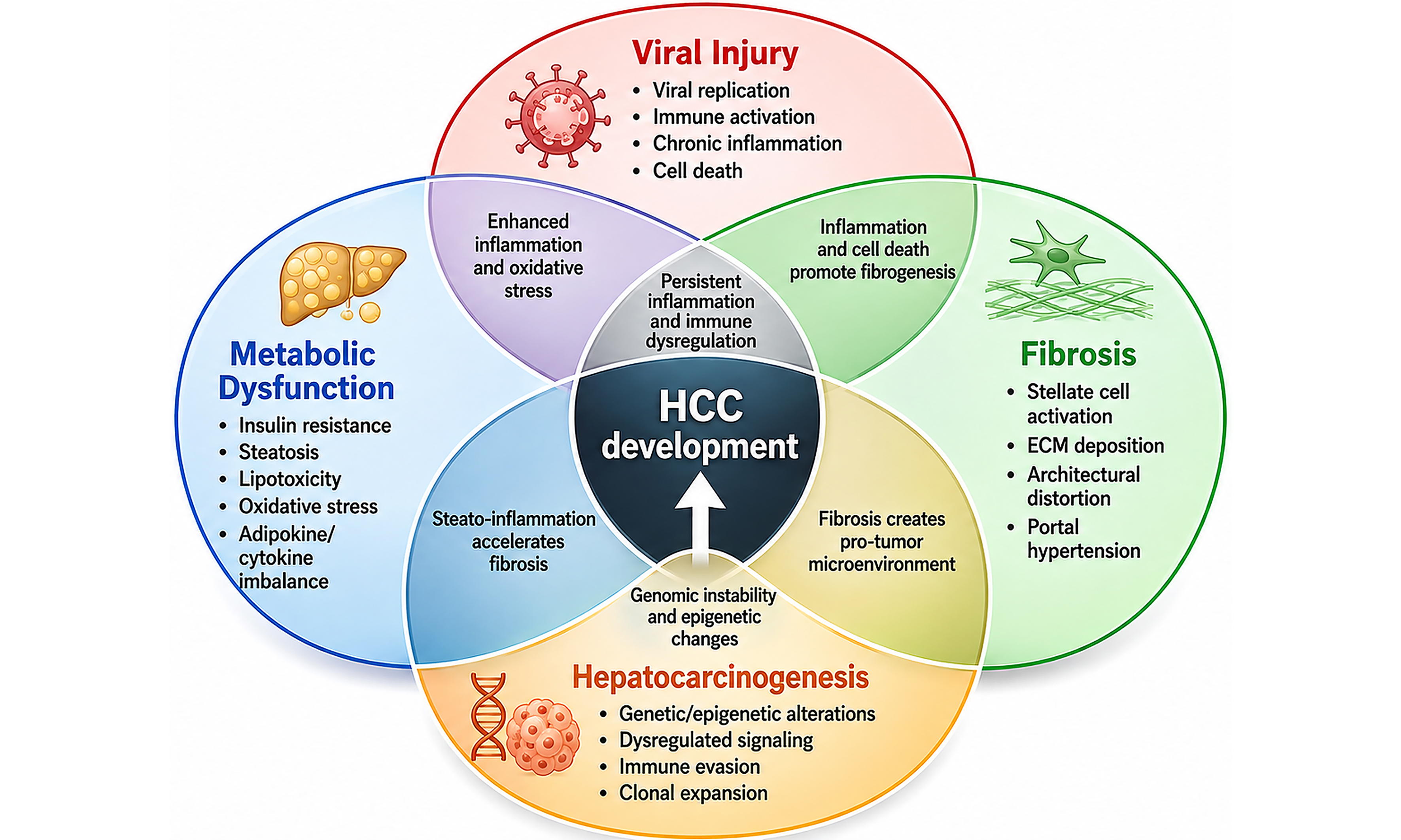
Graphical Abstract
Abstract
Against the background of the growing global burden of obesity and metabolic disease, metabolic dysfunction-associated steatotic liver disease (MASLD) has emerged as a leading cause of chronic liver disease worldwide. Simultaneously, many patients remain affected by chronic hepatitis C virus (HCV) infection or its long-term sequelae, reshaping the spectrum of liver disease in the direct-acting antiviral era. HCV may directly induce hepatic steatosis through genotype-specific mechanisms, whereas MASLD promotes steatosis through systemic metabolic dysregulation. Both pathways can independently cause liver injury, and their coexistence may accelerate fibrosis progression and increase the risk of hepatocellular carcinoma (HCC). Emerging evidence indicates that hepatic steatosis before and after sustained virologic response (SVR) is associated with suboptimal fibrosis regression and persistent HCC risk. These findings highlight the need for integrated clinical management incorporating metabolic optimisation and individualised surveillance. Patients with advanced fibrosis, persistent metabolic dysfunction, or residual steatosis remain at increased risk of HCC despite viral eradication and should not be uniformly classified as low risk. This review examines the biological and clinical intersections between chronic HCV infection and MASLD, focusing on fibrosis progression, hepatocarcinogenesis, and residual HCC risk after SVR. It also emphasises the importance of identifying and managing MASLD in patients with current or previous HCV infection to reduce long-term complications and improve clinical outcomes.
Keywords


 Download PDF
INTRODUCTION
The landscape of chronic liver disease is shifting from a predominantly viral hepatitis paradigm toward an era characterised by overlapping viral and metabolic liver disease[1]. Hepatitis C virus (HCV) exploits several host metabolic pathways to support viral replication, thereby contributing to hepatic metabolic dysregulation during chronic infection[1]. The natural history of chronic HCV infection is further modified by host factors, including obesity, insulin resistance, and hepatic steatosis, which may accelerate fibrosis progression and increase the risk of hepatocellular carcinoma (HCC)[1,2]. The introduction of direct-acting antivirals (DAAs) has substantially changed the treatment and natural history of chronic HCV infection. However, viral eradication does not necessarily reverse coexisting metabolic liver disease, particularly in patients with obesity, insulin resistance, or other persistent metabolic risk factors[2].
Chronic HCV infection is a multisystem disease associated with several extrahepatic manifestations, including mixed cryoglobulinaemic vasculitis, selected autoimmune disorders, lymphoma, and cardiometabolic disease[3]. HCV genotype may influence the metabolic and steatogenic phenotype of infection, although the available evidence varies considerably among genotypes[4]. Host factors, particularly insulin resistance, obesity, and hepatic steatosis, may further amplify inflammation and fibrogenesis[5-7]. These interactions provide the rationale for considering HCV infection and metabolic dysfunction-associated steatotic liver disease (MASLD) as potentially synergistic, rather than entirely independent, drivers of liver injury and HCC risk.
EPIDEMIOLOGICAL OVERLAP AND CLINICAL BURDEN
Hepatic steatosis is common in chronic HCV infection. Its occurrence and severity vary according to viral genotype and host metabolic characteristics, particularly obesity and insulin resistance[4-7]. The coexistence of fatty liver and chronic viral hepatitis has important clinical consequences, as patients with both conditions have higher risks of HCC and mortality than those without fatty liver[8].
Hepatic steatosis may persist or emerge after sustained virologic response (SVR) despite improvement in non-invasive fibrosis markers. Post-treatment steatosis appears to be driven predominantly by persistent host metabolic factors, particularly excess adiposity and insulin resistance, while changes in lipid metabolism may also occur after viral eradication[9-12]. Accordingly, virologic cure does not necessarily eliminate the metabolic drivers of steatotic liver disease, and MASLD may remain clinically relevant after DAA therapy[9-12].
The graphical abstract summarises the converging contributions of viral injury, metabolic dysfunction, chronic inflammation, immune dysregulation, fibrosis, and genomic alterations to hepatocarcinogenesis and HCC development.
PATHOBIOLOGICAL MECHANISMS LINKING HCV AND MASLD
Virus-driven lipid dysregulation
HCV alters hepatic lipid metabolism through several interconnected mechanisms. It enhances de novo lipogenesis by activating lipogenic transcription factors, thereby facilitating the formation of lipoviroparticles required for viral assembly[13-16]. HCV also promotes the production of phospholipids and sphingomyelins, increases the generation of lipotoxic ceramides, and suppresses mitochondrial fatty-acid oxidation, further contributing to intracellular lipid accumulation[17].
Another key mechanism involves interference with very-low-density lipoprotein assembly and secretion, as HCV co-opts the lipoprotein-export pathway to support lipoviroparticle production. HCV also modulates nuclear receptor-mediated transcriptional networks and alters activity within the pentose phosphate pathway. Collectively, these effects disrupt hepatic lipid homeostasis, promote intracellular fat accumulation, and contribute to hepatocellular dysfunction[18-23].
The relative contributions of direct viral effects and host metabolic factors differ according to HCV genotype. Genotype 3 has a distinctive direct steatogenic effect, supported by experimental evidence of genotype 3-specific intracellular triglyceride accumulation and HCV-mediated impairment of microsomal triglyceride transfer protein activity and very-low-density lipoprotein secretion[19,23]. By contrast, in non-genotype 3 infection, hepatic steatosis is more commonly associated with host cardiometabolic factors, including insulin resistance, obesity, and visceral adiposity, although viral and metabolic mechanisms may coexist and amplify liver injury[5,6,12]. Accordingly, steatosis in chronic HCV infection should not be considered uniformly virus-induced; rather, its pathogenesis reflects a genotype-dependent balance between direct viral effects and host metabolic susceptibility.
Metabolic-immune crosstalk
The combined contribution of chronic HCV infection and MASLD to HCC development remains incompletely defined. Nevertheless, several interconnected mechanisms may explain their synergistic effects. Disruption of hepatic metabolic homeostasis promotes the accumulation of toxic lipid species within hepatocytes, leading to lipotoxicity and cellular injury. HCV proteins, particularly the core protein, promote oxidative stress through direct mechanisms, including nicotinamide adenine dinucleotide phosphate (NADPH) oxidase activation, and indirect mechanisms involving mitochondrial dysfunction, endoplasmic reticulum stress, and impaired antioxidant defences. This lipotoxic and oxidative environment contributes to hepatocyte death and perpetuates chronic wound-healing responses, thereby facilitating progression toward HCC[24-27].
Effective control of HCV infection requires coordinated innate and adaptive immune responses[28]. In many patients, viral persistence leads to chronic infection and progressive immune dysfunction. Even after viral eradication with DAAs, immune restoration may remain incomplete. HCV-specific CD8+ T cells may retain features of functional exhaustion, including impaired cytotoxic and antitumour activity. Dysfunction of CD4+ T cells may further compromise CD8+ T-cell responses, while natural killer-cell activity may remain altered after treatment. Collectively, these abnormalities may contribute to defective immunosurveillance and continued susceptibility to malignant transformation[29-34].
Chronic oxidative stress induces repeated cycles of hepatocellular injury and regeneration, promoting telomere shortening and cellular senescence[35]. In the setting of impaired clearance, senescent cells may accumulate and contribute to genomic instability[36]. Cellular senescence can initially participate in tumour-suppressive surveillance of premalignant hepatocytes, whereas persistence of senescent cells may contribute to progressive tissue dysfunction[37,38]. The senescence-associated secretory phenotype creates a proinflammatory microenvironment that can induce senescence in neighbouring cells and amplify tissue injury[39]. Thus, although cellular senescence initially serves protective functions, persistent senescence may ultimately promote metabolic dysfunction, steatosis progression, and long-term HCC risk[35-39].
Liver fibrosis is a major determinant of disease progression and HCC risk. Persistent liver injury activates hepatic stellate cells and promotes their transformation into myofibroblasts, which produce extracellular matrix components. Activated stellate cells also secrete profibrogenic and protumourigenic mediators, including transforming growth factor-β, platelet-derived growth factor, and hepatocyte growth factor. These mediators promote angiogenesis, cellular proliferation, and tumour invasion while impairing antitumour immune responses[40,41].
Metabolic dysfunction further amplifies fibrogenesis through metabolic reprogramming[42]. Hyperinsulinaemia and steatohepatitis enhance fibrotic signalling by increasing the expression of profibrotic mediators, including transforming growth factor-β and connective tissue growth factor. Adipokine signalling, particularly through leptin receptors, also stimulates myofibroblast proliferation, collagen deposition, and resistance to apoptosis, thereby reinforcing fibrosis and creating a procarcinogenic microenvironment[43,44].
CLINICAL PHENOTYPE AND DISEASE PROGRESSION
Excess adiposity is common among patients with chronic HCV infection. In a large cohort of 11,469 US veterans initiating DAA therapy, 78.1% were overweight or obese, including 36.8% with obesity[45]. Obesity and insulin resistance are associated with hepatic steatosis and may contribute to disease progression in chronic HCV infection[5,6,12].
Obesity is a major driver of both simple steatosis and metabolic dysfunction-associated steatohepatitis (MASH). When the storage capacity of adipose tissue is exceeded, excess lipids, predominantly triglycerides, accumulate within hepatocytes. This ectopic lipid deposition promotes steatosis and activates inflammatory responses. In parallel, HCV infection independently induces hepatic inflammation, creating a sustained proinflammatory milieu when MASLD and chronic HCV infection coexist[46].
Hepatic inflammation initially serves a protective role by limiting cellular injury, promoting tissue repair, and maintaining homeostasis. However, persistent inflammation can cause hepatocellular injury, progressive liver dysfunction, and worsening metabolic disturbances. This chronic inflammatory state contributes to the transition from simple steatosis to MASH and may promote further progression to fibrosis, cirrhosis, and HCC[46,47].
The reported prevalence of hepatic steatosis among patients with chronic HCV infection ranges from approximately 40% to 86%, with the wide variation reflecting differences in HCV genotype distribution, host metabolic risk, population characteristics, and methods used to diagnose steatosis[48]. Genotype 3 is particularly associated with more frequent and severe hepatic steatosis, whereas host cardiometabolic factors generally make a greater relative contribution in other genotypes[49-52].
HEPATOCARCINOGENESIS IN THE DUAL DISEASE STATE
Disruption of hepatic metabolic pathways promotes the accumulation of toxic lipids and metabolic intermediates within hepatocytes, leading to lipotoxicity, cellular dysfunction, and injury[1].
Oxidative stress, metabolic dysfunction, and lipotoxicity can substantially alter immune responses to viral infection and malignant transformation[53]. Immunosurveillance may be compromised by HCV-related mechanisms that do not fully resolve after DAA therapy, as well as by systemic metabolic factors that increase HCC risk[53,54]. Metabolic impairment associated with chronic HCV infection may also contribute directly to carcinogenesis[55].
Hyperinsulinaemia may promote hepatocarcinogenesis through sustained activation of the insulin receptor and insulin-like growth factor-1 receptor signalling axes. Recruitment of insulin receptor substrate proteins activates downstream phosphatidylinositol 3-kinase/Akt/mammalian target of rapamycin and Ras/Raf/mitogen-activated protein kinase pathways, promoting protein synthesis, cell-cycle progression, cellular proliferation, and resistance to apoptosis[55]. In fatty liver, premalignant hepatocytes that escape cell death remain exposed to hyperglycaemia, hyperinsulinaemia, lipotoxicity, and oxidative stress. This metabolic environment may preserve mitogenic insulin and insulin-like growth factor signalling despite systemic metabolic insulin resistance, thereby conferring a survival and growth advantage on premalignant hepatocytes[55]. HCV may further perturb insulin receptor substrate-1 and insulin receptor substrate-2 signalling and alter phosphatidylinositol 3-kinase/Akt pathway activity, providing a plausible mechanistic link between HCV-associated insulin resistance, metabolic dysregulation, and oncogenic signalling[56-59].
The interaction between viral persistence, residual post-viral effects, and metabolic dysfunction may create a self-reinforcing cycle of lipotoxicity, immune impairment, senescence, and fibrosis, ultimately increasing HCC risk[60].
THE POST-SVR LANDSCAPE
After SVR, the relative contribution of the different drivers of liver injury changes substantially. Viral replication and the associated direct hepatic injury cease, whereas obesity, insulin resistance, type 2 diabetes mellitus (T2DM), dyslipidaemia, and other cardiometabolic abnormalities may persist or worsen[2,9,61,62]. Accordingly, persistent or incident hepatic steatosis after HCV eradication is generally more consistent with ongoing host metabolic dysfunction than with a residual direct effect of the eradicated virus[9,11,62]. Patients who fulfil the relevant metabolic and steatotic criteria after SVR should therefore be assessed and managed as having MASLD rather than assuming that their steatosis remains an HCV-related manifestation[2,61,62].
Most international guidelines recommend continued HCC surveillance in patients with cirrhosis after achieving SVR. Some guidelines also extend surveillance to selected individuals with advanced fibrosis, including METAVIR F3 disease[63-65]. However, the cost-effectiveness of this strategy remains debated, particularly in patients with advanced fibrosis. Improved methods for HCC risk stratification after SVR are therefore needed. Several studies have proposed risk scoring systems based on fibrosis markers, portal hypertension, liver function, alpha-fetoprotein levels, and high-risk demographic factors. However, these approaches have not yet been widely implemented in routine clinical practice[4,66]. The course of fibrosis after SVR is heterogeneous. Viral eradication removes an important profibrogenic stimulus and is generally followed by improvement in aminotransferases and non-invasive fibrosis markers. However, these early changes may partly reflect resolution of necroinflammation rather than true regression of established extracellular matrix and architectural distortion. Histological fibrosis regression may occur over longer follow-up, but it is not universal, particularly in patients with advanced baseline fibrosis or persistent non-viral drivers of liver injury. Thus, even among patients without overt MASLD after SVR, improvement in fibrosis is probable but should not be assumed in every individual, and serial assessment remains necessary[66].
A recent systematic review and meta-analysis evaluated the relationship between MASLD and HCC risk after SVR in patients with chronic HCV infection[60]. Across the included studies, the reported prevalence of steatotic liver disease ranged from 5.9% to 57.6%[60]. This wide variation likely reflects differences in the definitions used, including legacy non-alcoholic fatty liver disease (NAFLD) or metabolic dysfunction-associated fatty liver disease (MAFLD) criteria; the methods used to detect steatosis, such as ultrasonography, computed tomography, controlled attenuation parameter, or histology; and differences in ethnicity, geographic setting, HCV genotype distribution, baseline fibrosis severity, obesity, T2DM, and other metabolic risk factors[9-12,60].
The cumulative incidence of HCC also varied from 4.35% to 14.36%, partly because of differences in follow-up duration, baseline fibrosis stage, study design, and the prevalence of established metabolic risk factors[60]. This heterogeneity limits the use of a single pooled prevalence or absolute HCC-risk estimate for individual patients or different geographic populations. Nevertheless, the direction of association was consistent: metabolic dysfunction was associated with increased HCC risk after SVR[60]. Clinical application should therefore focus less on a universal numerical estimate and more on individualised post-SVR risk assessment incorporating fibrosis stage, persistent steatosis, obesity, T2DM, and other ongoing liver-injury cofactors[60,67-71].
Before viral eradication, HCV-related inflammation and metabolic liver injury may act synergistically to accelerate fibrogenesis[12,46,66]. Consequently, the fibrosis burden observed at the time of SVR may represent the cumulative effect of both viral and metabolic injury rather than either condition alone[12,66,67]. After SVR, patients without persistent metabolic or other hepatic injury may experience a more favourable fibrosis trajectory, whereas obesity, hepatic steatosis, T2DM, alcohol exposure, and other ongoing insults may attenuate fibrosis regression or contribute to further progression[66-68]. Persistent metabolic and liver-related risk factors are also associated with residual clinical events, including HCC and cirrhosis-related complications[67-71]. These factors should therefore be incorporated into post-SVR risk stratification rather than treating all patients with virologic cure as a homogeneous group.
In a study by Di Nardo et al. comparing MASLD and MAFLD criteria for estimating the 5 year risk of de novo HCC in patients with steatotic liver disease who achieved SVR after DAA therapy, the authors reported that MAFLD criteria provided a more accurate estimation of HCC risk[72].
Table 1 summarises the clinical implications before and after SVR in chronic HCV infection[4,12,63].
Clinical implications before and after sustained virologic response in chronic HCV infection
| Domain | Before SVR: active HCV infection | After SVR: virologic cure |
| Viral activity | Ongoing viral replication with detectable HCV RNA[65,86] | Undetectable HCV RNA 12 weeks or more after treatment completion, indicating virologic cure[65,86] |
| Liver inflammation | Persistent necroinflammation driven by active viral infection, with possible additional metabolic injury[12,46] | Inflammation generally decreases substantially, although residual metabolic or other non-viral inflammatory activity may persist[9,11,66] |
| Fibrosis progression | Progressive fibrosis may result from the cumulative effects of viral inflammation and coexisting metabolic liver injury[12,46,66] | Fibrosis may regress after viral eradication, but regression is heterogeneous and may be attenuated by persistent steatosis, obesity, T2DM, alcohol exposure, or other liver injury[66-68,102-106] |
| Cirrhosis | The risk of cirrhosis increases with the duration and severity of untreated infection and associated cofactors[65,66] | Progression risk decreases after SVR, but established cirrhosis may persist and should not be considered reversed solely because non-invasive markers improve[66,104,107] |
| HCC risk | Risk is increased, particularly in patients with advanced fibrosis, cirrhosis, or coexisting metabolic dysfunction[8,12,60] | Risk is reduced but not eliminated, particularly in patients with cirrhosis, advanced fibrosis, T2DM, obesity, or persistent steatosis[60,63-71] |
| Metabolic dysfunction | Insulin resistance, obesity, T2DM, dyslipidaemia, and hepatic steatosis may coexist with HCV infection and amplify liver injury[5-7,12] | Metabolic dysfunction may persist or emerge after viral eradication and may become the predominant driver of steatosis and ongoing liver injury[2,9,11,61,62] |
| Portal hypertension | Portal hypertension may develop in patients with advanced fibrosis or cirrhosis[66,107] | Portal hypertension may improve after SVR but can persist, particularly in patients with established advanced chronic liver disease or ongoing cofactors[107] |
| Extrahepatic manifestations | Cryoglobulinaemic vasculitis, renal disease, lymphoproliferative disorders, and cardiometabolic manifestations may occur[3] | Many HCV-related extrahepatic manifestations improve after virologic cure, although abnormalities in immune activation and immune-cell function may persist in some patients after DAA-induced SVR[3,29-31,34] |
| Liver function and decompensation | Liver function may deteriorate as fibrosis progresses, with an increasing risk of hepatic decompensation[65,66] | Liver function and decompensation risk generally improve, but clinically important risk may remain in patients with advanced chronic liver disease[66,70,107] |
| Mortality | Liver-related and all-cause mortality are increased, especially in advanced liver disease[8,70,71] | Mortality risk decreases after SVR but may not normalise in patients with cirrhosis, metabolic dysfunction, or other ongoing liver injury[8,70,71] |
| Surveillance requirements | Patients with cirrhosis require HCC surveillance and monitoring for complications of portal hypertension[63-65] | HCC surveillance should continue in patients with cirrhosis; selected patients with advanced fibrosis may also require surveillance according to the applicable guideline and individualised risk[63-65,107] |
| Management focus | Achieve viral eradication, assess fibrosis severity, and manage complications and metabolic cofactors[65,83,86] | Apply long-term fibrosis and HCC risk stratification, manage metabolic dysfunction, and continue appropriate surveillance[60,63-68,102-107] |
DIAGNOSTIC AND RISK STRATIFICATION STRATEGIES
Historically, liver fibrosis assessment relied primarily on liver biopsy as the reference standard for staging. However, biopsy is invasive and carries a risk of complications, reported in up to 3% of cases, with a small but notable mortality risk of approximately 0.03%. These limitations have driven a shift toward reliable non-invasive modalities for the diagnosis and staging of liver fibrosis[73].
Elastography is a non-invasive imaging modality that is now widely used in clinical practice to assess liver fibrosis. Most elastography techniques estimate liver stiffness by measuring shear wave propagation velocity. The main approaches include ultrasound-based elastography and magnetic resonance elastography[74,75].
BLOOD-BASED NON-INVASIVE TESTS
Routine laboratory tests support the non-invasive evaluation of chronic liver disease and include aspartate aminotransferase (AST), alanine aminotransferase (ALT), gamma-glutamyl transferase (GGT), total bilirubin, albumin, prothrombin time, and platelet count. These routinely available parameters can be incorporated into simple composite scores, whereas additional biomarkers may be combined in proprietary serum panels[74].
SIMPLE COMPOSITE SCORES
The aspartate aminotransferase-to-platelet ratio index (APRI) is calculated by dividing the ratio of AST to its upper limit of normal by the platelet count (×109/L) and multiplying the result by 100. Evidence from studies and meta-analyses in HCV- and hepatitis B virus-related liver disease supports the utility of APRI for estimating fibrosis severity. Because APRI is derived from routine laboratory parameters, it is a simple, accessible, and cost-effective tool for initial fibrosis assessment[76,77].
The fibrosis-4 index (FIB-4) was initially developed in the APRICOT study to estimate liver fibrosis in patients with human immunodeficiency virus (HIV)/HCV coinfection. It incorporates age, AST, ALT, and platelet count. Subsequent studies have supported its utility across different liver diseases and have proposed age-adjusted thresholds where appropriate. FIB-4 is therefore a practical and widely accessible first-line tool for fibrosis risk stratification, although its interpretation should account for age and the clinical context[76,78,79].
The interpretation of serial fibrosis estimates after HCV cure should also consider that histological fibrosis may regress after SVR, whereas age at infection and the duration of liver injury may influence long-term outcomes[80,81].
PROPRIETARY SERUM BIOMARKER PANELS
FibroTest® (Biopredictive, Paris, France) is a proprietary serum biomarker panel originally developed and validated in patients with chronic HCV infection. It combines α2-macroglobulin, haptoglobin, GGT, total bilirubin, and apolipoprotein A1, together with age and sex. A meta-analysis of 30 studies involving more than 3,000 patients demonstrated its diagnostic utility for liver fibrosis staging[82].
Risk stratification is essential for identifying patients with chronic liver disease who are at higher risk of advanced fibrosis, cirrhosis, or liver-related complications. Current approaches use stepwise algorithms that combine simple non-invasive scores, such as FIB-4 and APRI, with more advanced modalities, including elastography or proprietary serum panels such as FibroTest®. This approach improves diagnostic accuracy and reduces the need for liver biopsy. Low-risk patients can usually be monitored, intermediate-risk patients require further assessment with imaging-based elastography, and high-risk patients should be prioritised for specialist referral and management. This layered strategy enhances cost-effectiveness, minimises unnecessary invasive procedures, and enables earlier identification of patients who may benefit from surveillance and therapeutic intervention. Recent guidelines and studies support the use of integrated risk-based pathways in routine clinical practice, emphasising their role in improving patient outcomes and resource allocation[83].
INTEGRATED THERAPEUTIC AND MANAGEMENT APPROACHES
The introduction of DAAs has transformed chronic HCV management, with contemporary regimens achieving SVR in more than 95% of treated patients[84]. However, viral eradication does not eliminate non-viral determinants of long-term outcomes. Metabolic abnormalities may persist or emerge after SVR[9,62], while residual HCC risk remains clinically relevant, particularly in patients with advanced liver disease or persistent metabolic risk factors[67-71]. Cardiovascular risk also remains clinically important in this population; notably, DAA treatment and achievement of SVR have been associated with a lower incidence of cardiovascular events compared with untreated HCV infection or failure to achieve SVR[85]. DAA therapy currently achieves high cure rates in patients with HCV infection. Nevertheless, important clinical challenges remain because patients who achieve SVR may retain residual HCC risk, particularly in the presence of advanced liver disease, while individuals with undiagnosed active HCV infection remain exposed to ongoing HCV-related liver injury and HCC risk[63-71,86].
A recent study of 1,280 elderly patients who achieved HCV eradication with DAAs and had no prior history of HCC reported that 25.8% developed MASLD within 24 weeks after SVR. MASLD at this time point was associated with a 3.04-fold increased risk of subsequent HCC development. Overall, 6.7% of the cohort developed HCC. These findings suggest that patients with overlapping etiologies of liver injury may remain at increased risk of advanced fibrosis and HCC, even after successful viral clearance[87].
LIFESTYLE AND WEIGHT MANAGEMENT
Lifestyle-based intervention remains the foundation of MASLD management before and after HCV eradication. Management should combine an appropriate dietary pattern, regular physical activity, reduced sedentary behaviour, and sustained weight reduction when excess adiposity is present. These interventions may improve insulin sensitivity, reduce hepatic steatosis and inflammatory activity, and provide broader cardiometabolic benefits[88,89]. Nutritional and behavioural strategies should be individualised according to comorbidities, genetic background, environmental exposures, socioeconomic circumstances, and regional and cultural practices rather than applied uniformly across populations[88].
GENERAL CARDIOMETABOLIC TREATMENTS
The effect of pharmacological treatment of cardiometabolic comorbidities on fibrosis progression and HCC risk remains incompletely established in patients with current or previous HCV infection. Nevertheless, experimental and observational evidence suggests potential hepatic and extrahepatic benefits from metformin, statins, and aspirin[90-94].
Metformin may exert antiviral and metabolic effects through activation of adenosine monophosphate-activated protein kinase, enhancement of type I interferon-mediated antiviral pathways, and suppression of HCV core protein expression[95]. In patients with T2DM who achieved SVR, metformin use has also been associated with a lower incidence of HCC[96]. Lipophilic statins have been associated with reduced fibrosis progression and lower risks of HCC and liver-related mortality in patients with chronic viral hepatitis[93,94]. Combined metformin and statin use has similarly been associated with reduced HCC risk in patients with HCV infection, although these findings are derived mainly from observational data[97]. Aspirin use has also been associated with lower HCC incidence and liver-related mortality, but its potential benefits must be balanced against individual bleeding risk[92].
LIVER-TARGETED TREATMENT FOR MASH
Liver-targeted pharmacotherapy should be considered separately from treatment prescribed primarily for diabetes, obesity, dyslipidaemia, or cardiovascular risk. Vitamin E has demonstrated histological benefit in selected non-diabetic adults with non-cirrhotic steatohepatitis[98,99]. A small randomised study also reported reductions in liver enzyme levels among patients with HCV genotype 3 receiving vitamin E; however, the evidence is insufficient to support its routine use specifically in patients with overlapping HCV infection and MASLD[100].
Resmetirom, a liver-directed thyroid hormone receptor-β agonist, became the first pharmacological treatment approved by the US Food and Drug Administration for non-cirrhotic MASH with moderate-to-advanced fibrosis[101]. However, its efficacy and safety have not been specifically established in patients with concomitant active HCV infection, and evidence in patients with previous HCV infection remains limited. Treatment decisions should therefore be individualised according to the current fibrosis stage, metabolic phenotype, cirrhosis status, potential contraindications, and the locally approved indication.
TIMING AND ATTRIBUTION OF FIBROSIS ASSESSMENT AFTER SVR
In patients with coexisting chronic HCV infection and MASLD, fibrosis documented before antiviral treatment may reflect liver injury caused by HCV, MASH, or the cumulative contribution of both conditions. Furthermore, aminotransferase-based fibrosis scores and liver stiffness measurements may decrease rapidly during and after DAA therapy because of the resolution of necroinflammation, in addition to any subsequent regression of extracellular matrix fibrosis. Therefore, an early reduction in non-invasive fibrosis measurements after SVR should not automatically be interpreted as regression of established architectural fibrosis[102-105].
Management of obesity, T2DM, dyslipidaemia, hypertension, and other cardiometabolic risk factors should begin or continue irrespective of the timing of fibrosis reassessment. By contrast, when eligibility for MASH-targeted pharmacotherapy depends on documentation of significant fibrosis, fibrosis severity should preferably be reassessed after SVR has been confirmed and liver biochemistry has stabilised[102,103]. Current guidance does not specify a mandatory waiting interval after HCV eradication. Nevertheless, reassessment approximately 6-12 months after completion of DAA therapy represents a reasonable pragmatic approach because much of the early decrease in liver stiffness occurs during the first months after treatment and may partly reflect resolution of inflammatory activity rather than true fibrosis regression[104-106].
A prolonged delay in treatment is not necessary when significant non-cirrhotic fibrosis has already been established by a reliable pretreatment liver biopsy or by concordant validated non-invasive tests, particularly when the patient has a clear metabolic phenotype consistent with active MASLD. Conversely, an isolated elevated FIB-4 or liver stiffness measurement obtained before treatment or immediately after SVR should not be considered sufficient to attribute fibrosis specifically to MASH[102-104].
Post-SVR reassessment should follow a sequential non-invasive strategy. A simple blood-based test such as FIB-4 may be followed by vibration-controlled transient elastography or another validated imaging-based modality. The enhanced liver fibrosis test may be considered when elastography is unavailable, while magnetic resonance elastography can assist in selected patients with indeterminate or discordant results. Liver biopsy should be reserved for cases in which non-invasive tests remain discordant, an alternative liver disease is suspected, or histological confirmation would materially alter the decision to initiate MASH-targeted therapy[102-104].
Persistent concordant evidence of non-cirrhotic significant fibrosis in a patient with an appropriate metabolic phenotype may support consideration of MASH-targeted pharmacotherapy according to the approved indication. However, patients with established cirrhosis before antiviral treatment should continue cirrhosis-specific management and HCC surveillance despite subsequent improvement in aminotransferases, FIB-4, or liver stiffness. In patients with compensated advanced chronic liver disease, post-SVR non-invasive criteria may help refine the assessment of clinically significant portal hypertension and decompensation risk, but improvement in these measurements does not by itself establish reversal of cirrhosis[104,107].
HEPATIC AND EXTRAHEPATIC OUTCOMES OF CARDIOMETABOLIC THERAPIES
The selection of glucose-lowering and weight-management therapies in patients with coexisting HCV and MASLD should consider both the hepatic phenotype and major competing cardiometabolic risks, particularly atherosclerotic cardiovascular disease, heart failure, and chronic kidney disease. Incretin-based therapies are especially relevant because they provide substantial weight reduction and glycaemic improvement while also demonstrating potential liver-specific benefits. In the phase 3 ESSENCE trial, semaglutide improved both MASH resolution without worsening of fibrosis and fibrosis improvement without worsening of MASH in patients with non-cirrhotic F2-F3 fibrosis[108]. In the phase 2 SYNERGY-NASH trial, tirzepatide produced dose-dependent improvement in MASH resolution and a favourable, although less definitive, signal for fibrosis improvement[109].
Beyond their hepatic effects, glucagon-like peptide-1 receptor agonists may reduce cardiovascular risk. Semaglutide reduced major adverse cardiovascular events in patients with established cardiovascular disease and overweight or obesity, even in the absence of diabetes[110]. These findings support the use of incretin-based therapy when clinically indicated for obesity or T2DM in patients with MASLD. However, their hepatic efficacy should not be assumed to be identical in patients with active HCV infection or in dedicated post-SVR HCV-MASLD populations, which have not been specifically evaluated in the pivotal MASH trials.
Sodium-glucose cotransporter 2 inhibitors also provide important extrahepatic benefits. Empagliflozin reduces the risk of cardiovascular death or hospitalisation for heart failure in patients with heart failure with preserved ejection fraction and lowers the risk of kidney disease progression or cardiovascular death in patients with chronic kidney disease[111,112]. Although sodium-glucose cotransporter 2 inhibitors may improve body weight, glycaemic control, aminotransferase levels, and hepatic steatosis, evidence of histological MASH resolution or direct antifibrotic efficacy remains insufficient. They should therefore be prescribed according to established diabetes, heart-failure, or kidney-disease indications rather than as liver-targeted MASH therapy[102,111,112].
METABOLIC AND BARIATRIC SURGERY
Metabolic and bariatric surgery should be considered in appropriately selected patients with obesity and MASLD who meet contemporary surgical eligibility criteria, particularly when lifestyle and pharmacological interventions have not achieved durable weight reduction or adequate control of obesity-related complications[113]. Surgery should be viewed as an obesity and metabolic intervention with potential hepatic benefits rather than as a treatment directed specifically at previous HCV infection.
Evidence supports clinically relevant improvement in steatohepatitis after surgery. In the randomised BRAVES trial, both Roux-en-Y gastric bypass and sleeve gastrectomy were more effective than lifestyle intervention plus best medical care in achieving histological resolution of steatohepatitis without worsening of fibrosis at 1 year among patients with obesity and biopsy-confirmed non-cirrhotic steatohepatitis[114]. Observational evidence also suggests that bariatric surgery may reduce major adverse liver outcomes and cardiovascular events in patients with obesity and biopsy-proven fibrotic steatohepatitis[115]. However, these studies were not designed specifically for patients with previous or active HCV infection, and their findings should not be extrapolated uncritically to the HCV-MASLD overlap population.
Surgical candidacy should be determined through multidisciplinary assessment of liver fibrosis, portal hypertension, hepatic functional reserve, cardiometabolic comorbidities, nutritional status, and operative risk. Patients with established cirrhosis require particularly careful selection, and those with clinically significant portal hypertension or decompensated liver disease should be evaluated in experienced hepatology, bariatric surgery, and transplantation centres[102,107]. Long-term follow-up is essential to monitor nutritional deficiencies, sarcopenia, weight recurrence, metabolic control, and liver-related outcomes.
Therefore, effective management of MASLD before and after DAA therapy is essential to limit disease progression and reduce HCC-related mortality.
Overall, these findings suggest that addressing metabolic dysfunction in patients with coexisting MASLD and HCV infection may provide dual hepatic and extrahepatic benefits. Nevertheless, further research is needed to clarify the impact of these therapeutic strategies in patients with overlapping viral and metabolic liver disease.
RESEARCH GAPS AND FUTURE DIRECTIONS
Several specific research priorities should be addressed to improve the management of patients with overlapping HCV and MASLD. First, multicentre prospective cohorts should enrol patients before DAA therapy and perform standardised metabolic and fibrosis assessments at baseline, sustained virologic response at 12 weeks (SVR12), 6-12 months, and during longer-term follow-up. These studies should combine anthropometric and cardiometabolic phenotyping with FIB-4, vibration-controlled transient elastography, controlled attenuation parameter, and, where feasible, magnetic resonance-based measurements. Such longitudinal designs would help distinguish the early inflammation-related reduction in liver stiffness from subsequent fibrosis regression and determine how persistent obesity, T2DM, steatosis, and alcohol exposure modify the post-SVR fibrosis trajectory[9,62,66-68,102-106].
Second, mechanistic studies should investigate whether previous HCV infection leaves persistent molecular or immunological signatures that interact with ongoing metabolic liver injury after viral eradication. Studies incorporating paired liver tissue, circulating biomarkers, immune profiling, epigenetic analyses, and transcriptomic or metabolomic platforms could clarify the relative contributions of residual virus-associated alterations and active MASH to fibrosis progression and hepatocarcinogenesis[24-44,53-60].
Third, dedicated interventional trials are required in patients with MASLD after HCV cure. Trials should evaluate liver-targeted therapies, incretin-based treatments, sodium-glucose cotransporter 2 inhibitors, and structured weight-loss interventions using both hepatic and extrahepatic outcomes. Relevant endpoints should include change in liver fat, MASH resolution, fibrosis improvement, cardiometabolic events, kidney outcomes, treatment safety, and patient-reported outcomes[102,103,108-112]. Metabolic and bariatric surgery should also be evaluated prospectively in appropriately selected post-SVR patients, particularly those with severe obesity and non-cirrhotic fibrotic MASH[113-115].
Fourth, post-SVR HCC risk models should be developed using standardised variables that integrate baseline and longitudinal fibrosis measurements, steatosis, obesity, T2DM, alcohol exposure, age, sex, platelet count, portal hypertension, and alpha-fetoprotein. These models should undergo external validation across different ethnic, geographic, and HCV-genotype populations rather than being derived and tested within a single cohort[60,67-71].
Finally, pragmatic surveillance studies should determine whether selected non-cirrhotic patients with advanced fibrosis and substantial metabolic risk benefit from continued HCC surveillance after SVR. These studies should compare risk-based strategies with conventional fibrosis-stage-based surveillance and incorporate cost-effectiveness, feasibility, patient adherence, and regional resource availability[60,63-71,107].
CONCLUSION
Hepatic fat accumulation in HCV infection shares several pathophysiological features with MASLD, including insulin resistance, oxidative stress, metabolic dysfunction, and chronic inflammation. Each condition can independently contribute to liver injury; however, when they coexist, their interaction may amplify disease progression through overlapping mechanisms involving low-grade inflammation, metabolic dysregulation, oxidative stress, immune dysfunction, fibrosis, and the direct effects of viral proteins. This viral metabolic overlap should therefore be regarded as a distinct clinical phenotype rather than as the simple coexistence of two separate liver diseases.
The residual risk of HCC after SVR should remain a central consideration in both research and clinical practice. Patients who achieve SVR should undergo continued clinical follow-up and individualised HCC risk assessment, particularly when advanced fibrosis, obesity, T2DM, or persistent steatosis are present. Standard HCC surveillance should continue in patients with cirrhosis after SVR, whereas surveillance in non-cirrhotic patients with advanced fibrosis should be considered according to applicable guidelines and individualised risk[63-65,107]. Obesity, T2DM, or persistent steatosis alone should not be interpreted as an indication for routine HCC surveillance, although these factors may increase residual risk and should inform longitudinal risk assessment[60,67-71].
Future management strategies should integrate antiviral history, fibrosis stage, metabolic risk profile, and steatosis status into long-term care pathways. Addressing metabolic dysfunction should be considered a core component of hepatology practice in patients with current or prior HCV infection. This includes structured follow-up, individualised HCC risk assessment, guideline-based HCC surveillance when indicated, weight and cardiometabolic optimisation, and careful assessment of patients without overt fatty liver before antiviral therapy but who remain at risk of incident steatosis after SVR. A clinically translational perspective is needed, ensuring that mechanistic insights into lipotoxicity, immune dysregulation, senescence, inflammation, and fibrosis are directly connected to risk stratification and patient management.
At the same time, important uncertainties should be acknowledged. Evidence remains limited or heterogeneous regarding surveillance strategies in non-cirrhotic patients, the optimal incorporation of metabolic factors into HCC risk models, and the effect of metabolic therapies on long-term liver-related outcomes and HCC prevention in patients with overlapping HCV and MASLD. Future studies should therefore develop and validate risk models that incorporate metabolic comorbidities, define cost-effective surveillance strategies for this evolving population, and test whether targeted metabolic interventions can modify liver-related outcomes after SVR. Until such evidence is available, a comprehensive strategy combining viral cure, metabolic control, nutritional optimisation, longitudinal risk assessment, and guideline-concordant HCC surveillance when indicated remains essential to improve long-term outcomes in patients with coexisting MASLD and chronic HCV infection.
DECLARATIONS
Authors’ contributions
Conceptualised the idea and planned the manuscript: El-Kassas M
Wrote the initial draft: Awad A
Revised and edited the manuscript: AlNaamani KM, Alswat KA, Elzouki AN
All authors revised and approved the final version.
Availability of data and materials
Not applicable.
AI and AI-assisted tools statement
Not applicable.
Financial support and sponsorship
None.
Conflicts of interest
All authors declared that there are no conflicts of interest.
Ethical approval and consent to participate
Not applicable.
Consent for publication
Not applicable.
Copyright
© The Author(s) 2026.
REFERENCES
1. Leslie J, Geh D, Elsharkawy AM, Mann DA, Vacca M. Metabolic dysfunction and cancer in HCV: Shared pathways and mutual interactions. J Hepatol. 2022;77:219-36.
2. Huang CF, Huang JF, Yu ML, Chuang WL. Metabolic Dysfunction-Associated Steatotic Liver Disease After Hepatitis C Virus Cure. Kaohsiung J Med Sci. 2026;42:e70155.
3. Cacoub P, Saadoun D. Extrahepatic manifestations of chronic HCV infection. N Engl J Med. 2021;384:1038-52.
4. Leumi S, El Kassas M, Zhong J. Hepatitis C virus genotype 4: a poorly characterized endemic genotype. J Med Virol. 2021;93:6079-88.
5. Gaggini M, Carli F, Rosso C, et al. Altered metabolic profile and adipocyte insulin resistance mark severe liver fibrosis in patients with chronic liver disease. Int J Mol Sci. 2019;20:6333.
6. Pais R, Rusu E, Zilisteanu D, et al. Prevalence of steatosis and insulin resistance in patients with chronic hepatitis B compared with chronic hepatitis C and non-alcoholic fatty liver disease. Eur J Intern Med. 2015;26:30-6.
7. Fiore G, Fera G, Napoli N, Vella F, Schiraldi O. Liver steatosis and chronic hepatitis C: a spurious association? Eur J Gastroenterol Hepatol 1996;8:125-9.
8. Kim MN, Han K, Yoo J, Hwang SG, Ahn SH. Increased risk of hepatocellular carcinoma and mortality in chronic viral hepatitis with concurrent fatty liver. Aliment Pharmacol Ther. 2022;55:97-107.
9. Shengir M, Elgretli W, Cinque F, et al. Metabolic factors drive early increase in hepatic steatosis despite improvement in non-invasive fibrosis markers after hepatitis C eradication with direct-acting antivirals. Clin Res Hepatol Gastroenterol. 2025;49:102639.
11. Soliman H, Ziada D, Hamisa M, et al. The effect of HCV eradication after direct-acting antiviral agents on hepatic steatosis: a prospective observational study. Endocr Metab Immune Disord Drug Targets. 2022;22:100-7.
12. Adinolfi LE, Gambardella M, Andreana A, Tripodi MF, Utili R, Ruggiero G. Steatosis accelerates the progression of liver damage of chronic hepatitis C patients and correlates with specific HCV genotype and visceral obesity. Hepatology. 2001;33:1358-64.
13. Yamaguchi A, Tazuma S, Nishioka T, et al. Hepatitis C virus core protein modulates fatty acid metabolism and thereby causes lipid accumulation in the liver. Dig Dis Sci. 2005;50:1361-71.
14. Lin W, Tsai WL, Shao RX, et al. Hepatitis C virus regulates transforming growth factor beta1 production through the generation of reactive oxygen species in a nuclear factor kappaB-dependent manner. Gastroenterology. 2010;138:2509-18.
15. Sato C, Saito T, Misawa K, et al. Impaired mitochondrial β-oxidation in patients with chronic hepatitis C: relation with viral load and insulin resistance. BMC Gastroenterol. 2013;13:112.
16. Tanaka N, Moriya K, Kiyosawa K, Koike K, Gonzalez FJ, Aoyama T. PPARalpha activation is essential for HCV core protein-induced hepatic steatosis and hepatocellular carcinoma in mice. J Clin Invest. 2008;118:683-94.
17. Yang W, Hood BL, Chadwick SL, et al. Fatty acid synthase is up-regulated during hepatitis C virus infection and regulates hepatitis C virus entry and production. Hepatology. 2008;48:1396-403.
18. Tsutsumi T, Suzuki T, Shimoike T, et al. Interaction of hepatitis C virus core protein with retinoid X receptor alpha modulates its transcriptional activity. Hepatology. 2002;35:937-46.
19. Abid K, Pazienza V, de Gottardi A, et al. An in vitro model of hepatitis C virus genotype 3a-associated triglycerides accumulation. J Hepatol. 2005;42:744-51.
20. Levy G, Habib N, Guzzardi MA, et al. Nuclear receptors control pro-viral and antiviral metabolic responses to hepatitis C virus infection. Nat Chem Biol. 2016;12:1037-45.
21. Dharancy S, Malapel M, Perlemuter G, et al. Impaired expression of the peroxisome proliferator-activated receptor alpha during hepatitis C virus infection. Gastroenterology. 2005;128:334-42.
22. Gastaminza P, Cheng G, Wieland S, Zhong J, Liao W, Chisari FV. Cellular determinants of hepatitis C virus assembly, maturation, degradation, and secretion. J Virol. 2008;82:2120-9.
23. Perlemuter G, Sabile A, Letteron P, et al. Hepatitis C virus core protein inhibits microsomal triglyceride transfer protein activity and very low density lipoprotein secretion: a model of viral-related steatosis. FASEB J. 2002;16:185-94.
24. Kim SJ, Syed GH, Khan M, et al. Hepatitis C virus triggers mitochondrial fission and attenuates apoptosis to promote viral persistence. Proc Natl Acad Sci U S A. 2014;111:6413-8.
25. Moriya K, Miyoshi H, Shinzawa S, et al. Hepatitis C virus core protein compromises iron-induced activation of antioxidants in mice and HepG2 cells. J Med Virol. 2010;82:776-92.
26. Ripoli M, D'Aprile A, Quarato G, et al. Hepatitis C virus-linked mitochondrial dysfunction promotes hypoxia-inducible factor 1 alpha-mediated glycolytic adaptation. J Virol. 2010;84:647-60.
27. Kim SJ, Syed GH, Siddiqui A. Hepatitis C virus induces the mitochondrial translocation of Parkin and subsequent mitophagy. PLoS Pathog. 2013;9:e1003285.
28. Dustin LB. Innate and adaptive immune responses in chronic HCV infection. Curr Drug Targets. 2017;18:826-43.
29. Hengst J, Falk CS, Schlaphoff V, et al. Direct-acting antiviral-induced hepatitis C virus clearance does not completely restore the altered cytokine and chemokine milieu in patients with chronic hepatitis C. J Infect Dis. 2016;214:1965-74.
30. Osuch S, Metzner KJ, Caraballo Cortés K. Reversal of T cell exhaustion in chronic HCV infection. Viruses. 2020;12:799.
31. Luxenburger H, Neumann-Haefelin C, Thimme R, Boettler T. HCV-specific T cell responses during and after chronic HCV infection. Viruses. 2018;10:645.
32. Wherry E, Kurachi M. Molecular and cellular insights into T cell exhaustion. Nat Rev Immunol. 2015;15:486-99.
33. Gerlach JT, Diepolder HM, Jung MC, et al. Recurrence of hepatitis C virus after loss of virus-specific CD4+ T-cell response in acute hepatitis C. Gastroenterology. 1999;117:933-41.
34. Golden-Mason L, McMahan RH, Kriss MS, et al. Early and late changes in natural killer cells in response to ledipasvir/sofosbuvir treatment. Hepatol Commun. 2018;2:364-75.
35. Sekoguchi S, Nakajima T, Moriguchi M, et al. Role of cell-cycle turnover and oxidative stress in telomere shortening and cellular senescence in patients with chronic hepatitis C. J Gastroenterol Hepatol. 2007;22:182-90.
36. Maciejowski J, de Lange T. Telomeres in cancer: tumour suppression and genome instability. Nat Rev Mol Cell Biol. 2017;18:175-86.
37. Kang TW, Yevsa T, Woller N, et al. Senescence surveillance of pre-malignant hepatocytes limits liver cancer development. Nature. 2011;479:547-51.
38. Baker DJ, Wijshake T, Tchkonia T, et al. Clearance of p16Ink4a-positive senescent cells delays ageing-associated disorders. Nature. 2011;479:232-6.
39. Acosta JC, Banito A, Wuestefeld T, et al. A complex secretory program orchestrated by the inflammasome controls paracrine senescence. Nat Cell Biol. 2013;15:978-90.
40. Affo S, Yu LX, Schwabe RF. The role of cancer-associated fibroblasts and fibrosis in liver cancer. Annu Rev Pathol. 2017;12:153-86.
41. Baglieri J, Brenner DA, Kisseleva T. The role of fibrosis and liver-associated fibroblasts in the pathogenesis of hepatocellular carcinoma. Int J Mol Sci. 2019;20:1723.
42. Bates J, Vijayakumar A, Ghoshal S, et al. Acetyl-CoA carboxylase inhibition disrupts metabolic reprogramming during hepatic stellate cell activation. J Hepatol. 2020;73:896-905.
43. Manolakopoulos S, Bethanis S, Liapi C, et al. An assessment of serum leptin levels in patients with chronic viral hepatitis: a prospective study. BMC Gastroenterol. 2007;7:17.
44. Myers MG Jr, Leibel RL, Seeley RJ, Schwartz MW. Obesity and leptin resistance: distinguishing cause from effect. Trends Endocrinol Metab. 2010;21:643-51.
45. Do A, Esserman DA, Krishnan S, et al. Excess weight gain after cure of hepatitis C infection with direct-acting antivirals. J Gen Intern Med. 2020;35:2025-34.
47. Battineni G, Sagaro GG, Chintalapudi N, Amenta F, Tomassoni D, Tayebati SK. Impact of obesity-induced inflammation on cardiovascular diseases (CVD). Int J Mol Sci. 2021;22:4798.
48. Siphepho PY, Liu YT, Shabangu CS, et al. The impact of steatosis on chronic hepatitis C progression and response to antiviral treatments. Biomedicines. 2021;9:1491.
49. Sheridan DA, Shawa IT, Thomas EL, et al. Infection with the hepatitis C virus causes viral genotype-specific differences in cholesterol metabolism and hepatic steatosis. Sci Rep. 2022;12:5562.
50. Hourioux C, Patient R, Morin A, et al. The genotype 3-specific hepatitis C virus core protein residue phenylalanine 164 increases steatosis in an in vitro cellular model. Gut. 2007;56:1302-8.
51. Patra T, Sasaki R, Meyer K, Ray RB, Ray R. Transforming growth factor β acts as a regulatory molecule for lipogenic pathways among hepatitis C virus genotype-specific infections. J Virol. 2019;93:e00811-19.
52. Mirandola S, Realdon S, Iqbal J, et al. Liver microsomal triglyceride transfer protein is involved in hepatitis C liver steatosis. Gastroenterology. 2006;130:1661-9.
53. Dunn GP, Bruce AT, Ikeda H, Old LJ, Schreiber RD. Cancer immunoediting: from immunosurveillance to tumor escape. Nat Immunol. 2002;3:991-8.
54. Pfister D, Núñez NG, Pinyol R, et al. NASH limits anti-tumour surveillance in immunotherapy-treated HCC. Nature. 2021;592:450-6.
56. Shintani Y, Fujie H, Miyoshi H, et al. Hepatitis C virus infection and diabetes: direct involvement of the virus in the development of insulin resistance. Gastroenterology. 2004;126:840-8.
57. Kawaguchi T, Yoshida T, Harada M, et al. Hepatitis C virus down-regulates insulin receptor substrates 1 and 2 through up-regulation of suppressor of cytokine signaling 3. Am J Pathol. 2004;165:1499-508.
58. Miyamoto H, Moriishi K, Moriya K, et al. Involvement of the PA28gamma-dependent pathway in insulin resistance induced by hepatitis C virus core protein. J Virol. 2007;81:1727-35.
59. Aytug S, Reich D, Sapiro LE, Bernstein D, Begum N. Impaired IRS-1/PI3-kinase signaling in patients with HCV: a mechanism for increased prevalence of type 2 diabetes. Hepatology. 2003;38:1384-92.
60. Rahadini AAD, Rahadina A. Association between metabolic associated fatty liver disease and HCC risk after SVR in HCV patients: a systematic review and meta-analysis. Clin Exp Hepatol. 2025;11:129-36.
61. Hamed AE, Elwan N, Naguib M, et al. Diabetes association with liver diseases: an overview for clinicians. Endocr Metab Immune Disord Drug Targets. 2019;19:274-80.
62. Huang CF, Dai CY, Lin YH, et al. Dynamic change of metabolic dysfunction-associated steatotic liver disease in chronic hepatitis C patients after viral eradication: a nationwide registry study in Taiwan. Clin Mol Hepatol. 2024;30:883-94.
63. European Association for the Study of the Liver. EASL Clinical Practice Guidelines: management of hepatocellular carcinoma. J Hepatol. 2018;69:182-236.
64. Jelic S; ESMO Guidelines Working Group. Hepatocellular carcinoma: ESMO clinical recommendations for diagnosis, treatment and follow-up. Ann Oncol. 2009;20:41-5.
65. Ghany MG, Morgan TR; AASLD-IDSA Hepatitis C Guidance Panel. Hepatitis C Guidance 2019 update: American Association for the Study of Liver Diseases-Infectious Diseases Society of America recommendations for testing, managing, and treating hepatitis C virus infection. Hepatology. 2020;71:686-721.
66. Elsharkawy A, Samir R, El-Kassas M. Fibrosis regression following hepatitis C antiviral therapy. World J Hepatol. 2022;14:1120-30.
67. Peleg N, Issachar A, Sneh Arbib O, et al. Liver steatosis is a major predictor of poor outcomes in chronic hepatitis C patients with sustained virological response. J Viral Hepat. 2019;26:1257-65.
68. Benhammou JN, Moon AM, Pisegna JR, et al. Nonalcoholic fatty liver disease risk factors affect liver-related outcomes after direct-acting antiviral treatment for hepatitis C. Dig Dis Sci. 2021;66:2394-406.
69. Degasperi E, D’Ambrosio R, Iavarone M, et al. Factors associated with increased risk of de novo or recurrent hepatocellular carcinoma in patients with cirrhosis treated with direct-acting antivirals for HCV infection. Clin Gastroenterol Hepatol. 2019;17:1183-91.e7.
70. van der Meer AJ, Feld JJ, Hofer H, et al. Risk of cirrhosis-related complications in patients with advanced fibrosis following hepatitis C virus eradication. J Hepatol. 2017;66:485-93.
71. Wiese M, Fischer J, Löbermann M, et al.; East German HCV Study Group. Evaluation of liver disease progression in the German hepatitis C virus (1b)-contaminated anti-D cohort at 35 years after infection. Hepatology. 2014;59:49-57.
72. Di Nardo F, Dallio M, Romeo M, et al. A head-to-head comparison of MAFLD and MASLD diagnostic criteria in estimating the risk of hepatocellular carcinoma (HCC) in HCV patients achieving sustained virological response. Dig Liver Dis. 2026;58:S26-7.
74. Masuzaki R, Kanda T, Sasaki R, et al. Noninvasive assessment of liver fibrosis: current and future clinical and molecular perspectives. Int J Mol Sci. 2020;21:4906.
75. Sporea I, Gilja OH, Bota S, Şirli R, Popescu A. Liver elastography - an update. Med Ultrason. 2013;15:304-14.
76. Xiao G, Yang J, Yan L. Comparison of diagnostic accuracy of aspartate aminotransferase to platelet ratio index and fibrosis-4 index for detecting liver fibrosis in adult patients with chronic hepatitis B virus infection: a systemic review and meta-analysis. Hepatology. 2015;61:292-302.
77. Wai CT, Greenson JK, Fontana RJ, et al. A simple noninvasive index can predict both significant fibrosis and cirrhosis in patients with chronic hepatitis C. Hepatology. 2003;38:518-26.
78. Sterling RK, Lissen E, Clumeck N, et al.; APRICOT Clinical Investigators. Development of a simple noninvasive index to predict significant fibrosis in patients with HIV/HCV coinfection. Hepatology. 2006;43:1317-25.
79. Ishiba H, Sumida Y, Tanaka S, et al. ; Japan Study Group of Non-Alcoholic Fatty Liver Disease (JSG-NAFLD). The novel cutoff points for the FIB4 index categorized by age increase the diagnostic accuracy in NAFLD: a multi-center study. J Gastroenterol. 2018;53:1216-24.
80. Shiratori Y, Imazeki F, Moriyama M, et al. Histologic improvement of fibrosis in patients with hepatitis C who have sustained response to interferon therapy. Ann Intern Med. 2000;132:517-24.
81. Minola E, Prati D, Suter F, et al. Age at infection affects the long-term outcome of transfusion-associated chronic hepatitis C. Blood. 2002;99:4588-91.
82. Poynard T, Morra R, Halfon P, et al. Meta-analyses of FibroTest diagnostic value in chronic liver disease. BMC Gastroenterol. 2007;7:40.
83. Archer AJ, Belfield KJ, Orr JG, Gordon FH, Abeysekera KW. EASL clinical practice guidelines: non-invasive liver tests for evaluation of liver disease severity and prognosis. Frontline Gastroenterol. 2022;13:436-9.
84. Feld JJ, Jacobson IM, Hézode C, et al. ; ASTRAL-1 Investigators. Sofosbuvir and velpatasvir for HCV genotype 1, 2, 4, 5, and 6 infection. N Engl J Med. 2015;373:2599-607.
85. Butt AA, Yan P, Shuaib A, Abou-Samra AB, Shaikh OS, Freiberg MS. Direct-acting antiviral therapy for HCV infection is associated with a reduced risk of cardiovascular disease events. Gastroenterology. 2019;156:987-96.e8.
86. Di Marco L, La Mantia C, Di Marco V. Hepatitis C: standard of treatment and what to do for global elimination. Viruses. 2022;14:505.
87. Sano T, Amano K, Ide T, et al. ; SAKS Study Group. Metabolic management after sustained virologic response in elderly patients with hepatitis C virus: a multicenter study. Hepatol Res. 2024;54:326-35.
88. Roman S, Ojeda-Granados C, Ramos-Lopez O, Panduro A. Genome-based nutrition: an intervention strategy for the prevention and treatment of obesity and nonalcoholic steatohepatitis. World J Gastroenterol. 2015;21:3449-61.
89. Raza S, Rajak S, Upadhyay A, Tewari A, Anthony Sinha R. Current treatment paradigms and emerging therapies for NAFLD/NASH. Front Biosci. 2021;26:206-37.
90. Chen HP, Shieh JJ, Chang CC, et al. Metformin decreases hepatocellular carcinoma risk in a dose-dependent manner: population-based and in vitro studies. Gut. 2013;62:606-15.
91. Ma S, Zheng Y, Xiao Y, Zhou P, Tan H. Meta-analysis of studies using metformin as a reducer for liver cancer risk in diabetic patients. Medicine 2017;96:e6888.
92. Simon TG, Duberg AS, Aleman S, Chung RT, Chan AT, Ludvigsson JF. Association of aspirin with hepatocellular carcinoma and liver-related mortality. N Engl J Med. 2020;382:1018-28.
93. Simon TG, Duberg AS, Aleman S, et al. Lipophilic statins and risk for hepatocellular carcinoma and death in patients with chronic viral hepatitis: results from a nationwide Swedish population. Ann Intern Med. 2019;171:318-27.
94. Simon TG, King LY, Zheng H, Chung RT. Statin use is associated with a reduced risk of fibrosis progression in chronic hepatitis C. J Hepatol. 2015;62:18-23.
95. Tsai WL, Chang TH, Sun WC, et al. Metformin activates type I interferon signaling against HCV via activation of adenosine monophosphate-activated protein kinase. Oncotarget. 2017;8:91928-37.
96. Tsai PC, Kuo HT, Hung CH, et al. ; T-COACH Study Group. Metformin reduces hepatocellular carcinoma incidence after successful antiviral therapy in patients with diabetes and chronic hepatitis C in Taiwan. J Hepatol. 2023;78:281-92.
97. Tsai PC, Huang CF, Yeh ML, et al. ; T-COACH Study Group. Metformin and statins reduce hepatocellular carcinoma risk in chronic hepatitis C patients with failed antiviral therapy. Clin Mol Hepatol. 2024;30:468-86.
98. Rinella ME, Neuschwander-Tetri BA, Siddiqui MS, et al. AASLD Practice Guidance on the clinical assessment and management of nonalcoholic fatty liver disease. Hepatology. 2023;77:1797-835.
99. Sanyal AJ, Chalasani N, Kowdley KV, et al.; NASH CRN. Pioglitazone, vitamin E, or placebo for nonalcoholic steatohepatitis. N Engl J Med. 2010;362:1675-85.
100. Bunchorntavakul C, Wootthananont T, Atsawarungruangkit A. Effects of vitamin E on chronic hepatitis C genotype 3: a randomized, double-blind, placebo-controlled study. J Med Assoc Thai. 2014;97:S31-40.
101. US. Food and Drug Administration. Novel drug approvals for 2024. Available from https://www.fda.gov/drugs/novel-drug-approvals-fda/novel-drug-approvals-2024. [accessed 10 September 2026].
102. European Association for the Study of the Liver (EASL), European Association for the Study of Diabetes (EASD), European Association for the Study of Obesity (EASO). EASL-EASD-EASO Clinical Practice Guidelines on the management of metabolic dysfunction-associated steatotic liver disease (MASLD). J Hepatol. 2024;81:492-542.
103. Chen VL, Morgan TR, Rotman Y, et al. Resmetirom therapy for metabolic dysfunction-associated steatotic liver disease: October 2024 updates to AASLD Practice Guidance. Hepatology. 2025;81:312-20.
104. European Association for the Study of the Liver. EASL Clinical Practice Guidelines on non-invasive tests for evaluation of liver disease severity and prognosis - 2021 update. J Hepatol. 2021;75:659-89.
105. Singh S, Facciorusso A, Loomba R, Falck-Ytter YT. Magnitude and kinetics of decrease in liver stiffness after antiviral therapy in patients with chronic hepatitis C: a systematic review and meta-analysis. Clin Gastroenterol Hepatol. 2018;16:27-38.e4.
106. Pietsch V, Deterding K, Attia D, et al. Long-term changes in liver elasticity in hepatitis C virus-infected patients with sustained virologic response after treatment with direct-acting antivirals. United European Gastroenterol J. 2018;6:1188-98.
107. de Franchis R, Bosch J, Garcia-Tsao G, Reiberger T, Ripoll C; Baveno VII Faculty. Baveno VII - renewing consensus in portal hypertension. J Hepatol. 2022;76:959-74.
108. Sanyal AJ, Newsome PN, Kliers I, et al. ; ESSENCE Study Group. Phase 3 trial of semaglutide in metabolic dysfunction-associated steatohepatitis. N Engl J Med. 2025;392:2089-99.
109. Loomba R, Hartman ML, Lawitz EJ, et al. ; SYNERGY-NASH Investigators. Tirzepatide for metabolic dysfunction-associated steatohepatitis with liver fibrosis. N Engl J Med. 2024;391:299-310.
110. Lincoff AM, Brown-Frandsen K, Colhoun HM, et al. ; SELECT Trial Investigators. Semaglutide and cardiovascular outcomes in obesity without diabetes. N Engl J Med. 2023;389:2221-32.
111. Anker SD, Butler J, Filippatos G, et al. ; EMPEROR-Preserved Trial Investigators. Empagliflozin in heart failure with a preserved ejection fraction. N Engl J Med. 2021;385:1451-61.
112. Herrington WG, Staplin N, Wanner C, et al. ; The EMPA-KIDNEY Collaborative Group. Empagliflozin in patients with Chronic Kidney Disease. N Engl J Med. 2023;388:117-27.
113. Eisenberg D, Shikora SA, Aarts E, et al. 2022 American Society for Metabolic and Bariatric Surgery (ASMBS) and International Federation for the Surgery of Obesity and Metabolic Disorders (IFSO): indications for metabolic and bariatric surgery. Surg Obes Relat Dis. 2022;18:1345-56.
114. Verrastro O, Panunzi S, Castagneto-Gissey L, et al. Bariatric-metabolic surgery versus lifestyle intervention plus best medical care in non-alcoholic steatohepatitis (BRAVES): a multicentre, open-label, randomised trial. Lancet. 2023;401:1786-97.
Cite This Article
 Open Access
How to Cite
El-Kassas M, Awad A, AlNaamani KM, Alswat KA, Elzouki AN. Chronic hepatitis C and metabolic dysfunction-associated steatotic liver disease: converging pathobiology, hepatocarcinogenesis, and clinical implications in the post-SVR era. Hepatoma Res. 2026;12:56. https://dx.doi.org/10.20517/2394-5079.2026.57
Download Citation
If you have the appropriate software installed, you can download article citation data to the citation manager of your choice. Simply select your manager software from the list below and click on download.
Export Citation File
Type of Import
Tips on Downloading Citation
Citation Manager File Format
Type of Import
Direct Import: When the Direct Import option is selected (the default state), a dialogue box will give you the option to Save or Open the downloaded citation data. Choosing Open will either launch your citation manager or give you a choice of applications with which to use the metadata. The Save option saves the file locally for later use.
Indirect Import: When the Indirect Import option is selected, the metadata is displayed and may be copied and pasted as needed.
About This Article
Special Topic
Copyright

Data & Comments
Data
0










Comments
Comments must be written in English. Spam, offensive content, impersonation, and private information will not be permitted. If any comment is reported and identified as inappropriate content by OAE staff, the comment will be removed without notice. If you have any queries or need any help, please contact us at [email protected].