



Clinical applications and advances in noninvasive prognostic assessment of metabolic dysfunction-associated steatotic liver disease
 0
0 Abstract
Metabolic dysfunction-associated steatotic liver disease (MASLD) represents a growing global health burden, with prognosis determined by dual adverse outcomes: hepatic progression to liver-related events (LREs) and extrahepatic complications, particularly cardiovascular disease (CVD), chronic kidney disease (CKD), type 2 diabetes mellitus (T2DM), and extrahepatic malignancies. Accurate noninvasive prognostic assessment is therefore essential for risk stratification and individualized management. This review summarizes recent advances in noninvasive tools for predicting outcomes in MASLD. Serum-based models, spanning from established indices to emerging multi-omics and genetic markers, show robust performance for risk stratification. Imaging techniques, notably vibration-controlled transient elastography (VCTE) and magnetic resonance elastography (MRE), along with their derivative models, provide quantitative liver stiffness measurements with established prognostic value. The two-step sequential approach has been validated for integrated risk assessment. Importantly, the utility of these tools extends beyond hepatic risk stratification to the evaluation of extrahepatic complications. Despite this progress, key challenges remain: population-related heterogeneity in model performance, limited clinical applicability of complex models, and a lack of dynamic monitoring strategies. Future research should focus on multi-ethnic validation, the development of dynamic prediction models incorporating longitudinal data, and the integration of artificial intelligence (AI) to enable precision management across the full disease course.
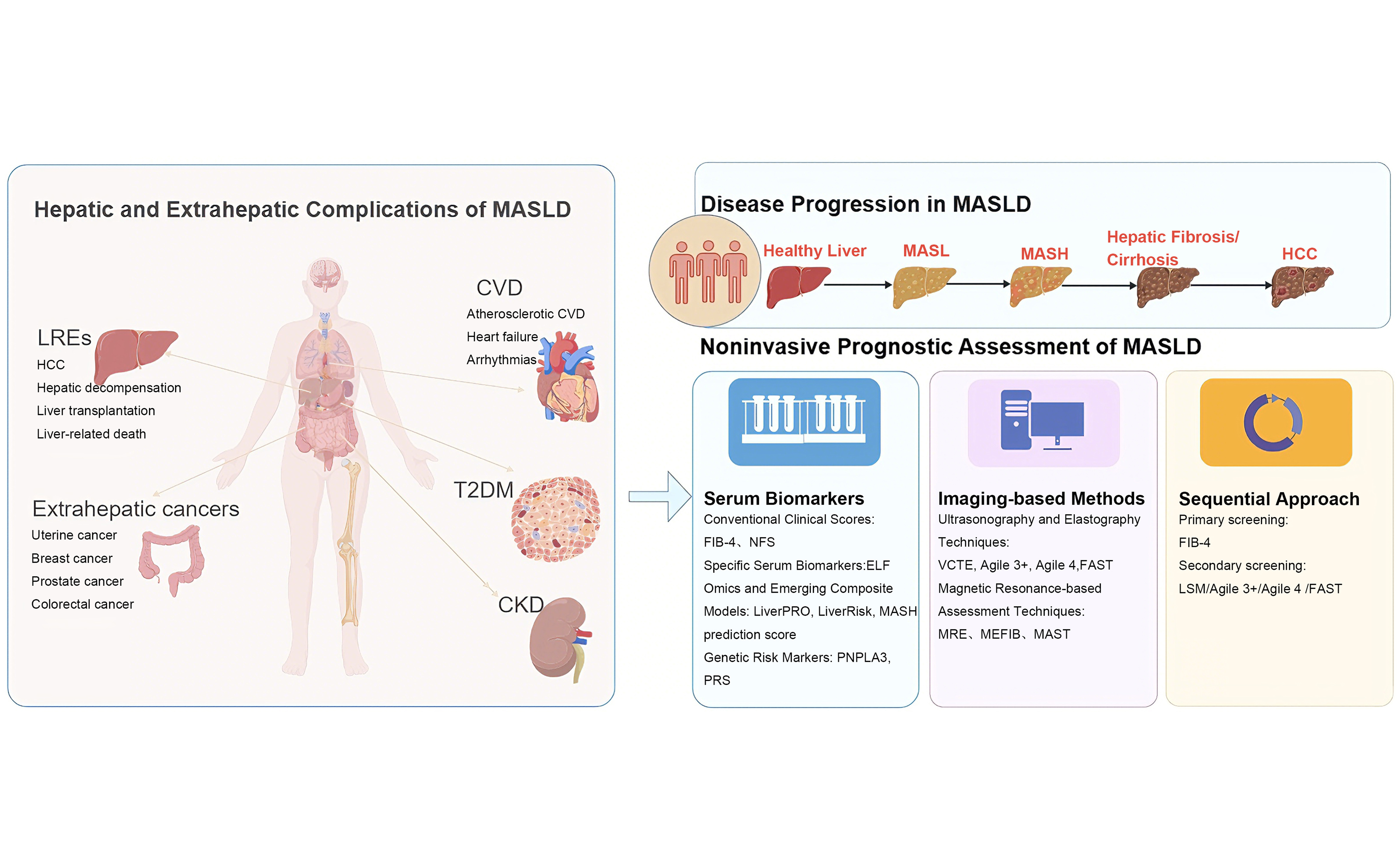
Keywords
INTRODUCTION
Non-alcoholic fatty liver disease (NAFLD) is one of the most prevalent chronic liver diseases worldwide. In 2020, an international expert panel renamed NAFLD as metabolic dysfunction-associated fatty liver disease (MAFLD) to emphasize the central role of metabolic disorders in disease initiation and progression, and adopted a positive diagnostic criterion instead of the previous exclusionary criteria[1,2]. Its definition was updated by an international expert consensus in 2023, and the nomenclature “metabolic dysfunction-associated steatotic liver disease (MASLD)” was proposed in the multisociety Delphi consensus[3]. Once regarded as a “benign” liver condition, metabolic dysfunction-associated steatotic liver (MASL) can progress in some patients to metabolic dysfunction-associated steatohepatitis (MASH) and liver fibrosis, leading to liver-related events (LREs). Meanwhile, MASLD significantly increases the risk of extrahepatic complications such as cardiovascular disease (CVD), type 2 diabetes mellitus (T2DM), and chronic kidney disease (CKD)[4-7]. These events are the core contributors to poor prognosis in MASLD patients. This dual adverse outcome not only severely reduces quality of life but also imposes a substantial burden on healthcare systems.
Therefore, developing tools that accurately predict the risks of both hepatic and extrahepatic complications is crucial for risk-stratified management of MASLD. By early identifying high-risk populations, timely interventions can be initiated to delay or prevent disease progression, and to achieve comprehensive management of the overall prognosis of MASLD patients. This review summarizes recent advances in noninvasive technologies in MASLD prognostic prediction and risk stratification, and explores the key areas that urgently require improvement.
DATABASES AND SEARCH STRATEGY
A comprehensive literature search was performed across internationally authoritative medical databases (including PubMed, Embase, and Web of Science) and evidence-based medicine resources. The search was conducted using the following keywords: “metabolic dysfunction-associated steatotic liver disease”, “MASLD”, “metabolic dysfunction-associated fatty liver disease”, “MAFLD”, “non-alcoholic fatty liver disease”, “NAFLD”, “liver-related events”, “noninvasive prognostic assessment”, “liver fibrosis”, and “extrahepatic complications”. The search timeframe spanned from the inception of each database to February 2026, and only English-language publications were considered. Following the initial identification of potentially relevant records, irrelevant literature was excluded by reviewing titles and abstracts; the remaining studies were then assessed in full text. Further screening and evaluation were carried out according to the quality, reliability, and practical applicability of the literature.
HEPATIC ADVERSE OUTCOMES
LREs are the core hepatic adverse outcomes in MASLD, including hepatocellular carcinoma (HCC), liver decompensation (e.g., ascites, esophagogastric variceal bleeding, hepatic encephalopathy, hepatorenal syndrome), liver transplantation, and liver-related mortality[8-10]. These events directly determine patient prognosis and clinical intervention priorities. Scoring models based on serum markers or clinical parameters are currently the most widely used key tools in clinical practice [Table 1][11-13].
The role of noninvasive assessment methods in LREs of MASLD
| Main category | Subcategory | Model/Tool name | Components | Study population | Median follow-up | Outcome | Validation type | AUROC | Limitations |
| Serum biomarkers | Routine serum biomarkers | FIB-4[18] | Age, AST, ALT, platelets | 2,518 MASLD patients | 4.8 years | LREs | - | 0.74 | Basic screening; modest accuracy |
| NFS[18] | Age, BMI, albumin, platelets, AST/ALT, hyperglycaemia | 2,518 MASLD patients | 4.8 years | LREs | - | 0.70 | Basic screening; modest accuracy | ||
| Omics and emerging composite models | MASH prediction score[20] | BMI, AST, tyrosine, phospholipid, total lipid | 311 suspected MASH patients | 7.2 years | Liver-related mortality | External validation | 0.83 | Only externally validated in the Finnish population; applicability to other ethnic groups remains to be determined | |
| Fibulin-3[21] | Fibulin-3 | 50 MASLD patients | 6.2 years | LREs | External validation | 0.761 | High cost; limited accessibility | ||
| LiverRisk[23] | AST, ALT, GGT, glucose, sex, cholesterol, age, platelets | 462 SLD patients | 4.4 years | LREs | External validation | 0.80 | Requires further validation | ||
| LiverPRO[23] | Age, AST, ALP, GGT, INR, albumin, sodium, platelets, bilirubin, cholesterol | 462 SLD patients | 4.4 years | LREs | External validation | 0.78 | Developed based on European population data; applicability to Asian populations requires further validation | ||
| Specific serum biomarkers | ELF[28] | HA, PIIINP, TIMP-1 | 457 CLD patients | 7.0 years | LREs | - | 0.87 | High cost; limited accessibility; susceptible to interference from ageand extrahepatic fibroinflammatory diseases | |
| Genomics | PNPLA3[37] | PNPLA3 | 471 MASLD patients | 5.4 years | Liver-related mortality | - | 0.833 | High cost; limited accessibility; modest predictive accuracy with single-gene assays; requires combination with other genetic variants | |
| Sequential approach | - | Sequential approach[73] | Primary screening: FIB-4; Secondary screening: LSM/Agile3+/Agile4/FAST | 8,131 MASLD patients | 3.9 years | LREs | - | 0.776-0.815 | Requires integration of two detection modalities; workflow more complex than that of single models |
| Imaging | Ultrasound-based | VCTE[53] | LSM | 1,057 MASLD patients | 3.1 years | LREs | - | 0.878 | Limited applicability to patients with severe obesity or massive ascites |
| Agile[8] | Agile3+: LSM, platelets, sex, hyperglycaemia, age, AST/ALT Agile4: LSM, platelets, sex, hyperglycaemia, AST/ALT | 16,603 MASLD patients | 4.3 years | LREs | - | 0.87-0.91 | Influenced by age and sex; limited applicability to patients with severe obesity; requires further external validation | ||
| acFibroMASH[58] | LSM, AST, Scr | 9,034 MASLD patients | - | LREs | - | 0.835 | Scr levels influenced by multiple factors; lacks validation in external cohorts | ||
| FAST[58] | LSM, CAP, AST | 9,034 MASLD patients | - | LREs | -- | 0.750 | Limited applicability to patients with severe obesity or massive ascites | ||
| MRE-based | MEFIB[67] | MRE, FIB-4 | 297 MASLD patients | 4.0 years | Hepatic decompensation | - | 0.89 | High cost; limited accessibility;high technical threshold | |
| MAST[67] | MRE, AST, MRI-PDFF | 297 MASLD patients | 4.0 years | Hepatic decompensation | - | 0.81 | High cost; limited accessibility; high technical threshold |
Serum biomarkers
Noninvasive serum-based biomarkers are the cornerstone of risk stratification and clinical management for MASLD. They have evolved from early-generation simple fibrosis scores derived from routine clinical parameters to a variety of advanced technologies: multi-omics assays (proteomics, metabolomics, lipidomics), polygenic risk markers, and machine learning models.
Conventional clinical scores
Among validated serumbased prognostic tools, the Fibrosis-4 (FIB-4) index is the most widely used for routine practice[14-16]. It is calculated as: FIB-4 = [age × aspartate aminotransferase (AST)]/[platelet count × √alanine aminotransferase (ALT)][17]. A large meta-analysis of 2,518 biopsy-proven MASLD patients confirmed the prognostic utility of FIB-4: patients with FIB-4 > 2.67 had a significantly higher risk of adverse outcomes (20.8%) than those with FIB-4 < 1.3 (1.3%). For 5-year outcome prediction, the time-dependent area under the receiver operating characteristic curve (AUC) of FIB-4 (0.74) was comparable to that of histological fibrosis stage on biopsy (0.72), suggesting that FIB-4 can substitute for biopsy in selected situations[18]. MASLD prognosis is influenced by dynamic factors, including lifestyle modifications and pharmacotherapy, and longitudinal changes in FIB-4 can reflect intervention effects and prognostic trends. Specifically, dynamic elevation of FIB-4 is significantly associated with increased risks of all-cause mortality, CVD, and LREs in MASLD patients - those with high and persistently elevated FIB-4 levels have nearly a 7-fold higher risk of LREs than patients with low baseline FIB-4[19]. In contrast to static risk stratification, monitoring FIB-4 changes guides clinical decision-making and allows timely adjustment of treatment strategies.
Omics and emerging composite models
Emerging composite models integrating multi-dimensional indicators have been developed in recent years. These models combine multi-level data including clinical characteristics, routine biochemical parameters, and omics to achieve more refined prognostic risk stratification. A study by Chinese researchers developed a metabolomics-based MASH prediction score using machine learning algorithms. This score incorporates four indicators: body mass index (BMI), AST, tyrosine, and the ratio of phospholipids to total lipids in very low-density lipoprotein. It can not only identify MASH and stratify its risk, with AUCs of 0.87 [95% confidence interval (CI): 0.83-0.91] and 0.81 (95%CI: 0.75-0.88) in Chinese and European populations, respectively, but also identify individuals at high risk of liver-related mortality (AUC 0.83, 95%CI: 0.79-0.86). Its prognostic performance is significantly superior to the NAFLD Fibrosis Score (NFS)[20]. This score was developed in a Chinese patient cohort and only externally validated in the Finnish population; its applicability in other ethnic groups requires further verification in multicenter, multi-ethnic cohorts. In addition to metabolomics-based models, proteomic profiling has also identified novel biomarkers for MASLD prognostic assessment. Another study using proteomic profiling found that Fibulin-3 is not only significantly associated with progressive liver fibrosis in MASLD, but also an independent predictor of LREs. The AUC of Fibulin-3 concentrations for predicting 5-year LREs was 0.761. Using a cutoff value of
Specific serum biomarkers
Models based on specific fibrosis-related biomarkers are also used for MASLD prognostic assessment. Among them, the ELF score is a well- validated proprietary assay[24-26]. It combines three biomarkers: hyaluronic acid (HA), N-terminal propeptide of type III collagen (PIIINP), and tissue inhibitor of metalloproteinases 1 (TIMP-1), and performs well in fibrosis staging[27]. A multicenter study from the UK confirmed its prognostic value: Cox proportional hazards model analysis showed that compared with the reference group with ELF score < 8.34, the HRs for LREs in the ELF score ranges of 8.34-10.425, 10.426-12.51, and 12.52-16.67 were 5.0, 20.0, and 75.0, respectively, indicating a clear dose-response relationship between ELF score and adverse outcome risk[28]. However, as a proprietary assay, ELF has key limitations: high cost and interference from age and extrahepatic fibroinflammatory conditions, which restrict its widespread clinical use. Other models based on specific serum markers have also demonstrated potential clinical value: for example, the ADAPT score [age, diabetes status, procollagen III peptide (PRO-C3), platelet count] and the FIBROSpect test (α2-macroglobulin, HA, TIMP-1), both perform well in identifying advanced MASLD fibrosis[29,30]. However, current research on the ability of these two models to predict long-term clinical outcomes such as LREs and all-cause mortality is relatively scarce, and their prognostic value requires further validation in large prospective multicenter cohorts.
Genetic risk markers
With the continuous rise in the global prevalence of metabolic diseases, the incidence of MASLD-related HCC has increased significantly. Its pathogenesis involves interactions between metabolic disorders, chronic inflammation, and genetic susceptibility[31]. Genetic variations in lipid metabolism genes are key drivers of MASLD progression to HCC[32]. The patatin-like phospholipase domain-containing protein 3 (PNPLA3) variant is the best-established genetic risk factor, driving progression to cirrhosis and HCC[33-35]. A meta-analysis of 109 studies including 118,302 MASLD patients showed that compared with non-carriers, carriers of PNPLA3 gene variations had more severe liver injury and histological changes. Meanwhile, the PNPLA3 GG genotype was significantly associated with higher mortality and LREs in MASLD patients[36].
These emerging serum-based prognostic models, developed using omics, genetic, and machine learning approaches, remain largely in the early stages of investigation. Most generally lack large-scale, multicenter external validation, prospective evidence of clinical utility, and feasibility for widespread clinical implementation, and their translational value requires confirmation in additional high-quality studies.
Imaging-based methods
Ultrasonography and elastography techniques
Conventional ultrasonography is the first-line imaging modality for initial evaluation of MASLD, with well-recognized advantages of low cost, widespread equipment availability, and safety profile. It can detect mild or greater degrees of hepatic steatosis based on typical features such as hepatic parenchymal hyper-echogenicity and hepatorenal echo contrast[43,44]. However, its diagnostic performance is limited by inter-device variability and operator-dependent subjectivity, which leads to suboptimal reproducibility. Semi-quantitative ultrasound indices, such as the hepatorenal ratio (HRR), have demonstrated considerable clinical utility for MASLD, particularly hepatic steatosis. One study has demonstrated a strong correlation between HRR and histological steatosis severity (r = 0.80, P < 0.01), with a cut-off of ≥ 1.24 for diagnosing steatosis (sensitivity 92.7%, specificity 92.5%)[45]. HRR is the ratio of echogenic intensity between regions of interest placed in hepatic and renal parenchyma at the same depth, which may partially reduce operator-dependent bias[46]. However, it cannot be reliably used in patients with acute or chronic kidney injury or those who have undergone right nephrectomy[47]. In addition to assessing hepatic steatosis, conventional ultrasonography can reliably exclude focal hepatic lesions and identify indirect signs of portal hypertension, including splenomegaly, ascites, and portal vein dilatation, thus providing preliminary evidence for identifying patients at high risk of adverse hepatic outcomes[48]. Despite its central role in first-line evaluation, conventional ultrasonography has limited ability to accurately quantify liver fibrosis and fails to enable precise risk stratification for long-term adverse outcomes. To overcome this limitation, elastography techniques represented by vibration-controlled transient elastography (VCTE) have been developed and widely used in clinical practice.
Among imaging modalities, VCTE is the most widely used noninvasive tool for assessing liver fibrosis[49-53]. With technical advantages such as ease of use, short time consumption, high patient tolerance, and high repeatability, it avoids the invasive risks and sampling errors of liver biopsy, and has become a core auxiliary tool for the diagnosis, staging, and prognostic assessment of MASLD patients. Its core detection indicator - liver stiffness measurement (LSM) - is not only a key quantitative indicator for assessing liver fibrosis staging and identifying high-risk populations in MASLD patients but can also dynamically track changes in liver stiffness after treatment, providing real-time evidence for disease prognosis monitoring[54,55].
Multiple high-quality studies have confirmed the predictive value of LSM for adverse outcomes in MASLD[18]. A large US cohort study enrolled 30,414 MASLD participants with a total follow-up of 69,974 person-years. The results showed that for each 5 kPa increase in LSM, the risk of HCC increased by 18%; and the annual incidence of HCC increased gradually with increasing LSM intervals. Annual incidence rates were 0.34, 0.45, 0.78, and 0.94 per 100 person-years in the LSM 10-14.9, 15-19.9, 20-24.9, and ≥ 25 kPa groups, respectively[56]. The composite scoring system derived from LSM has further improved the accuracy of prognostic prediction. A multicenter study involving 16 centers and 16,603 MASLD patients (median follow-up of 51.7 months) showed that the VCTE-based Agile score (integrating LSM with clinical parameters such as platelet count, transaminases, diabetes status, age, and sex) was significantly associated with the risk of LREs. Its time-dependent AUC was not only superior to traditional noninvasive models such as FIB-4, NFS, and LSM but also significantly better than histological fibrosis staging by liver biopsy. Moreover, patients with improved Agile scores from baseline had a significantly lower subsequent risk of LREs, providing a reliable basis for evaluating treatment response[8]. Meanwhile, the study has also verified the diagnostic value of Agile3+ for at-risk MASH, with an AUC of 0.708 (0.672-0.744)[57]. Recently, the VCTE-derived acFibroMASH index was proposed based on data from 3,004 biopsy-proven MASLD patients in 29 Chinese and 9 international cohorts. In terms of diagnostic performance, the index had an AUC of 0.808 (95%CI: 0.748-0.869) for identifying at-risk MASH with significant fibrosis, which was significantly higher than that of the conventional FibroScan-AST (FAST) score (0.764; 95%CI: 0.694-0.834, P = 0.040). For clinical use, the index uses dual cut-offs: < 0.15 rules out at-risk MASH with 90% sensitivity and 93% negative predictive value (NPV), while > 0.39 rules in at-risk MASH with 90% specificity and 60% positive predictive value (PPV). This addresses the dual clinical needs of screening and confirmatory diagnosis. Further validation of its prognostic value showed that, consistent with its diagnostic stratification performance, patients with an acFibroMASH index > 0.39 had an HR of 11.23 for LREs, compared with those with an index < 0.15. The index also achieved an AUC of 0.835 for predicting the 5-year risk of LREs, with prognostic performance that was again significantly superior to that of the FAST score (0.750). These findings confirm that the acFibroMASH index is a reliable tool for identifying at-risk MASH and predicting LREs risk[58]. However, a core parameter of acFibroMASH is serum creatinine, which is affected by non-hepatic factors including renal function, dehydration, muscle mass, and drug use. Failure to adjust for these confounders may reduce model accuracy in specific populations, such as patients with CKD, emaciation, or obesity[59,60]. Dynamic changes in LSM also have prognostic value in MASLD. The 2022 Baveno VII criteria first incorporated baseline LSM and longitudinal changes into clinical outcome prediction[61]. A multicenter prospective study involving 1,403 MASLD patients showed that patients with LSM progressing to ≥ 10 kPa had a significantly higher cumulative incidence of LREs (16%) than non-progressors (4%), while patients with LSM improving to < 10 kPa had a significantly lower incidence of LREs (7%) than non-regressors (32%), confirming the close association between dynamic changes in LSM and LREs risk[55]. However, recent evidence challenges this conclusion: a global multicenter cohort study across 5 countries found that both previous and current LSM values were associated with LREs, but current LSM better reflects real-time liver status. When the latest LSM data are available, the additional predictive value of baseline LSM and dynamic changes is negligible[62]. This conclusion differs from previous studies, indicating that larger, longer longitudinal studies are needed to clarify the predictive performance of baseline and most recent LSM and the magnitude of dynamic changes for MASLD clinical outcomes. Such studies will inform the development of clinical monitoring strategies.
Magnetic resonance-based assessment techniques
As a noninvasive assessment technology, magnetic resonance elastography (MRE) has shown high diagnostic accuracy for MASLD fibrosis staging[63-65]. Liver stiffness measured by MRE is associated with adverse outcomes such as LREs and all-cause mortality[66]. A recent multicenter individual-patient-data meta-analysis of 454 patients evaluated the prognostic value of MRE-derived tools. The results showed that positivity for the MEFIB index (combination of MRE and FIB-4) (HR = 49.22, 95%CI: 6.23-388.64, P < 0.001) and positivity for the MAST score [combination of AST, MRE, and magnetic resonance imaging proton density fat fraction (MRI-PDFF)] (HR = 3.86, 95%CI: 1.46-10.17, P < 0.001) were both independent predictors of new-onset liver decompensation, and the discriminative power of the MEFIB index was significantly higher than the MAST score[67]. One study also validated the diagnostic value of the MAST score and MEFIB index for identifying at-risk MASH, with corresponding AUCs of 0.79 and 0.68, respectively[68]. These MRI-based tools offer additional options for identifying high-risk patients with MASLD. However, available data are limited to single-time-point MRE assessments, and the association between longitudinal changes in MRE-based measures and adverse outcomes remains unexplored. Further longitudinal studies are needed to address this gap. MRE also has practical limitations: high cost and limited availability, which restrict its routine use in primary care and resource-limited settings.
Sequential approach
International guidelines and clinical studies recommend a sequential approach as the core strategy for liver fibrosis assessment in MASLD patients [Figure 1]. This approach uses a two-step model - initial screening followed by refined assessment - to balance noninvasiveness, cost, and accuracy, and it is now the standard pathway for fibrosis risk stratification in routine practice[69-71]. A growing body of evidence confirms that the sequential approach performs well not only for fibrosis staging but also for longterm prognosis, particularly prediction of LREs[15,72]. A large Korean cohort study used the sequential approach recommended by the Korean Association for the Study of the Liver (FIB-4 for initial screening followed by VCTE for abnormal results) to follow MASLD patients and assess LREs risk. This method had an overall accuracy of 67.7%-99.8% for predicting LREs. Replacing LSM in the second step with Agile 3+, Agile 4, or FAST scores resulted in comparable prognostic performance[73]. This approach has also been prospectively validated in MASLD patients with type 2 diabetes in multicenter cohorts: the approach combining FIB-4 and LSM effectively stratifies the risk of advanced fibrosis and LREs, and optimized liver stiffness cut-offs further improve risk stratification[74]. Another multicenter longitudinal study of 16 tertiary centers in the United States, Europe, and Asia focused on HCC, a key adverse outcome, and refined sequential approach risk thresholds. Patients with FIB-4 ≥ 3.25 or baseline LSM ≥ 20 kPa had an annual HCC incidence > 1% and require prioritized HCC surveillance. Among patients with elevated initial FIB-4, LSM ≥ 15 kPa identified an extremely high HCC risk subgroup requiring intensified surveillance[15]. From a health economics perspective, the sequential approach combining FIB-4 and VCTE offers potential cost-saving benefits by efficiently ruling out low-risk individuals at the initial screening stage and limiting further detailed assessment exclusively to high-risk subgroups. While this algorithm has established clinical utility for prognostic stratification and continuity of care in MASLD, and is recommended by multiple international guidelines for risk stratification in MASLD populations, robust health economic evidence remains limited. Specifically, large, long-term studies evaluating its cost-effectiveness and cost-utility are lacking, and its long-term health economic value requires further validation in prospective real-world cohorts.
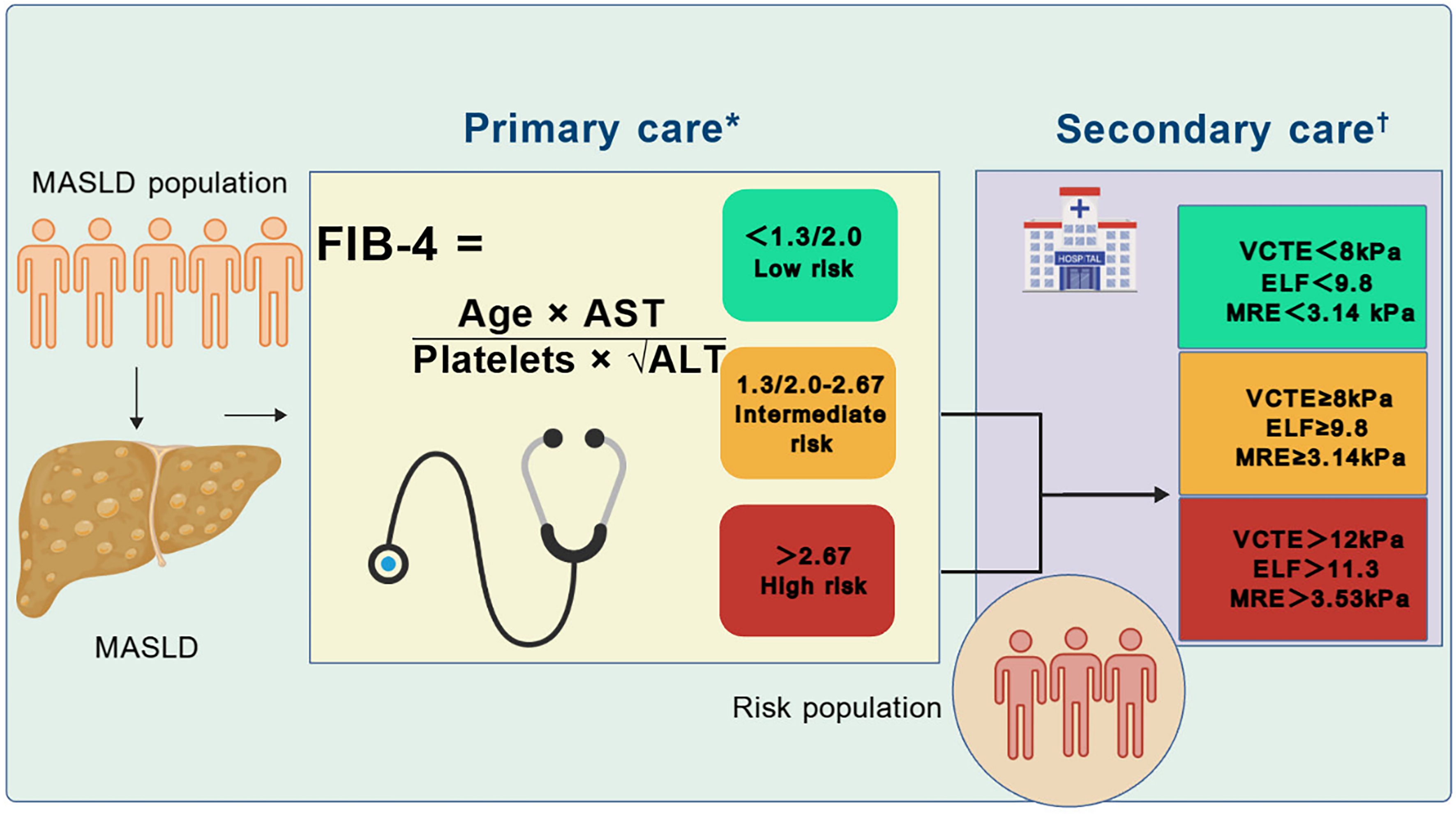
Figure 1. The sequential approach in MASLD. Created with BioGDP.com[105]. *FIB-4 thresholds are recommended by clinical practice guidelines[106,107]; †VCTE thresholds are recommended by clinical practice guidelines[106,107]. ELF and MRE thresholds are exploratory values derived from meta-analyses and cohort studies[108-110]. MASLD: Metabolic dysfunction-associated steatotic liver disease; FIB-4: Fibrosis-4 index; AST: aspartate aminotransferase; ALT: alanine aminotransferase; VCTE: vibration-controlled transient elastography; ELF: enhanced liver fibrosis; MRE: magnetic resonance elastography.
EXTRAHEPATIC COMPLICATIONS
Although assessing hepatic outcomes is central to the prognostic management of MASLD, an approach focused exclusively on the liver has inherent limitations. MASLD is a systemic metabolic disorder rather than an isolated liver disease. Its pathophysiology extends along a “metabolic disturbance-chronic inflammation-oxidative stress” axis, producing remote effects on extrahepatic organs and ultimately contributing to multisystem complications, including CVD, CKD, T2DM, and extrahepatic malignancies[6,31,75,76]. Extrahepatic complication risk assessment has long been a clinical gap, as most conventional tools have focused exclusively on liver outcomes. In recent years, noninvasive tools targeting affected organs including the cardiovascular and renal systems have gradually advanced. These advances address the need for dual hepatic-extrahepatic outcome assessment in MASLD and support the development of individualized prognostic management that spans the full range of affected organ systems throughout the disease course.
CVD
MASLD and CVD share a complex bidirectional relationship driven by common metabolic risk factors, including obesity and insulin resistance, which together worsen both hepatic and cardiovascular damage[77-79]. A large Swedish prospective study found that patients with biopsy-proven MASLD had a higher incidence of CVD than controls, and that this risk persisted across the disease course and increased with disease severity, particularly fibrosis stage. These findings highlight the importance of cardiovascular risk stratification in MASLD[80]. Pooled data showed an all cause mortality rate of 12.6 per 1,000 person-years in MASLD, with cardiovascular mortality (4.2 per 1,000 person-years) exceeding liver-related mortality and representing the leading cause of death[81]. Cardiovascular risk assessment is therefore essential in the management of MASLD to optimize treatment and improve long-term outcomes.
Several noninvasive liver fibrosis assessment methods have shown utility for CVD risk prediction in MASLD, providing practical tools for identifying high-risk individuals. A meta-analysis of 19 cohorts (1,481,875 adults) found that FIB-4 [odds ratio (OR) = 1.77, 95%CI: 1.58-1.99] and NFS (OR = 2.40, 95%CI: 1.83-3.14) were associated with higher CVD risk, independent of MASLD status, suggesting their potential as universal CVD risk indicators[82]. Beyond serum markers, imaging tools have also demonstrated prognostic value; elevated LSM was associated with higher total CVD events and all-cause mortality, whereas elevated controlled attenuation parameter (CAP) showed a protective effect. These associations persisted in dichotomized analyses. Both LSM and CAP improved risk discrimination [C-statistic increment 0.037, integrated discrimination improvement (IDI) 52%][83]. Integrated metabolic risk factor models have also shown prognostic value. Patients with MASLD often have a higher burden of metabolic comorbidities, including obesity and T2DM, which increase susceptibility to CVD. A retrospective study of 2,962 MASLD patients demonstrated that a combination of age ≥ 60 years and ≥ 4 metabolic conditions (overweight/obesity, dysglycemia, hypertension, dyslipidemia) identified the subgroup with the highest CVD risk. This algorithm demonstrated comparable performance to well-validated cardiovascular risk scores, such as the Framingham Risk Score and the Atherosclerotic Cardiovascular Disease Risk Score[84]. However, the model had high NPV but relatively low PPV, suggesting that its primary clinical utility lies in identifying low-risk individuals and thereby reducing unnecessary interventions. Reduced muscle strength has also emerged as an independent predictor[85-87]. In a MASLD sub-cohort of the UK Biobank, CVD risk increased as handgrip strength (HGS) decreased, and low HGS was independently associated with higher CVD risk[88]. These findings suggest that interventions to improve muscle strength, such as resistance training and high-quality protein supplementation, may reduce CVD risk in MASLD. However, interventional evidence remains limited, and randomized trials are needed to confirm this hypothesis.
Atherosclerosis, a chronic progressive inflammatory disease, is the core pathological basis of CVD. Therefore, atherosclerotic vascular lesions may serve as key targets for CVD monitoring in MASLD[89]. A study of 153 MASLD patients who underwent carotid ultrasound found that both FIB-4 and LSM were associated with carotid atherosclerosis and moderate-to-severe coronary artery stenosis; the association between LSM and severe coronary stenosis was particularly strong, providing a reference for CVD risk assessment[90]. Peripheral arterial disease, a key manifestation of atherosclerosis, has also been studied in relation to metabolic risk in MASLD. A recent clinical study found that the prevalence of peripheral arterial disease in MASLD patients increased with increasing FIB-4 levels: 51.3% in the FIB-4 < 1.33 group versus 86.5% in the FIB-4 > 2.66 group (P < 0.001). In multivariable regression, each 1-unit increase in FIB-4 was associated with a 66% higher risk of peripheral arterial disease. However, subgroup analyses showed no significant interaction, and larger, longer cohort studies are needed to verify the stability and specificity of this association[91]. Collectively, these studies show that noninvasive assessment tools such as FIB-4 are associated with a higher burden of vascular complications - including coronary, carotid, and peripheral arterial disease - in patients with MASLD, supporting their potential to predict cardiovascular complications and expanding their value for multiorgan outcome assessment.
CKD
CKD is a common extrahepatic complication of MASLD, affecting approximately 20%-55% of MASLD patients[92,93]. Clinical evidence shows that MASLD patients have a significantly increased risk of developing CKD, and this risk increases with the severity of liver disease, particularly in patients with MASH or liver fibrosis[93]. MASLD-related CKD also imposes a substantial healthcare burden, driven largely by the need for renal replacement therapy in those who progress to endstage renal disease[94,95].
Noninvasive liver fibrosis scores and metabolic factors both demonstrate prognostic value for CKD risk in MASLD. A cross-sectional study found that MASLD fibrosis and T2DM had an additive interaction on CKD risk. Both FIB-4 and NFS predicted CKD incidence and progression, with FIB-4 showing better discriminative performance[96]. In patients with MASLD, the number of metabolic syndrome (MetS) components and liver fibrosis severity were all positively associated with CKD risk, and MetS also increased the risk of incident endstage renal disease[97]. Notably, there are still relatively few risk prediction models for MASLD-related CKD, and clinical practice mostly relies on the combined assessment model of “liver fibrosis score + renal function indicators”. Given that MASLD patients have a significant risk of developing CKD and related adverse outcomes, incorporating renal function monitoring into the routine assessment system of MASLD patients is of great significance for optimizing clinical management strategies[92].
T2DM
MASLD is essentially a hepatic manifestation of systemic metabolic abnormalities and shares a bidirectional relationship with T2DM[98]. A recent meta-analysis of 1,832,125 patients with T2DM reported a MASLD prevalence of 65.04%[99]. The bidirectional interaction mechanism between MASLD and T2DM is well characterized. T2DM promotes MASLD progression - from simple steatosis to MASH and cirrhosis - through insulin resistance, disordered lipid metabolism, and chronic inflammation, thereby accelerating the development of HCC and extrahepatic complications such as CVD and CKD. Conversely, MASLD increases the risk of incident T2DM and impairs glycemic control in patients with established T2DM by reducing insulin sensitivity[98].
A large meta-analysis of 33 studies including 501,022 individuals further found that incident T2DM risk was higher in patients with MASLD than in those without (HR = 2.19) and increased with liver fibrosis severity[100]. Another study from Finland showed that the coexistence of MASLD and MetS was associated with higher risks of CVD and T2DM, whereas MASLD alone was not, suggesting that MetS status may help identify patients at elevated risk[101]. Similarly, it is worth noting that when MASLD improves or resolves on ultrasound, the incidence of T2DM declines over time[75]. This finding suggests that established noninvasive MASLD assessment tools may have value in predicting T2DM risk, although this hypothesis requires validation in prospective cohort studies across different populations to define clinical applicability.
Extrahepatic malignancies
MASLD is frequently accompanied by metabolic risk factors linked to tumorigenesis. Consequently, extrahepatic malignancies represent the second leading cause of death in patients with MASLD[75]. A meta-analysis of 64 observational cohort studies has quantified the association between MASLD and cancer incidence. The results showed that the overall incidence of HCC was 1.25 per 1,000 person-years in MASLD, rising to 14.5 per 1,000 person-years in patients with advanced fibrosis or cirrhosis - a rate substantially higher than in those without advanced disease. Notably, the overall incidence of extrahepatic cancers in MASLD patients was 10.58 per 1,000 person-years, exceeding that of HCC. The most common types were uterine, breast, prostate, colorectal, and lung cancer, in that order[76]. In contrast to HCC, extrahepatic malignancy incidence did not rise with advanced fibrosis or cirrhosis. This finding indicates that alongside MASLD-related HCC surveillance, equal emphasis should be placed on extrahepatic cancer screening: all adult patients with MASLD, regardless of fibrosis stage, may benefit from routine extrahepatic malignancy screening. Another meta-analysis of 8 observational studies (56,745 patients with MASLD, 704 gastrointestinal cancer cases) found that compared with non-lean MASLD patients, lean MASLD patients had significantly higher risks of developing HCC [risk ratio (RR) = 1.77, 95%CI: 1.15-2.73], pancreatic cancer (RR = 1.97, 95%CI: 1.01-3.86), and colorectal cancer (RR = 1.53, 95%CI: 1.12-2.09). However, no significant differences were observed for esophageal, biliary tract, or small intestinal cancer incidence between the two groups[102]. Nevertheless, current data are insufficient to confirm whether extrahepatic cancer incidence rises with liver disease severity, meaning that risk stratification based solely on fibrosis scores may have limitations for extrahepatic malignancies in MASLD[76]. Accordingly, new risk models that incorporate metabolic features of MASLD are therefore needed; a prediction system based on metabolic indicators represents a promising research direction.
LIMITATIONS
This review summarizes the clinical applications and advances in noninvasive prognostic assessment for MASLD. Several limitations should be noted. First, population heterogeneity remains a major limitation. The performance of noninvasive prognostic tools may vary significantly by age, ethnicity, and comorbidities - particularly diabetes, obesity, and CKD - as discussed in relevant sections above. Several models were developed and validated in mono-ethnic cohorts, and their generalizability to other populations requires further confirmation in larger, multi‑ethnic studies. With respect to study endpoints, the ascertainment criteria and observation time windows for LREs and extrahepatic complications are not fully standardized across studies, which may introduce bias in head-to-head comparisons and interpretation of prognostic performance across tools. Furthermore, the subclassification of MASLD not only influences clinical outcome risk, but also leads to differential performance thresholds of the same noninvasive prognostic tool across subgroups. This is another reason for the limited generalizability of current prognostic models. In addition, no systematic quality appraisal or risk-of-bias assessment was performed for the included primary studies. Finally, the discussion of emerging tools is based largely on studies reporting positive findings, which may confer an inherent risk of publication bias.
SUMMARY AND OUTLOOK
MASLD is characterized by a core prognostic signature of concurrent hepatic progression and extrahepatic involvement. Its noninvasive prognostic assessment framework - a well-established system spanning the full disease course - serves as a key alternative to invasive liver biopsy and enables early, precise risk stratification to guide patient management. Built on the combined use of serum marker panels and imaging modalities, this framework integrates simple clinical scores, novel fibrosis-specific markers, omics-derived biomarkers, and genetic risk tools, with VCTE and MRE as representative imaging techniques, forming a multidimensional assessment system. This multidimensional system plays a pivotal role in risk stratification and long-term prognostication, and underpins standardized diagnosis, treatment, and full-spectrum disease management in MASLD.
Despite these advances, clinical translation and widespread adoption of these tools face three key practical challenges. First, substantial heterogeneity across study cohorts and endpoint definitions limits model generalizability. Second, clinical utility is limited: complex multi-omics and machine learning models are cumbersome to implement and are associated with high costs, making them difficult to popularize in primary care settings. Finally, most existing tools provide static assessments and lack the dynamic predictive capacity to capture real-time prognostic changes driven by lifestyle interventions or pharmacotherapy. Future research should advance the field in three core directions: first, developing cross-population universal models using global multicenter, multi-ethnic cohort data to address heterogeneity-related limitations in generalizability; second, building dynamic prediction models incorporating longitudinal follow-up data to enable real-time monitoring of treatment response; third, leveraging artificial intelligence (AI) to enable integrated, precision clinical care. AI has already yielded important breakthroughs in MASLD diagnosis and management. AI-based measurement tool has been validated as an adjunct to support pathologists in the pathological assessment of MASH[103]. Quantitative techniques such as qFibrosis have also emerged as key histological endpoints in clinical trials of investigational agents, enabling quantitative evaluation of pathological features including liver fibrosis and precise prognostic risk stratification[104]. Building on these advances, future AI applications should prioritize refining the population-specific performance of noninvasive tools, developing multimodal dynamic prognostic models, enabling AI-driven end-to-end MASH drug development, and facilitating real-time monitoring of therapeutic responses to approved therapies. The goal is to deliver precise risk stratification, individualized treatment guidance, and continuum-of-care management for patients with MASLD, ultimately improving long-term clinical outcomes.
DECLARATIONS
Acknowledgments
The Graphical Abstract was created in BioGDP.
Authors’ contributions
Wrote the manuscript: He XL, Yang RX
Designed the project and revised the manuscript: Fan JG
Availability of data and materials
Not applicable.
AI and AI-assisted tools statement
Not applicable.
Financial support and sponsorship
This work was supported by the Noncommunicable Chronic Diseases-National Science and Technology Major Project (2023ZD0508700). Additional support was provided by the National Natural Science Foundation of China (82470600), and the Construction Project of the “Discipline Peak-Climbing Plan” of Xinhua Hospital Affiliated to Shanghai Jiao Tong University School of Medicine (XKPF2024B401).
Conflicts of interest
Fan JG is an Editorial Board Member of Hepatoma Research and also serves as a Guest Editor for the Special Issue “Novel Molecular Mechanisms and Therapeutic Targets in Metabolic Dysfunction-Associated Steatohepatitis (MASLD): From Bench to Bedside”. Fan JG had not involved in any steps of editorial processing, notably including reviewers’ selection, manuscript handling and decision making. The other authors declare that there are no conflicts of interest.
Ethical approval and consent to participate
Not applicable.
Consent for publication
Not applicable.
Copyright
© The Author(s) 2026.
REFERENCES
1. Eslam M, Newsome PN, Sarin SK, et al. A new definition for metabolic dysfunction-associated fatty liver disease: an international expert consensus statement. J Hepatol. 2020;73:202-9.
2. Eslam M, Sanyal AJ, George J; International Consensus Panel. MAFLD: a consensus-driven proposed nomenclature for metabolic associated fatty liver disease. Gastroenterology. 2020;158:1999-2014.e1.
3. Rinella ME, Lazarus JV, Ratziu V, et al. ; NAFLD Nomenclature consensus group. A multisociety Delphi consensus statement on new fatty liver disease nomenclature. J Hepatol. 2023;79:1542-56.
4. Huang DQ, Wong VWS, Rinella ME, et al. Metabolic dysfunction-associated steatotic liver disease in adults. Nat Rev Dis Primers. 2025;11:14.
5. Kim Y, Park Y, Rho H, Yao T, Gao B, Hwang S. Inflammation in MASLD progression and cancer. JHEP Rep. 2025;7:101414.
6. Younossi ZM, Kalligeros M, Henry L. Epidemiology of metabolic dysfunction-associated steatotic liver disease. Clin Mol Hepatol. 2025;31:S32-50.
7. Zhang X, Lau HC, Yu J. Pharmacological treatment for metabolic dysfunction-associated steatotic liver disease and related disorders: current and emerging therapeutic options. Pharmacol Rev. 2025;77:100018.
8. Lin H, Lee HW, Yip TC, et al. ; VCTE-Prognosis Study Group. Vibration-controlled transient elastography scores to predict liver-related events in steatotic liver disease. JAMA. 2024;331:1287-97.
9. Zhou XD, Kim SU, Yip TC, et al. ; VCTE-Prognosis Study Group. Long-term liver-related outcomes and liver stiffness progression of statin usage in steatotic liver disease. Gut. 2024;73:1883-92.
10. Liu WY, Huang S, Ji H, et al. ; VCTE-Prognosis Study Group. From “Burnt-Out” to “Burning-Out”: capturing liver fat loss in patients with advanced metabolic dysfunction-associated steatotic liver disease from a dynamic perspective. Gastroenterology. 2025;169:326-36.e4.
11. Feng G, Wong VW, Targher G, Byrne CD, Zheng MH. Non-invasive tests of fibrosis in the management of MASLD: revolutionising diagnosis, progression and regression monitoring. Gut. 2025;74:1741-50.
12. Lam SM, Wang Z, Song JW, et al. Non-invasive lipid panel of MASLD fibrosis transition underscores the role of lipoprotein sulfatides in hepatic immunomodulation. Cell Metab. 2025;37:69-86.e7.
13. Younossi ZM, de Avila L, Petta S, et al. ; Global NASH/MASH Council (GNC). Global performance of non-invasive tests in MASLD: insights from the G-MASLD study. Hepatology. 2026;84:161-74.
14. Choi J, Nguyen VH, Przybyszewski E, et al. Statin use and risk of hepatocellular carcinoma and liver fibrosis in chronic liver disease. JAMA Intern Med. 2025;185:522-30.
15. Lai JC, Yang B, Lee HW, et al. Non-invasive risk-based surveillance of hepatocellular carcinoma in patients with metabolic dysfunction-associated steatotic liver disease. Gut. 2025;74:2050-7.
16. van Kleef LA, Strandberg R, Pustjens J, et al. FIB-4-based referral pathways have suboptimal accuracy to identify increased liver stiffness and incident advanced liver disease. Clin Gastroenterol Hepatol. 2026;24:733-42.
17. Chang KC, Lin TT, Wang TC, et al. Fibrosis-4 index reflects the severity of tricuspid regurgitation and patient prognosis in atrial fibrillation. Eur J Heart Fail. 2025;27:2292-300.
18. Mózes FE, Lee JA, Vali Y, et al. ; LITMUS investigators. Performance of non-invasive tests and histology for the prediction of clinical outcomes in patients with non-alcoholic fatty liver disease: an individual participant data meta-analysis. Lancet Gastroenterol Hepatol. 2023;8:704-13.
19. Zhou XD, Li YT, Kim SU, et al. ; VCTE-Prognosis Study Group, Paired Liver Biopsy Group. Longitudinal changes in fibrosis markers: monitoring stiffness/fibrosis progression and prognostic outcomes in MASLD. Clin Gastroenterol Hepatol. 2026;24:394-406.
20. Huang Q, Qadri SF, Bian H, et al. A metabolome-derived score predicts metabolic dysfunction-associated steatohepatitis and mortality from liver disease. J Hepatol. 2025;82:781-93.
21. Sakane S, Hikita H, Shirai K, et al. Proteomic analysis of serum extracellular vesicles reveals Fibulin-3 as a new marker predicting liver-related events in MASLD. Hepatol Commun. 2024;8:e0448.
22. Serra-Burriel M, Juanola A, Serra-Burriel F, et al. ; LiverScreen Consortium Investigators. Development, validation, and prognostic evaluation of a risk score for long-term liver-related outcomes in the general population: a multicohort study. Lancet. 2023;402:988-96.
23. Lindvig KP, Thorhauge KH, Hansen JK, et al. Development, validation, and prognostic evaluation of LiverPRO for the prediction of significant liver fibrosis in primary care: a prospective cohort study. Lancet Gastroenterol Hepatol. 2025;10:55-67.
24. Cheung CY, Wan P, Wan H, et al. Development and clinical validation of a novel protein biomarkers-based algorithm for risk prediction and diagnosis of advanced liver fibrosis: a multi-centre study. J Adv Res. 2026;84:871-81.
25. Ishiba H, Fujii H, Kamada Y, et al. ; Japan Study Group of Nonalcoholic Fatty Liver Disease (JSG-NAFLD). Accuracy of type IV collagen 7S versus Enhanced Liver Fibrosis score for diagnosing fibrosis in patients with metabolic dysfunction-associated steatotic liver disease. Hepatol Commun. 2025;9:e0563.
26. Younossi ZM, Felix S, Jeffers T, et al. Performance of the enhanced liver fibrosis test to estimate advanced fibrosis among patients with nonalcoholic fatty liver disease. JAMA Netw Open. 2021;4:e2123923.
27. Kjaergaard M, Lindvig KP, Thorhauge KH, et al. Using the ELF test, FIB-4 and NAFLD fibrosis score to screen the population for liver disease. J Hepatol. 2023;79:277-86.
28. Parkes J, Roderick P, Harris S, et al. Enhanced liver fibrosis test can predict clinical outcomes in patients with chronic liver disease. Gut. 2010;59:1245-51.
29. Eslam M, Wong GL, Hashem AM, et al. A sequential algorithm combining ADAPT and liver stiffness can stage metabolic-associated fatty liver disease in hospital-based and primary care patients. Am J Gastroenterol. 2021;116:984-93.
30. Tang LJ, Ma HL, Eslam M, et al. Among simple non-invasive scores, Pro-C3 and ADAPT best exclude advanced fibrosis in Asian patients with MAFLD. Metabolism. 2022;128:154958.
31. Targher G, Byrne CD, Tilg H. MASLD: a systemic metabolic disorder with cardiovascular and malignant complications. Gut. 2024;73:691-702.
32. Gellert-Kristensen H, Richardson TG, Davey Smith G, Nordestgaard BG, Tybjaerg-Hansen A, Stender S. Combined effect of PNPLA3, TM6SF2, and HSD17B13 variants on risk of cirrhosis and hepatocellular carcinoma in the general population. Hepatology. 2020;72:845-56.
33. Urias E, Tedesco NR, Burkholder DA, et al. PNPLA3 risk allele is associated with risk of hepatocellular carcinoma but not decompensation in compensated cirrhosis. Hepatol Commun. 2024;8:e0441.
34. Petta S, Armandi A, Bugianesi E. Impact of PNPLA3 I148M on clinical outcomes in patients with MASLD. Liver Int. 2025;45:e16133.
35. Kozlitina J, Sookoian S. Global epidemiological impact of PNPLA3 I148M on liver disease. Liver Int. 2025;45:e16123.
36. Souza M, Al-Sharif L, Diaz I, Mantovani A, Villela-Nogueira CA. Global epidemiology and implications of PNPLA3 I148M variant in metabolic dysfunction-associated steatotic liver disease: a systematic review and meta-analysis. J Clin Exp Hepatol. 2025;15:102495.
37. Grimaudo S, Pipitone RM, Pennisi G, et al. Association between PNPLA3 rs738409 C>G variant and liver-related outcomes in patients with nonalcoholic fatty liver disease. Clin Gastroenterol Hepatol. 2020;18:935-44.e3.
38. Kanda T, Sasaki-Tanaka R, Abe H, et al. Polygenic risk score for metabolic dysfunction-associated steatotic liver disease and steatohepatitis: a narrative review. Int J Mol Sci. 2025;26:5164.
39. Vilar-Gomez E, Yates KP, Kleiner DE, et al. Genetic and non-genetic drivers of histological progression and regression in MASLD. J Hepatol. 2026;84:502-16.
40. Xiao L, Li Y, Hong C, et al. Polygenic risk score of metabolic dysfunction-associated steatotic liver disease amplifies the health impact on severe liver disease and metabolism-related outcomes. J Transl Med. 2024;22:650.
41. Bianco C, Jamialahmadi O, Pelusi S, et al. Non-invasive stratification of hepatocellular carcinoma risk in non-alcoholic fatty liver using polygenic risk scores. J Hepatol. 2021;74:775-82.
42. Pelusi S, Baselli G, Pietrelli A, et al. Rare pathogenic variants predispose to hepatocellular carcinoma in nonalcoholic fatty liver disease. Sci Rep. 2019;9:3682.
43. Dasarathy S, Dasarathy J, Khiyami A, Joseph R, Lopez R, McCullough AJ. Validity of real time ultrasound in the diagnosis of hepatic steatosis: a prospective study. J Hepatol. 2009;51:1061-7.
44. Hernaez R, Lazo M, Bonekamp S, et al. Diagnostic accuracy and reliability of ultrasonography for the detection of fatty liver: a meta-analysis. Hepatology. 2011;54:1082-90.
45. Borges VF, Diniz AL, Cotrim HP, Rocha HL, Andrade NB. Sonographic hepatorenal ratio: a noninvasive method to diagnose nonalcoholic steatosis. J Clin Ultrasound. 2013;41:18-25.
46. Hamadouche M, Vilgrain V, Hobeika C, et al. Ultrasound hepatorenal index for the diagnosis of steatosis in patients with type 2 diabetes: a prospective validation study. Eur Radiol. 2025;35:7927-39.
47. Marshall RH, Eissa M, Bluth EI, Gulotta PM, Davis NK. Hepatorenal index as an accurate, simple, and effective tool in screening for steatosis. AJR Am J Roentgenol. 2012;199:997-1002.
48. Hidaka H, Uojima H. Ultrasonography in the diagnosis of complications in patients with portal hypertension. J Med Ultrason. 2022;49:347-58.
49. Caussy C, Vergès B, Leleu D, et al. Screening for metabolic dysfunction-associated steatotic liver disease-related advanced fibrosis in diabetology: a prospective multicenter study. Diabetes Care. 2025;48:877-86.
50. Marti-Aguado D, Carot-Sierra JM, Villalba-Ortiz A, et al. Identification of candidates for MASLD treatment with indeterminate vibration-controlled transient elastography. Clin Gastroenterol Hepatol. 2025;23:1183-93.e5.
51. Zhou XD, Chen QF, Kim SU, et al. Long-term glycemic control and the risk of liver stiffness progression and liver-related events in MASLD. Clin Gastroenterol Hepatol. 2026;24:1332-43.
52. Feng G, Boursier J, de Saint Loup M, et al. ; VCTE-Prognosis Study Group, Paired Liver Biopsy Group. Fibrotic MASH resolutionVCTE index to diagnose fibrotic MASH resolution and predict LREs: a global multicenter study. Med. 2026;7:100929.
53. Boursier J, Hagström H, Ekstedt M, et al. Non-invasive tests accurately stratify patients with NAFLD based on their risk of liver-related events. J Hepatol. 2022;76:1013-20.
54. Castera L, Friedrich-Rust M, Loomba R. Noninvasive assessment of liver disease in patients with nonalcoholic fatty liver disease. Gastroenterology. 2019;156:1264-81.e4.
55. Gawrieh S, Vilar-Gomez E, Wilson LA, et al. ; NASH Clinical Research Network. Increases and decreases in liver stiffness measurement are independently associated with the risk of liver-related events in NAFLD. J Hepatol. 2024;81:600-8.
56. John BV, Bastaich DR, Deng Y, Singal AG, Dahman B; Veterans Analysis of Liver Disease (VALID) group of investigators. Use of liver stiffness measurement for HCC risk stratification in metabolic dysfunction-associated steatotic liver disease. Hepatology. 2026;83:1456-68.
57. Fichez J, Mouillot T, Vonghia L, et al. Non-invasive tests for fibrotic MASH for reducing screen failure in therapeutic trials. JHEP Rep. 2025;7:101351.
58. Feng G, Mózes FE, Ji D, et al. acFibroMASH index for the diagnosis of fibrotic MASH and prediction of liver-related events: an international multicenter study. Clin Gastroenterol Hepatol. 2025;23:785-96.
59. Kirsztajn GM, Samaan F, Calice-Silva V, Pecoits-Filho R. Critical analysis of the estimated glomerular filtration rate. J Bras Nefrol. 2025;47:e20250107.
60. Levey AS, Titan SM, Powe NR, Coresh J, Inker LA. Kidney disease, race, and GFR estimation. Clin J Am Soc Nephrol. 2020;15:1203-12.
61. de Franchis R, Bosch J, Garcia-Tsao G, Reiberger T, Ripoll C; Baveno VII Faculty. Baveno VII - renewing consensus in portal hypertension. J Hepatol. 2022;76:959-74.
62. Wong YJ, Chen VL, Abdulhamid A, et al. Comparing serial and current liver stiffness measurements to predict decompensation in compensated advanced chronic liver disease patients. Hepatology. 2025;81:523-31.
63. Ajmera VH, Liu A, Singh S, et al. Clinical utility of an increase in magnetic resonance elastography in predicting fibrosis progression in nonalcoholic fatty liver disease. Hepatology. 2020;71:849-60.
64. Park CC, Nguyen P, Hernandez C, et al. Magnetic resonance elastography vs transient elastography in detection of fibrosis and noninvasive measurement of steatosis in patients with biopsy-proven nonalcoholic fatty liver disease. Gastroenterology. 2017;152:598-607.e2.
65. Hsu C, Caussy C, Imajo K, et al. Magnetic resonance vs transient elastography analysis of patients with nonalcoholic fatty liver disease: a systematic review and pooled analysis of individual participants. Clin Gastroenterol Hepatol. 2019;17:630-7.e8.
66. Ajmera V, Kim BK, Yang K, et al. Liver stiffness on magnetic resonance elastography and the MEFIB index and liver-related outcomes in nonalcoholic fatty liver disease: a systematic review and meta-analysis of individual participants. Gastroenterology. 2022;163:1079-89.e5.
67. Noureddin N, Ajmera V, Bergstrom J, et al. MEFIB-Index and MAST-Score in the assessment of hepatic decompensation in metabolic dysfunction-associated steatosis liver disease-Individual participant data meta-analyses. Aliment Pharmacol Ther. 2023;58:856-65.
68. Castera L, Garteiser P, Laouenan C, et al. ; QUID NASH investigators. Prospective head-to-head comparison of non-invasive scores for diagnosis of fibrotic MASH in patients with type 2 diabetes. J Hepatol. 2024;81:195-206.
69. Eslam M, Fan JG, Yu ML, et al. The Asian Pacific association for the study of the liver clinical practice guidelines for the diagnosis and management of metabolic dysfunction-associated fatty liver disease. Hepatol Int. 2025;19:261-301.
70. Kanwal F, Shubrook JH, Adams LA, et al. Clinical care pathway for the risk stratification and management of patients with nonalcoholic fatty liver disease. Gastroenterology. 2021;161:1657-69.
71. Wattacheril JJ, Abdelmalek MF, Lim JK, Sanyal AJ. AGA clinical practice update on the role of noninvasive biomarkers in the evaluation and management of nonalcoholic fatty liver disease: expert review. Gastroenterology. 2023;165:1080-8.
72. Yip TC, Lee HW, Lin H, et al. Prognostic performance of the two-step clinical care pathway in metabolic dysfunction-associated steatotic liver disease. J Hepatol. 2025;83:304-14.
73. Lee HW, Lee JS, Kim MN, et al. Risk stratification by noninvasive tests in patients with metabolic dysfunction-associated steatotic liver disease. Clin Mol Hepatol. 2025;31:1018-31.
74. Chen Y, Dong B, Jin X, et al. ; DiabetesLiver Integrated Management (DLIM) Consortium. Two-step clinical care pathway to predict MASLD-related advanced fibrosis and long-term outcomes in type 2 diabetes. Gut. 2026;75:576-87.
75. Adams LA, Anstee QM, Tilg H, Targher G. Non-alcoholic fatty liver disease and its relationship with cardiovascular disease and other extrahepatic diseases. Gut. 2017;66:1138-53.
76. Thomas JA, Kendall BJ, Dalais C, Macdonald GA, Thrift AP. Hepatocellular and extrahepatic cancers in non-alcoholic fatty liver disease: a systematic review and meta-analysis. Eur J Cancer. 2022;173:250-62.
77. Lee HH, Lee HA, Kim EJ, et al. Metabolic dysfunction-associated steatotic liver disease and risk of cardiovascular disease. Gut. 2024;73:533-40.
78. Moon JH, Jeong S, Jang H, Koo BK, Kim W. Metabolic dysfunction-associated steatotic liver disease increases the risk of incident cardiovascular disease: a nationwide cohort study. EClinicalMedicine. 2023;65:102292.
79. Zheng H, Sechi LA, Navarese EP, Casu G, Vidili G. Metabolic dysfunction-associated steatotic liver disease and cardiovascular risk: a comprehensive review. Cardiovasc Diabetol. 2024;23:346.
80. Simon TG, Roelstraete B, Hagström H, Sundström J, Ludvigsson JF. Non-alcoholic fatty liver disease and incident major adverse cardiovascular events: results from a nationwide histology cohort. Gut. 2022;71:1867-75.
81. Younossi ZM, Golabi P, Paik JM, Henry A, Van Dongen C, Henry L. The global epidemiology of nonalcoholic fatty liver disease (NAFLD) and nonalcoholic steatohepatitis (NASH): a systematic review. Hepatology. 2023;77:1335-47.
82. Zhang J, Li L, Lin L, et al. Prognostic value of FIB-4 and NFS for cardiovascular events in patients with and without NAFLD. BMC Public Health. 2025;25:2747.
83. Cardoso CRL, Villela-Nogueira CA, Leite NC, Salles GF. Prognostic impact of liver fibrosis and steatosis by transient elastography for cardiovascular and mortality outcomes in individuals with nonalcoholic fatty liver disease and type 2 diabetes: the Rio de Janeiro Cohort Study. Cardiovasc Diabetol. 2021;20:193.
84. Shibata N, Morita Y, Ito T, et al. A machine learning algorithm for stratification of risk of cardiovascular disease in metabolic dysfunction-associated steatotic liver disease. Eur J Intern Med. 2024;129:62-70.
85. Damluji AA, Alfaraidhy M, AlHajri N, et al. Sarcopenia and cardiovascular diseases. Circulation. 2023;147:1534-53.
86. López-Bueno R, Andersen LL, Koyanagi A, et al. Thresholds of handgrip strength for all-cause, cancer, and cardiovascular mortality: a systematic review with dose-response meta-analysis. Ageing Res Rev. 2022;82:101778.
87. Wu Y, Wang W, Liu T, Zhang D. Association of grip strength with risk of all-cause mortality, cardiovascular diseases, and cancer in community-dwelling populations: a meta-analysis of prospective cohort studies. J Am Med Dir Assoc. 2017;18:551.e17-35.
88. Lim TS, Kwon S, Bae SA, et al. Association between handgrip strength and cardiovascular disease risk in MASLD: a prospective study from UK biobank. J Cachexia Sarcopenia Muscle. 2025;16:e13757.
89. Nielsen RV, Fuster V, Bundgaard H, et al. Personalized intervention based on early detection of atherosclerosis: JACC state-of-the-art review. J Am Coll Cardiol. 2024;83:2112-27.
90. Kubotsu Y, Sakamoto Y, Tago M, et al. FIB-4 index and liver stiffness measurement are potential predictors of atherosclerosis in metabolic dysfunction-associated steatotic liver disease. J Atheroscler Thromb. 2025;32:239-52.
91. Zhang C, Ying Y, Ru Y, et al. Association between FIB-4 index and lower extremity arterial disease in MASLD patients: a cross-sectional study. Lipids Health Dis. 2025;24:103.
92. Bilson J, Mantovani A, Byrne CD, Targher G. Steatotic liver disease, MASLD and risk of chronic kidney disease. Diabetes Metab. 2024;50:101506.
94. Elshahat S, Cockwell P, Maxwell AP, Griffin M, O’Brien T, O’Neill C. The impact of chronic kidney disease on developed countries from a health economics perspective: a systematic scoping review. PLoS One. 2020;15:e0230512.
95. Hydes TJ, Kennedy OJ, Buchanan R, et al. The impact of non-alcoholic fatty liver disease and liver fibrosis on adverse clinical outcomes and mortality in patients with chronic kidney disease: a prospective cohort study using the UK Biobank. BMC Med. 2023;21:185.
96. Sun Y, Hong L, Huang Z, et al. Fibrosis risk in nonalcoholic fatty liver disease is related to chronic kidney disease in older type 2 diabetes patients. J Clin Endocrinol Metab. 2022;107:e3661-9.
97. Bilson J, Hydes TJ, McDonnell D, et al. Impact of metabolic syndrome traits on kidney disease risk in individuals with MASLD: a UK biobank study. Liver Int. 2025;45:e16159.
98. Qi X, Li J, Caussy C, Teng GJ, Loomba R. Epidemiology, screening, and co-management of type 2 diabetes mellitus and metabolic dysfunction-associated steatotic liver disease. Hepatology. 2026;83:661-78.
99. En Li Cho E, Ang CZ, Quek J, et al. Global prevalence of non-alcoholic fatty liver disease in type 2 diabetes mellitus: an updated systematic review and meta-analysis. Gut. 2023;72:2138-48.
100. Mantovani A, Petracca G, Beatrice G, Tilg H, Byrne CD, Targher G. Non-alcoholic fatty liver disease and risk of incident diabetes mellitus: an updated meta-analysis of 501 022 adult individuals. Gut. 2021;70:962-9.
101. Käräjämäki AJ, Bloigu R, Kauma H, et al. Non-alcoholic fatty liver disease with and without metabolic syndrome: Different long-term outcomes. Metabolism. 2017;66:55-63.
102. Souza M, Diaz I, Barchetta I, Mantovani A. Gastrointestinal cancers in lean individuals with non-alcoholic fatty liver disease: a systematic review and meta-analysis. Liver Int. 2024;44:6-14.
103. Iyer JS, Juyal D, Le Q, et al. AI-based automation of enrollment criteria and endpoint assessment in clinical trials in liver diseases. Nat Med. 2024;30:2914-23.
104. Liu F, Goh GB, Tiniakos D, et al. qFIBS: an automated technique for quantitative evaluation of fibrosis, inflammation, ballooning, and steatosis in patients with nonalcoholic steatohepatitis. Hepatology. 2020;71:1953-66.
105. Jiang S, Li H, Zhang L, et al. Generic diagramming platform (GDP): a comprehensive database of high-quality biomedical graphics. Nucleic Acids Res. 2025;53:D1670-6.
106. European Association for the Study of the Liver, European Association for the Study of Diabetes, European Association for the Study of Obesity. EASL-EASD-EASO Clinical Practice Guidelines on the management of metabolic dysfunction-associated steatotic liver disease (MASLD): executive summary. Diabetologia. 2024;67:2375-92.
107. Rinella ME, Neuschwander-Tetri BA, Siddiqui MS, et al. AASLD Practice Guidance on the clinical assessment and management of nonalcoholic fatty liver disease. Hepatology. 2023;77:1797-835.
108. Liang JX, Ampuero J, Niu H, et al. ; LITMUS Consortium Investigators. An individual patient data meta-analysis to determine cut-offs for and confounders of NAFLD-fibrosis staging with magnetic resonance elastography. J Hepatol. 2023;79:592-604.
109. Castera L, Rinella ME, Tsochatzis EA. Noninvasive assessment of liver fibrosis. N Engl J Med. 2025;393:1715-29.
Cite This Article
 Open Access
How to Cite
Download Citation
Export Citation File:
Type of Import
Tips on Downloading Citation
Citation Manager File Format
Type of Import
Direct Import: When the Direct Import option is selected (the default state), a dialogue box will give you the option to Save or Open the downloaded citation data. Choosing Open will either launch your citation manager or give you a choice of applications with which to use the metadata. The Save option saves the file locally for later use.
Indirect Import: When the Indirect Import option is selected, the metadata is displayed and may be copied and pasted as needed.
About This Article
Special Topic
Copyright

Data & Comments
Data
0










Comments
Comments must be written in English. Spam, offensive content, impersonation, and private information will not be permitted. If any comment is reported and identified as inappropriate content by OAE staff, the comment will be removed without notice. If you have any queries or need any help, please contact us at [email protected].