

The transverse myocutaneous gracilis flap for breast reconstruction: current evidence, technical refinements and institutional experience
 0
0 
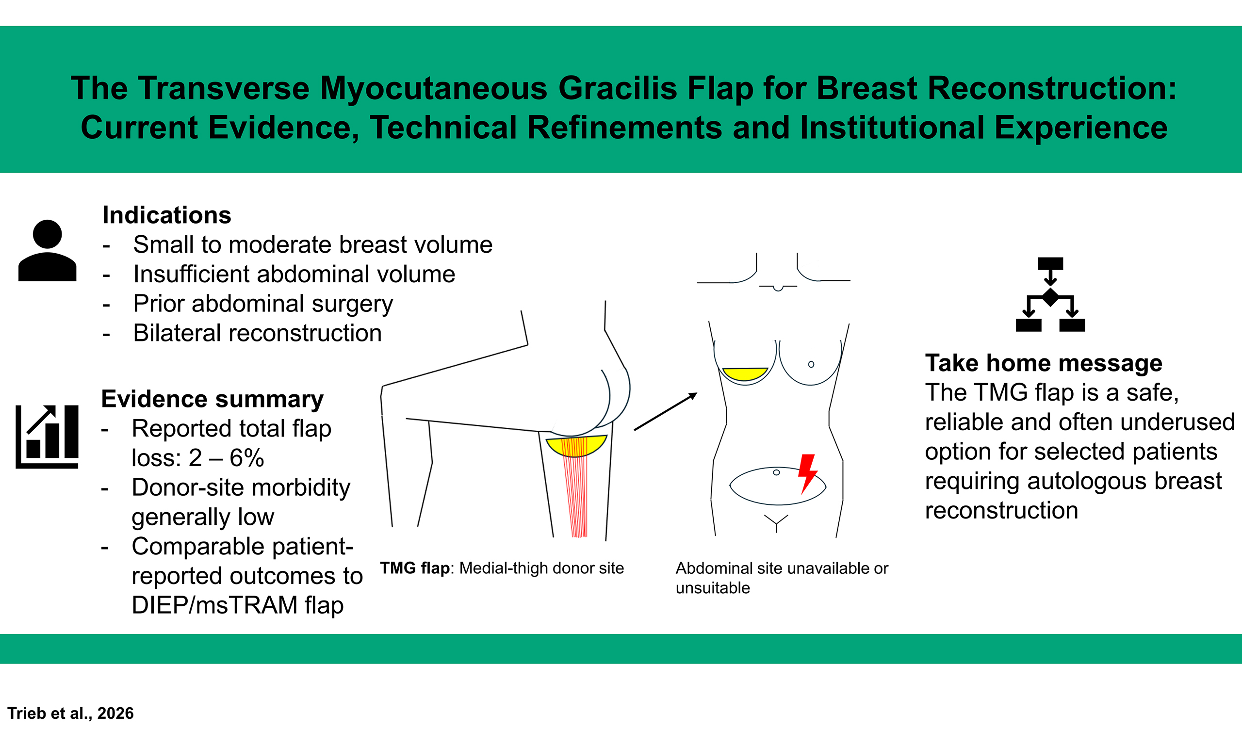
Graphical Abstract
Abstract
The deep inferior epigastric perforator (DIEP) flap and muscle-sparing transverse rectus abdominis myocutaneous (msTRAM) flap remain the gold standard for autologous breast reconstruction. However, a significant proportion of patients are not suitable candidates for abdominal-based flaps due to insufficient tissue, prior abdominal surgery, or patient preference. The transverse myocutaneous gracilis (TMG) flap, first described for breast reconstruction in 2004, has matured into a reliable workhorse flap, yet it remains underutilized in many reconstructive practices. Here, we provide a narrative review of the current literature on the TMG flap for breast reconstruction and present two cases from our institution for illustration. A narrative review of the literature was performed. Additionally, two patients who underwent TMG flap breast reconstruction at our hospital are presented as illustrative cases. The literature demonstrates total flap loss rates of 2.0%-6.3%, low donor-site morbidity, high patient satisfaction (BREAST-Q scores comparable to abdominal-based flaps), and preserved lower extremity function. The TMG flap is a safe, reliable, and often overlooked alternative for autologous breast reconstruction that deserves broader adoption, particularly in patients with small-to-moderate breast size, insufficient abdominal tissue, or when bilateral reconstruction is planned.
Keywords


 Download PDF
INTRODUCTION
Autologous breast reconstruction has evolved considerably over the past four decades. The introduction of the free transverse rectus abdominis myocutaneous (TRAM) flap, followed by the deep inferior epigastric perforator (DIEP) flap, established the lower abdomen as the preferred donor site, offering large tissue volume, favorable skin color match, and a well-concealed donor scar[1-3]. However, not all patients are suitable candidates for abdominal-based reconstruction. Prior abdominoplasty, previous abdominal flap harvest, insufficient abdominal adiposity, or patient reluctance to accept abdominal donor-site morbidity may preclude these options[4,5]. In this context, the medial thigh has emerged as a valuable alternative donor site. The gracilis myocutaneous flap has a long history in reconstructive surgery, initially described by Harii et al. in 1976 with a longitudinal skin paddle orientation[6]. However, the unreliable distal skin perfusion of the longitudinal design limited its clinical utility[7]. A paradigm shift occurred in 2004 when Wechselberger and Schoeller described the transverse myocutaneous gracilis (TMG) flap, repositioning the skin paddle transversely in the proximal medial thigh to capture the most reliable vascular territory of the gracilis pedicle[8]. This modification transformed the gracilis flap into a dependable option for breast reconstruction.
More recently, the profunda artery perforator (PAP) flap has gained increasing popularity as a muscle-sparing alternative among thigh-based reconstructive options. While the growing use of the PAP flap has further expanded the reconstructive repertoire, it may also have contributed to the TMG flap becoming comparatively overlooked.
Despite over two decades of favorable clinical experience and a growing body of supportive literature, the TMG flap remains underutilized and is relegated to a “second-line” option[4,5]. This review revisits the TMG flap and synthesizes current evidence on its anatomy, surgical technique, outcomes, and complications.
HISTORICAL EVOLUTION
Harii et al. first used the gracilis muscle as a free functional muscle transfer in 1976. Early myocutaneous flap designs used a longitudinal skin paddle, but the distal skin territory proved unreliable and was associated with high rates of partial flap necrosis[7,9]. Whetzel and Lechtman (1997) later clarified the fasciocutaneous vascular anatomy, demonstrating arterial connections between the main pedicle and superficial femoral artery perforators through a fascial network. Based on these findings, they proposed a myofasciocutaneous modification to improve skin paddle viability[7]. A key development followed in 2004 when Wechselberger and Schoeller described the TMG flap with a transversely oriented skin paddle in the proximal medial thigh. This design captured the dominant vascular territory of the ascending branch of the medial circumflex femoral artery (MCFA)[8]. The transverse design offered several advantages, including a more reliable blood supply, a well-concealed donor-site scar resembling that of a medial thigh lift, and soft subcutaneous tissue with characteristics well suited for breast reconstruction[10].
Since then, the flap has undergone numerous refinements. Schoeller et al. (2008) published guidelines for flap and patient selection based on 111 patients and 154 flaps[11]. Fansa et al. (2008) demonstrated the TMG as a “fast and reliable” method with operative times of 220 min for unilateral reconstructions[10]. Fattah et al. (2010) introduced technical refinements to increase volume and optimize inset[4]. Saint-Cyr et al. (2012) described extended and vertical extended modifications to increase flap volume[12]. Park et al. (2015) introduced the vertical upper gracilis (VUG) and bilateral upper gracilis (BUG) configurations, further expanding the flap’s versatility[13]. Most recently, Weitgasser et al. published a large dual-center retrospective series of 300 TMG flap breast reconstructions in 2021, summarizing three decades of experience with this technique[14].
ANATOMY AND VASCULAR SUPPLY
The gracilis muscle is a thin, flat muscle of the medial thigh compartment, originating from the inferior pubic ramus and inserting on the medial tibial condyle via the pes anserinus. It functions as a hip adductor and knee flexor. Although functional impairment after its harvest is often reported to be limited, sensory and motor abnormalities have been described[15-17]. The dominant vascular pedicle is the ascending branch of the MCFA, which typically arises from the profunda femoris artery. The pedicle enters the muscle on its deep surface at a mean distance of approximately 8-10 cm from the pubic tubercle[18,19]. Anatomical studies report a mean pedicle length of 6.7-7.0 cm and a mean arterial diameter of 1.9-2.5 mm, with venous diameters of 2.0-3.5 mm[12,19,20]. The accompanying venae comitantes drain into the deep venous system.
Zaussinger et al. (2019) performed a head-to-head anatomical comparison of the TMG, PAP, and fasciocutaneous infragluteal (FCI) flaps, reporting a mean TMG angiosome area of 74.1 ± 32.1 cm2, a pedicle diameter of 2.9 ± 0.6 mm, and a pedicle length of 6.7 ± 1.0 cm[20]. While the PAP flap offered a larger angiosome and longer pedicle, the TMG demonstrated consistent and reliable anatomy.
Whitaker et al. (2012) quantified the fasciocutaneous blood supply in 27 cadaver dissections, demonstrating a T-shaped angiosome with a maximum cutaneous territory of 35 cm × 19 cm for the main pedicle, supporting the feasibility of extended flap designs[9]. Wong et al. (2011) confirmed that the vascular territory extends more posteriorly than anteriorly and includes a vertical component, with perfused tissue volumes averaging 617 mL in cadaveric studies[21].
INDICATIONS AND PATIENT SELECTION
The TMG flap is indicated for autologous breast reconstruction in the following clinical scenarios:
· Primary reconstruction in patients with small-to-moderate breast size and insufficient abdominal tissue[4,8,11];
· Bilateral reconstruction, where the TMG may offer advantages over the DIEP flap due to a better-concealed donor scar and easier harvest[11,22];
· Reconstruction in patients with unavailable abdominal donor sites due to prior abdominoplasty, previous TRAM/DIEP harvest, or extensive abdominal scarring[13,23];
· Reconstruction following complications of implant-based breast reconstruction, particularly capsular contracture[24,25];
· Salvage reconstruction after prior flap failure[4].
Patient selection criteria have evolved over time. Initially, the TMG was considered suitable only for slim patients with low body mass index (BMI). However, Schwaiger et al. (2021) demonstrated in a 300-patient cohort that neither age nor overweight (BMI > 25) significantly increased complication rates, expanding the indication spectrum[26]. The key selection criterion remains adequate tissue availability at the medial thigh, which can be assessed clinically by a pinch test and, if needed, by preoperative imaging.
SURGICAL TECHNIQUE AND PEARLS
Based on the historical evolution of the gracilis flap and the accumulated clinical experience reported in the literature, the TMG flap has now reached a level of technical maturity that allows for standardized planning, reliable harvest, and predictable donor-site closure. The following section therefore summarizes the key operative principles and practical refinements that have become part of contemporary TMG flap breast reconstruction. It provides an overview of current practice rather than a description of a single institutional preference, while also highlighting modifications that have expanded the flap’s indications and improved its versatility.
Preoperative planning
The patient is marked in the standing position to mark the inferior gluteal fold, while the width of the skin paddle is best determined with the patient in the supine position and the leg abducted, allowing accurate assessment of tissue laxity and primary donor-site closure. The skin paddle is designed as a transverse ellipse centered over the proximal medial thigh, with the anterior border at the level of the greater saphenous vein and the posterior border at the midline of the inferior gluteal fold[10]. Skin paddle dimensions of up to 30 cm in width and 10 cm in height have been reported[10]. Preoperative computed tomography (CT) angiography can delineate posterior thigh perforator anatomy and assist with the planning and dissection of PAP flaps, although many experienced centers rely on clinical assessment alone[27].
Flap harvest
The patient is positioned supine with the hip abducted and externally rotated (frog-leg position), allowing simultaneous two-team surgery. The anterior incision is made first, and the gracilis muscle is identified deep to the adductor longus. The dominant pedicle is identified entering the deep surface of the muscle and dissected to its origin for maximum length. The muscle is then divided distally, and the flap is elevated from distal to proximal, including the gracilis muscle, overlying fascia, and the transverse skin paddle. Operative times average 220 min for unilateral and 325 min for bilateral cases[10].
Flap modifications to increase volume
The primary limitation of the TMG flap is its relatively modest volume (mean flap weight 320 g)[14]. During flap harvest, beveling of the subcutaneous tissue beyond the inferior border of the skin paddle allows additional adipose tissue to be incorporated without increasing the width of the skin paddle, thereby maximizing the available flap volume[28]. Additional strategies have been described to overcome this limitation. One approach is the extended TMG flap, in which the skin paddle is posteriorly extended beyond the gluteal fold to harvest additional tissue[12,21]. A further modification is the vertical extended TMG flap, which incorporates an additional vertical skin paddle component and has been shown to achieve mean flap weights of approximately 470 g[12]. Another option is the use of stacked or double TMG flaps, in which bilateral thigh tissue is harvested, and two flaps are stacked for unilateral breast reconstruction[4,11,13]. In addition, combined flaps such as the combined transverse upper gracilis and profunda artery perforator (TUGPAP) flap have been described, which merge the TMG flap with a PAP flap from the same thigh to increase available volume while maintaining a single donor site[29]. Finally, autologous fat grafting, either performed primarily or secondarily, can be used as an adjunct to further augment flap volume and improve contour[30,31].
Flap inset and recipient vessels
The flap is typically shaped into a cone by bringing the tips of the ellipse together. The internal mammary vessels are the preferred recipient vessels. The flap is inset and shaped to match the contralateral breast or the desired breast mound[11,32].
Donor-site closure
The donor site is closed primarily in layers, resulting in a scar that resembles a medial thigh lift. Patients report an initial “tight feeling” that resolves within 2-3 weeks[10].
OUTCOMES AND COMPLICATIONS
Flap survival
The TMG flap demonstrates excellent reliability. In the largest published series of 300 flaps, Weitgasser et al. reported a total flap loss rate of 6.3%[14]. A systematic review and meta-analysis by Siegwart et al. (2021), pooling 843 TMG flaps from 19 studies, reported a total flap loss rate of 2% [95% confidence interval (CI): 1%-3%][16]. These rates are comparable to those reported for DIEP flaps in the literature.
Donor-site morbidity
Reported donor-site morbidity following TMG flap harvest is generally manageable, although its frequency and severity vary substantially among studies because of heterogeneous definitions, assessment methods, and follow-up periods. The most common complication include wound dehiscence: 8% (95%CI: 4%-16%), seroma: 4% (95%CI: 2%-7%), hematoma: 2% (95%CI: 1%-4%), infection: 0%-5%, sensory disturbance: 0%-74%, and motor deficits: 0%-50%. Most reported sensory and motor symptoms were temporary[16]. Mahrhofer et al. (2023) analyzed 225 patients (288 flaps) and reported 14.9% minor and 2.7% major donor-site complications. Active tobacco use [relative risk (RR) 1.71, 95%CI: 1.02-2.88] and lower BMI were identified as risk factors. Importantly, most complications were managed conservatively, and only 1.4% of patients required donor-site refinement surgery[33]. Notably, the TMG flap avoids the abdominal wall morbidity (hernia, bulge) associated with TRAM flaps, especially when a bilateral reconstruction is performed. Existing studies suggest that persistent major functional deficits are uncommon. However, prospective studies incorporating preoperative baseline assessments and long-term follow-up remain necessary[16,17].
Comparison with DIEP flap
Weitgasser et al. (2020) compared 238 flaps used for simultaneous bilateral breast reconstruction: 152 DIEP flaps in 76 patients and 86 TMG flaps in 43 patients[22]. At the patient level, donor-site complications occurred in 7 of 43 TMG patients (16.3%) and 18 of 76 DIEP patients (23.7%). Postoperative lipofilling was required in 65.1% of TMG patients and 38.2% of DIEP patients (P = 0.005). At the flap level, total flap loss occurred in 3 of 86 TMG flaps (3.5%) and 4 of 152 DIEP flaps (2.6%; P = 0.71). Although the overall complication profile slightly favored the DIEP, donor-site morbidity was lower and less severe in the TMG group. The significantly higher need for secondary lipofilling in the TMG group reflects the volume limitation of the flap. Mahrhofer et al. (2024) performed the first photometric comparison of skin color match between DIEP and TMG flaps, demonstrating that the DIEP flap provided a better color match at all time points, though both flaps showed improved color matching by 12-20 months postoperatively[34].
Patient-reported outcomes
Mahrhofer et al. (2024) reported BREAST-Q outcomes in 82 TMG patients with a median follow-up of 5.9 years[17]. They reported satisfaction scores of 66/100 for breast, 78/80 for Lower Extremity Function Scale (LEFS), and 8/11 for donor-site satisfaction. These scores are comparable to those reported for DIEP and other autologous reconstruction methods in the literature[2,3]. Active smoking was significantly associated with lower LEFS scores (P = 0.049)[17].
THE VUG AND STACKED CONFIGURATIONS
Park et al. (2015) introduced the VUG flap as an alternative skin paddle orientation, arguing that the vertical pattern avoids some of the problems associated with transverse designs, including wound dehiscence at the posterior extent of the incision and labial distortion[13]. In their series of 22 patients (33 flaps), 88% utilized a vertical skin paddle. The VUG can be combined in bilateral stacked configurations (BUG) for unilateral reconstruction of larger breasts, with a reported flap loss rate of only 3%[13]. These modifications have transformed the gracilis territory from a limited donor site into what some authors consider “the best secondary breast reconstructive option”[13].
ADJUNCTIVE FAT GRAFTING
Autologous fat grafting has become an integral component of TMG-based breast reconstruction. Russe et al. (2018) reported on 145 fat grafting sessions in 83 patients following TMG reconstruction, with an average of 1.8 sessions per patient and 30 cc per breast per session[30]. Fat grafting was used to improve contour, shape, and volume, and could be combined with other pending reconstructive procedures in 72% of cases. Sharp et al. (2023) described primary lipofilling at the index TMG procedure, injecting a mean of 42 mL of fat into the pectoralis major muscle. This approach reduced the need for secondary lipofilling from 21.5% to 12.5% of patients[31]. The combination of TMG flap with adjunctive fat grafting may expand indications to include even very lean patients who would otherwise be considered poor candidates for autologous reconstruction[30].
INSTITUTIONAL ILLUSTRATIVE CASES
Case 1: staged bilateral prophylactic mastectomy and TMG reconstruction in a BRCA1 carrier
A 51-year-old woman with a significant family history of breast cancer and a confirmed BRCA1 germline mutation presented for risk-reducing bilateral mastectomy and autologous breast reconstruction. After thorough counseling regarding reconstructive options, the patient selected staged bilateral prophylactic mastectomy with autologous reconstruction using TMG flaps. The staged approach was chosen to allow recovery between procedures and optimize the aesthetic outcome of each reconstruction.
The patient underwent a prophylactic right mastectomy with immediate autologous reconstruction using a free TMG flap harvested from the right medial thigh. The flap was anastomosed to the internal mammary vessels.
Three months later, the patient underwent the contralateral procedure: prophylactic left mastectomy with immediate autologous reconstruction using a free TMG flap harvested from the left medial thigh.
To complete the reconstruction, the patient subsequently underwent three-dimensional medical tattooing for nipple-areola complex (NAC) reconstruction, achieving a natural and symmetric appearance [Figure 1].
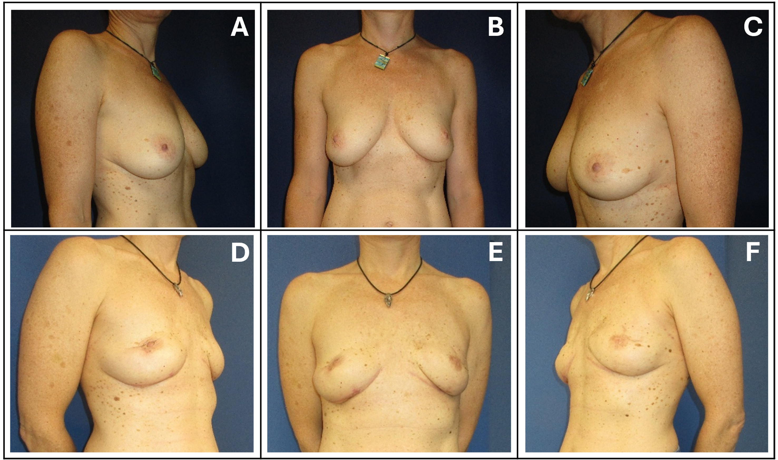
Figure 1. Pre- (A-C) and postoperative (D-F) views of a 51-year-old BRCA1 mutation carrier who underwent staged bilateral prophylactic mastectomy with bilateral TMG flap reconstruction. The postoperative photographs of this illustrative case were obtained 9 months after completion of the reconstruction and 3 months after three-dimensional NAC tattooing. TMG: Transverse myocutaneous gracilis; NAC: nipple-areola complex.
Case 2: salvage autologous breast reconstruction with TMG flap after implant-based reconstruction and severe capsular contracture
A 55-year-old woman was referred to our department with severe capsular contracture of the right breast following implant-based breast reconstruction performed at an external institution. Her surgical history included subcutaneous mastectomy for right breast cancer and multiple implant exchanges.
Clinically, she presented with significant pain, marked capsular fibrosis, and visible breast asymmetry. Examination revealed periareolar and inframammary fold scars, as well as a cranially displaced right nipple compared to the contralateral side. The reconstructive situation and asymmetry, including the high nipple position, were discussed in detail with the patient. She explicitly wished to preserve her native nipple despite its asymmetrical position.
Given the severity of capsular contracture and associated pain, the indication was made for implant removal and autologous breast reconstruction. Due to the patient’s slender body habitus and insufficient abdominal tissue volume, reconstruction using a DIEP or ms-TRAM flap was not feasible. Multiple pre-existing scars in the lower abdomen and periumbilical region also limited the suitability of abdominal donor sites. Therefore, reconstruction using a free TMG flap harvested from the left medial thigh was planned.
The implant was removed, and the right breast was reconstructed using a free TMG flap harvested from the left thigh. In addition, a tightening of the inframammary fold and lipofilling from the abdominal region to the medial upper quadrant of the right breast were performed to optimize contour and symmetry [Figure 2].
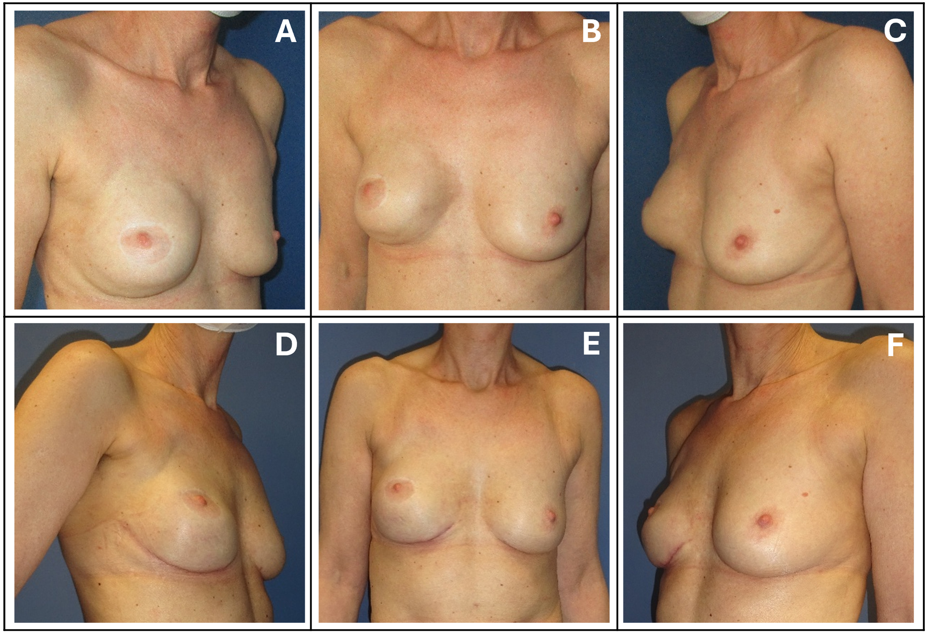
Figure 2. Pre- (A-C) and postoperative (D-F) views of a 55-year-old woman with severe capsular contracture and breast asymmetry following implant-based reconstruction of the right breast who underwent TMG reconstruction of the right breast. The postoperative photographs of this illustrative case were obtained 6 months after secondary lipofilling and inframammary-fold revision. TMG: Transverse myocutaneous gracilis.
DISCUSSION
The TMG flap has come a long way since its initial description for breast reconstruction in 2004[8]. Over two decades of clinical experience, supported by anatomical studies, large case series, systematic reviews, and patient-reported outcome data, have established the TMG flap as a safe, reliable, and versatile option for autologous breast reconstruction. Yet the TMG remains an “often forgotten” flap. Several factors may contribute to its underutilization. First, the widespread adoption of the DIEP flap as the preferred option for autologous breast reconstruction may reduce familiarity with alternative donor sites. Second, the perceived volume limitation of the TMG has historically restricted its application to small-breasted patients, though extended designs, stacking techniques, and adjunctive fat grafting have substantially expanded its volumetric capabilities[12,30,31]. Third, the learning curve for TMG harvest, while arguably shorter than for DIEP perforator dissection, requires dedicated training and case volume[10,32].
The advantages of the TMG flap deserve emphasis. The vascular anatomy is consistent and reliable, with the ascending branch of the MCFA providing a dependable pedicle[19,20]. Flap harvest is expedient, with operative times shorter than those typically reported for DIEP flaps[10,35]. The donor site scar is well concealed and mimics a medial thigh lift[16,33]. Importantly, the TMG entirely avoids abdominal fascial incision and therefore eliminates the abdominal donor-site risk of hernia or bulging associated with abdominally based flap harvest[16]. Lower extremity function is preserved, with LEFS scores of 78/80 at long-term follow-up[17]. For bilateral reconstruction, the TMG may offer particular advantages, as bilateral thigh harvest avoids splitting the abdominal flap and produces symmetric, well-concealed donor scars[11,22]. However, donor-site morbidity was limited in many published series, with reported sensory disturbances and measurable functional abnormalities. Lower-extremity function therefore cannot be considered uniformly preserved, particularly because prospective studies using standardized functional assessments remain limited. The primary limitation remains volume. With a mean flap weight of 320 g in the largest series, the TMG is best suited for small-to-moderate breast reconstruction[14]. However, extended flap designs can yield weights of 470-750 g, and stacked configurations or adjunctive fat grafting can further augment volume[12,13,30,31]. The need for secondary lipofilling is higher than with DIEP flaps (65% vs. 38%), which should be discussed with patients during preoperative counseling[22]. The skin color match of the TMG flap is inferior to that of the DIEP flap, particularly in the early postoperative period, though both improve over time[34]. This is a relevant consideration for delayed reconstruction with a skin-bearing flap but is less significant in immediate reconstruction after skin-sparing mastectomy, where the flap is largely deepithelialized.
A further important consideration is the comparison between the TMG and PAP flaps. In recent years, the PAP flap has gained increasing popularity as a muscle-sparing alternative for patients who are not suitable candidates for abdominally based reconstruction. Recent comparative evidence does not establish the universal superiority of either thigh-based flap. A comparative cohort study found similarly low functional donor-site morbidity after PAP and TMG reconstruction, but patients undergoing TMG reconstruction reported greater satisfaction with the scar and its position. Donor-site wound complications were also more frequent in the PAP cohort; however, the study included relatively small groups and used a historical TMG comparison cohort[36]. Available functional outcome data suggest that gracilis muscle harvest in TMG reconstruction results in minimal functional impairment, with long-term lower-extremity function remaining largely preserved[20,36,37]. Although the PAP flap preserves the muscle, its harvest often requires technically demanding intramuscular perforator dissection through the adductor musculature. As a result, some degree of muscular trauma may still occur despite formal muscle preservation. In addition, because PAP perforators are located more distally, the PAP skin paddle and resulting donor-site scar are typically positioned further inferiorly on the posterior or medial thigh[20,36]. Consequently, the scar may be more visible and is usually less easily concealed within the groin crease or inferior gluteal fold than the TMG donor scar. By contrast, the TMG flap allows placement of the donor-site scar in a more cranial and discreet position. For many patients, particularly younger women undergoing prophylactic reconstruction, this aesthetic consideration may represent a meaningful advantage of the TMG flap.
A recent meta-analysis found significantly lower rates of vascular complications and acute unplanned reoperations following PAP reconstruction than following gracilis-based thigh-flap reconstruction, while rates of flap loss and hematoma were similar[38]. Substantial heterogeneity prevented reliable pooled comparisons of several other clinically relevant outcomes.
Rather than viewing the TMG and PAP flaps as competing procedures, they should be regarded as complementary options within the spectrum of thigh-based autologous breast reconstruction. Ideally, reconstructive centers should be familiar with and able to offer both techniques. Preoperative computed tomography angiography (CTA)-based assessment of perforator anatomy, vascular caliber, pedicle course, soft-tissue distribution, and patient body habitus can then guide individualized flap selection and help tailor the reconstructive strategy to the anatomical and aesthetic requirements of each patient[27].
Recent publications have increasingly shifted the focus from basic flap feasibility toward technical optimization and management of the reconstructive pathway. A multicenter analysis found that the choice of ipsilateral or contralateral thigh harvest and differences in flap shaping and inset did not significantly influence the final aesthetic breast outcome; nevertheless, secondary refinement procedures remained common and should be anticipated as part of the reconstructive strategy[39]. Similarly, closed-incision negative-pressure therapy has been evaluated as a means of reducing medial-thigh donor-site complications, but the available retrospective evidence has not demonstrated a statistically significant reduction in surgical-site complications[40]. These findings illustrate that technical adjuncts may facilitate reconstruction and donor-site management but do not eliminate the need for careful flap design, tension-free closure, complication surveillance, and secondary correction when indicated.
Despite the growing body of literature supporting the use of the TMG flap for breast reconstruction, several important evidence gaps remain. Most published studies are retrospective, single-center case series, and high-level evidence remains scarce. Moreover, a considerable proportion of the published evidence originates from a limited number of specialized high-volume centers, with partially overlapping study periods and patient populations. This may limit the generalizability of the reported outcomes and potentially overrepresent results achieved after substantial institutional experience. Differences in terminology, particularly the partly interchangeable use of TMG and TUG, as well as variability in flap design, outcome definitions, follow-up duration, and reporting of secondary procedures further complicate comparisons. Patient-reported outcome studies are also limited by the frequent absence of preoperative baseline measurements and by the lack of a validated BREAST-Q donor-site module specifically designed for thigh-based flaps. Comparative studies directly evaluating the TMG flap against alternative reconstructive options, particularly the DIEP and PAP flaps, remain limited and are often subject to selection bias. Although available data suggest generally favorable patient-reported outcomes and minimal long-term functional impairment following gracilis harvest, prospective studies with standardized assessment of lower-extremity function and quality of life are still lacking. Similarly, long-term aesthetic outcomes, donor-site satisfaction, and the need for secondary revision procedures have not been evaluated consistently across institutions. The role of emerging modifications, including extended TMG designs, stacked configurations, and primary lipofilling techniques, also requires further investigation in larger patient cohorts. Future research should focus on prospective multicenter studies with standardized outcome measures, including BREAST-Q and functional assessment tools, to facilitate meaningful comparisons between reconstructive techniques. In addition, further studies evaluating patient selection algorithms, cost-effectiveness, and long-term outcomes relative to PAP and abdominally based flaps would help define the optimal role of the TMG flap within the modern reconstructive repertoire.
CONCLUSIONS
The TMG flap is a well-established, reliable, and versatile option for autologous breast reconstruction, yet it remains underutilized in everyday reconstructive practice. With reported low flap-loss rates, generally moderate donor-site morbidity, and favorable patient-reported outcomes, the TMG flap represents an important component of the contemporary reconstructive algorithm.
In this context, the TMG flap may be considered an important reconstructive option in selected patients. This is particularly relevant for women with small-to-moderate breast volume, patients undergoing bilateral reconstruction, and those who wish to avoid abdominal donor-site morbidity. In these settings, the TMG flap offers a favorable combination of reliability, aesthetic donor-site scars, and soft tissue characteristics well suited for breast reconstruction.
Moreover, continued technical refinements, including extended flap designs, stacked flap configurations, and adjunctive fat grafting, have expanded its volumetric potential and broadened its indications, reinforcing its role as a valuable option within contemporary autologous breast reconstruction.
DECLARATIONS
Authors’ contributions
Conceptualised the study: Trieb M
Contributed to the study design: Trieb M, Arkudas A
Led the drafting of the manuscript and analysis: Trieb M
Contributed to the interpretation of the data: Trieb M
Critically revised the manuscript for important intellectual content: Trieb M, Arkudas A, Horch RE
Supervised the project: Arkudas A, Horch RE
Provided administrative support: Horch RE
Availability of data and materials
Not applicable.
AI and AI-assisted tools statement
During the preparation of this manuscript, the AI tool ChatGPT (version 5.3, released 2026-02-05) was used solely for language editing. The tool did not influence the study design, data collection, analysis, interpretation, or the scientific content of the work. All authors take full responsibility for the accuracy, integrity, and final content of the manuscript.
Financial support and sponsorship
None.
Conflicts of interest
Arkudas A is the Guest Editor of the special issue “Advances and Trends in Modern Breast Surgery” of the journal Plastic and Aesthetic Research. Horch RE is an Associate Editor of Plastic and Aesthetic Research. They were not involved in any steps of editorial processing, notably including reviewers’ selection, manuscript handling and decision making. Trieb M declares no conflicts of interest.
Ethical approval and consent to participate
This manuscript is a narrative review supplemented by two illustrative patient cases from our institution. The cases were managed as part of routine clinical care and are included solely for educational and illustrative purposes. All patient data have been fully anonymized. The project was formally reviewed by the Ethics Committee of the Faculty of Medicine at Friedrich-Alexander-Universität Erlangen-Nürnberg and received ethics opinion No. 26-286-Br. Written informed consent was obtained from both patients.
Consent for publication
Written informed consent for the publication of clinical information and images was obtained from both patients.
Copyright
© The Authors 2026.
REFERENCES
1. Ludolph I, Arkudas A, Schmitz M, et al. Cracking the perfusion code?: Laser-assisted Indocyanine Green angiography and combined laser Doppler spectrophotometry for intraoperative evaluation of tissue perfusion in autologous breast reconstruction with DIEP or ms-TRAM flaps. J Plast Reconstr Aesthet Surg. 2016;69:1382-8.
2. Macadam SA, Zhong T, Weichman K, et al. Quality of life and patient-reported outcomes in breast cancer survivors: a multicenter comparison of four abdominally based autologous reconstruction methods. Plast Reconstr Surg. 2016;137:758-71.
3. Erdmann-Sager J, Wilkins EG, Pusic AL, et al. Complications and patient-reported outcomes after abdominally based breast reconstruction: results of the mastectomy reconstruction outcomes consortium study. Plast Reconstr Surg. 2018;141:271-81.
4. Fattah A, Figus A, Mathur B, Ramakrishnan VV. The transverse myocutaneous gracilis flap: technical refinements. J Plast Reconstr Aesthet Surg. 2010;63:305-13.
5. Blough JT, Saint-Cyr MH. Modern approaches to alternative flap-based breast reconstruction: transverse upper gracilis flap. Clin Plast Surg. 2023;50:313-23.
6. Harii K, Ohmori K, Sekiguchi J. The free musculocutaneous flap. Plast Reconstr Surg. 1976;57:294-303.
7. Whetzel TP, Lechtman AN. The gracilis myofasciocutaneous flap: vascular anatomy and clinical application. Plast Reconstr Surg. 1997;99:1642-52.
8. Wechselberger G, Schoeller T. The transverse myocutaneous gracilis free flap: a valuable tissue source in autologous breast reconstruction. Plast Reconstr Surg. 2004;114:69-73.
9. Whitaker IS, Karavias M, Shayan R, et al. The gracilis myocutaneous free flap: a quantitative analysis of the fasciocutaneous blood supply and implications for autologous breast reconstruction. PLoS One. 2012;7:e36367.
10. Fansa H, Schirmer S, Warnecke IC, Cervelli A, Frerichs O. The transverse myocutaneous gracilis muscle flap: a fast and reliable method for breast reconstruction. Plast Reconstr Surg. 2008;122:1326-33.
11. Schoeller T, Huemer GM, Wechselberger G. The transverse musculocutaneous gracilis flap for breast reconstruction: guidelines for flap and patient selection. Plast Reconstr Surg. 2008;122:29-38.
12. Saint-Cyr M, Wong C, Oni G, et al. Modifications to extend the transverse upper gracilis flap in breast reconstruction: clinical series and results. Plast Reconstr Surg. 2012;129:24e-36.
13. Park JE, Alkureishi LWT, Song DH. TUGs into VUGs and friendly BUGs: transforming the gracilis territory into the best secondary breast reconstructive option. Plast Reconstr Surg. 2015;136:447-54.
14. Weitgasser L, Mahrhofer M, Schwaiger K, et al. Lessons learned from 30 years of transverse myocutaneous gracilis flap breast reconstruction: historical appraisal and review of the present literature and 300 cases. J Clin Med. 2021;10:3629.
15. Ehni FREH, Arkudas A, Beier JP. Die freie mikrochirurgische TMG lappenplastik zur brustrekonstruktion. Plastische Chirurgie 2014;03:159-64.
16. Siegwart LC, Bolbos A, Tapking C, et al. Safety and donor site morbidity of the transverse musculocutaneous gracilis (TMG) flap in autologous breast reconstruction-a systematic review and meta-analysis. J Surg Oncol. 2021;124:492-509.
17. Mahrhofer M, Schwaiger K, Fierdel F, et al. Patient-reported outcomes following breast reconstruction with the transverse myocutaneous gracilis flap using the BREAST-Q and lower extremity function scale. Aesthet Surg J. 2024;44:NP168-76.
18. Juricic M, Vaysse P, Guitard J, Moscovici J, Becue J, Juskiewenski S. Anatomic basis for use of a gracilis muscle flap. Surg Radiol Anat. 1993;15:163-8.
19. Macchi V, Vigato E, Porzionato A, et al. The gracilis muscle and its use in clinical reconstruction: an anatomical, embryological, and radiological study. Clin Anat. 2008;21:696-704.
20. Zaussinger M, Tinhofer IE, Hamscha U, et al. A head-to-head comparison of the vascular basis of the transverse myocutaneous gracilis, profunda artery perforator, and fasciocutaneous infragluteal flaps: an anatomical study. Plast Reconstr Surg. 2019;143:381-90.
21. Wong C, Mojallal A, Bailey SH, Trussler A, Saint-Cyr M. The extended transverse musculocutaneous gracilis flap: vascular anatomy and clinical implications. Ann Plast Surg. 2011;67:170-7.
22. Weitgasser L, Schwaiger K, Medved F, Hamler F, Wechselberger G, Schoeller T. Bilateral simultaneous breast reconstruction with DIEP- and TMG flaps: head to head comparison, risk and complication analysis. J Clin Med. 2020;9:2031.
23. Wechselberger G, Traintinger H, Larcher L, Russe E, Edelbauer M, Ensat F. Clinical applications of the transverse musculocutaneous gracilis flap for secondary breast reconstruction after simple mastectomy. Plast Reconstr Surg. 2016;137:19-28.
24. Pülzl P, Huemer GM, Schoeller T. Transverse musculocutaneous gracilis flap for treatment of capsular contracture in tertiary breast reconstruction. Ann Plast Surg. 2015;74:167-72.
25. Bach AD, Morgenstern IH, Horch RE. Secondary “hybrid reconstruction” concept with silicone implants after autologous breast reconstruction - is it safe and reasonable? Med Sci Monit 2020;26:e921329.
26. Schwaiger K, Weitgasser L, Mahrhofer M, et al. Age and overweight are not contraindications for a breast reconstruction with a TMG-flap-a risk and complication analysis of a retrospective double center study including 300 patients. J Clin Med. 2021;10:926.
27. Sonda R, Atzeni M, Martini F, et al. The profunda artery perforators: anatomical study and radiological findings using computed tomography angiography in patients undergoing PAP flap breast reconstruction. J Plast Reconstr Aesthet Surg. 2024;89:164-73.
28. Weitgasser L, Mahrhofer M, Wechselberger G, Schoeller T. Letter comments on: “preoperative volume estimation in transverse upper gracilis flap surgery: a pilot study”. J Plast Reconstr Aesthet Surg. 2022;75:1523-4.
29. Ciudad P, Maruccia M, Orfaniotis G, et al. The combined transverse upper gracilis and profunda artery perforator (TUGPAP) flap for breast reconstruction. Microsurgery. 2016;36:359-66.
30. Russe E, Kholosy H, Weitgasser L, et al. Autologous fat grafting for enhancement of breast reconstruction with a transverse myocutaneous gracilis flap: a cohort study. J Plast Reconstr Aesthet Surg. 2018;71:1557-62.
31. Sharp O, Johal KS, Morgan M, Ramakrishnan VV. Primary lipofilling as an adjunct in transverse upper gracilis flap breast reconstruction. J Plast Reconstr Aesthet Surg. 2023;86:1-7.
32. Nickl S, Nedomansky J, Radtke C, Haslik W, Schroegendorfer KF. Optimization of breast reconstruction results using TMG flap in 30 cases: evaluation of several refinements addressing flap design, shaping techniques, and reduction of donor site morbidity. Microsurgery. 2018;38:489-97.
33. Mahrhofer M, Russe E, Schoeller T, Wechselberger G, Weitgasser L. Management of donor-site complications after breast reconstruction with the transverse musculocutaneous gracilis flap: proposal of a treatment algorithm. Microsurgery. 2023;43:537-45.
34. Mahrhofer M, Reichert R, Fierdel F, et al. Photometric comparison of skin color match between abdominal and thigh-based free flaps for breast reconstruction in white patients. Plast Reconstr Surg. 2024;154:1076e-83.
35. Promny T, Huberth P, Müller-Seubert W, et al. The impact of technical innovations and donor-site mesh repair on autologous abdominal-based breast reconstruction-a retrospective analysis. J Clin Med. 2024;13:2165.
36. Augustin A, Pülzl P, Morandi EM, et al. Donor-site morbidity and quality of life after autologous breast reconstruction with PAP versus TMG flap. Curr Oncol. 2022;29:5682-97.
37. Siegwart LC, Bolbos A, Haug VF, Diehm YF, Kneser U, Kotsougiani-Fischer D. Donor site morbidity in unilateral and bilateral transverse musculocutaneous gracilis (TMG) flap breast reconstruction: sensation, function, aesthesis and patient-reported outcomes. J Clin Med. 2021;10:5066.
38. Borrelli MR, Spake CSL, Rao V, et al. A systematic review and meta-analysis comparing the clinical outcomes of profunda artery perforator versus gracilis thigh flap as a second choice for autologous breast reconstruction. Ann Plast Surg. 2023;90:S256-67.
39. Vollbach FH, Neuss C, Siegwart LC, et al. The transverse myocutaneous gracilis flap (TMG) for breast reconstruction: ipsi vs. contralateral harvest-aesthetic outcome and refinement procedures. Breast Cancer. 2023;30:845-55.
Cite This Article
 Open Access
How to Cite
Trieb M, Horch RE, Arkudas A. The transverse myocutaneous gracilis flap for breast reconstruction: current evidence, technical refinements and institutional experience. Plast Aesthet Res. 2026;13:29. https://dx.doi.org/10.20517/2347-9264.2026.65
Download Citation
If you have the appropriate software installed, you can download article citation data to the citation manager of your choice. Simply select your manager software from the list below and click on download.
Export Citation File
Type of Import
Tips on Downloading Citation
Citation Manager File Format
Type of Import
Direct Import: When the Direct Import option is selected (the default state), a dialogue box will give you the option to Save or Open the downloaded citation data. Choosing Open will either launch your citation manager or give you a choice of applications with which to use the metadata. The Save option saves the file locally for later use.
Indirect Import: When the Indirect Import option is selected, the metadata is displayed and may be copied and pasted as needed.
About This Article
Special Topic
Copyright

Data & Comments
Data
0










Comments
Comments must be written in English. Spam, offensive content, impersonation, and private information will not be permitted. If any comment is reported and identified as inappropriate content by OAE staff, the comment will be removed without notice. If you have any queries or need any help, please contact us at [email protected].