
fig5
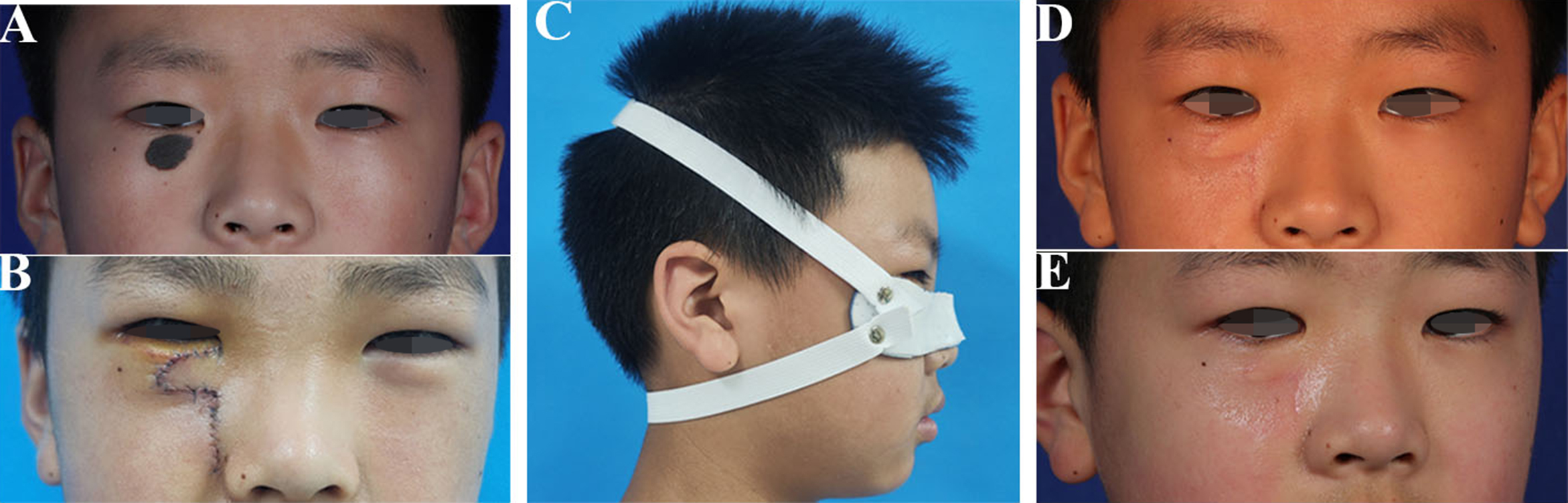
Figure 5. Comprehensive management of a congenital melanocytic nevus of the right lower eyelid in a 12-year-old male patient. (A) Preoperative appearance of a congenital melanocytic nevus (19 mm × 12 mm) on the right lower eyelid; (B) Intraoperative view after nevus excision and local flap reconstruction, highlighting the vertical (auxiliary) incision that conferred a high risk for cicatricial ectropion; (C) The custom low-temperature thermoplastic splint was applied on postoperative day 7 as part of the prophylactic protocol; (D) At the 8-week interim assessment, resolution of edema and controlled scar remodeling were observed without ectropion, with EGS grade 0 maintained; (E) Outcome at 6-month follow-up, demonstrating maintained normal eyelid position, contour, and function, confirming successful prevention of ectropion. EGS: Ectropion grading scale.