


fig1
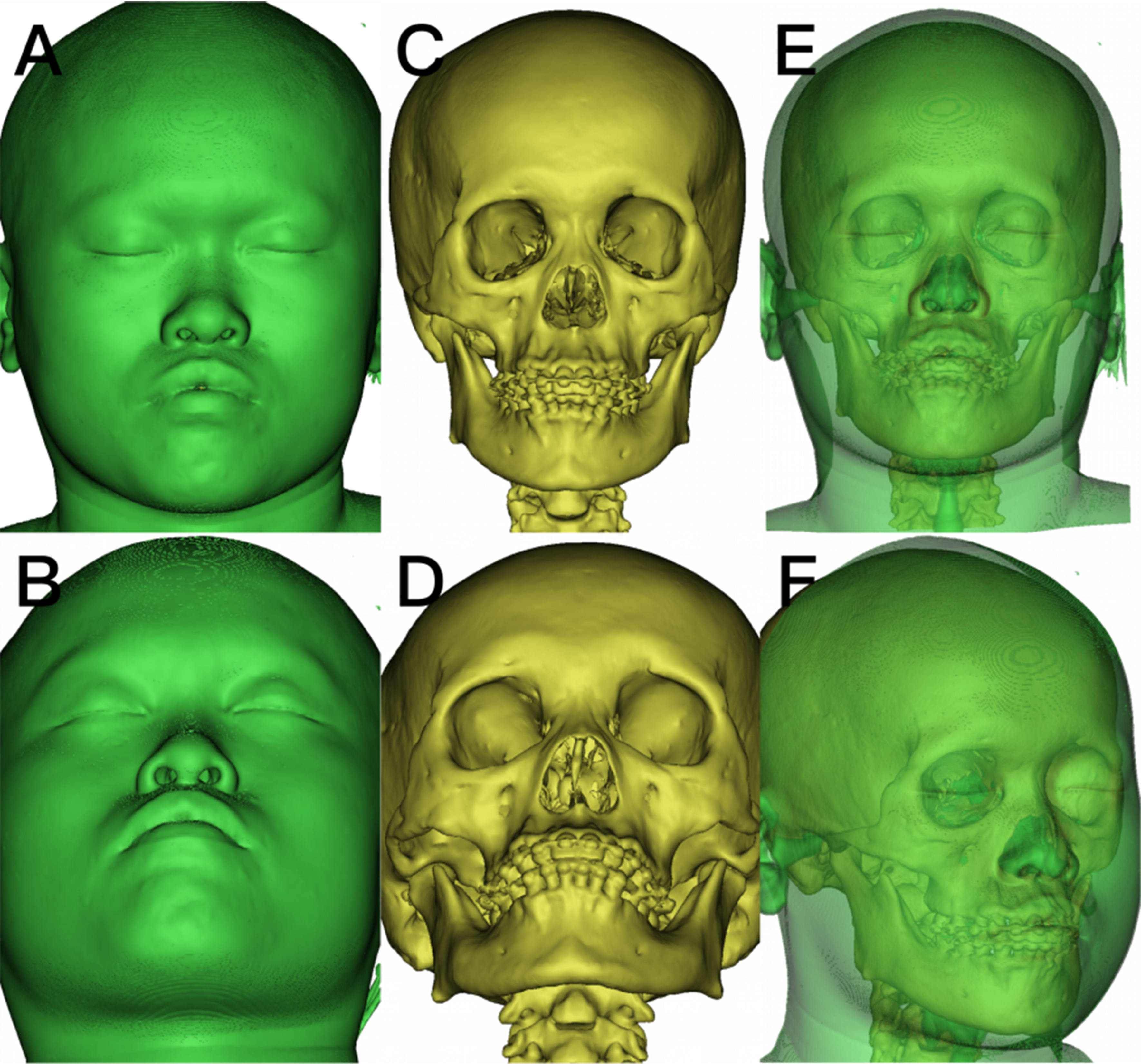
Figure 1. 3D reconstruction models used for layered teaching in the experimental group. (A and B) Frontal and upward-tilted views of the skin surface model; (C and D) Frontal and upward-tilted views of the skeletal model; (E and F) Frontal and lateral views of the combined skin-skeletal model. All models were generated using 3D reconstruction software to visualize the spatial relationships between soft tissue and bone for instructional purposes. All images are original and de-identified. 3D: Three-dimensional.


