


fig4
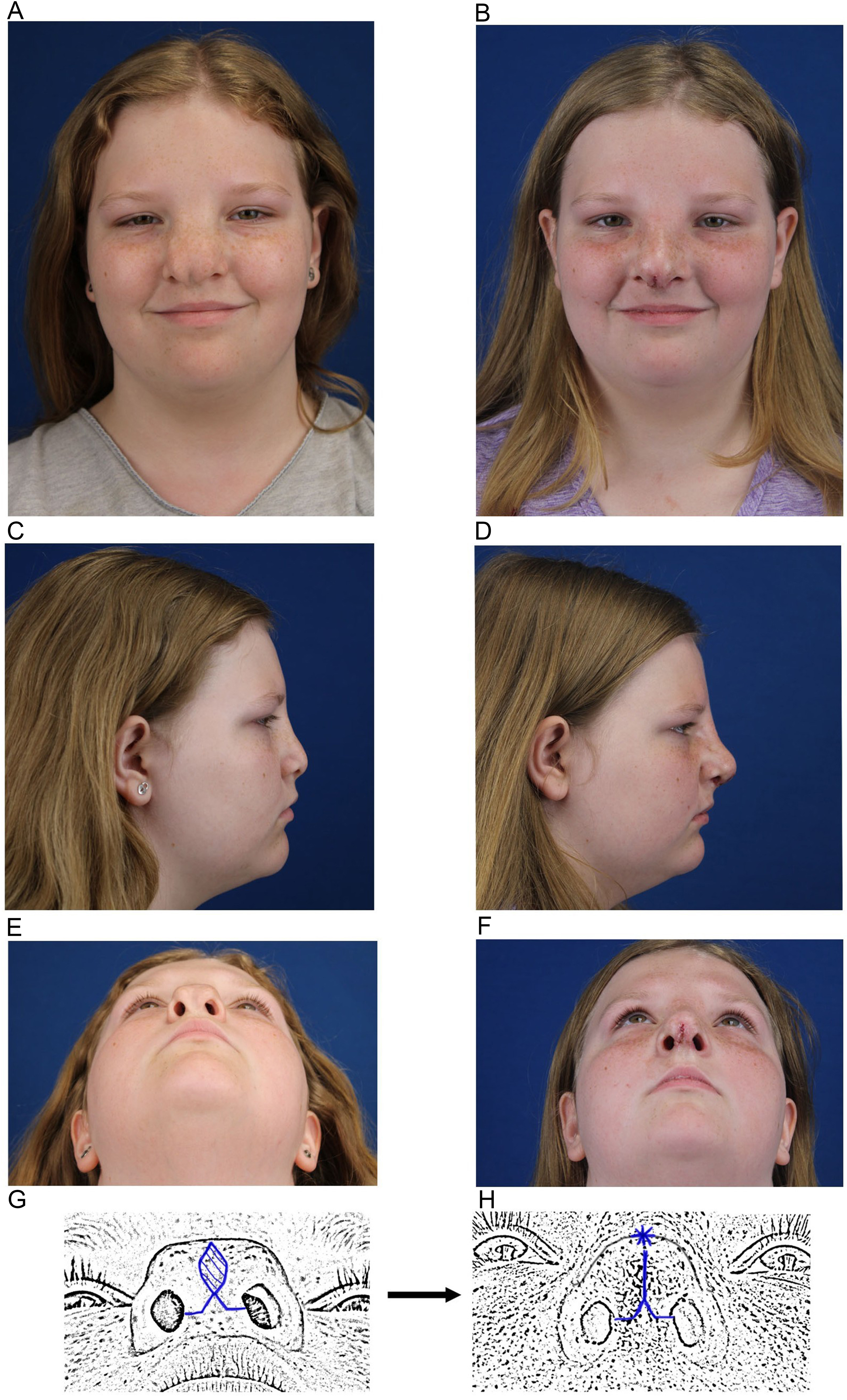
Figure 4. An example of techniques employed for a Type 0 Tessier. (A) Preoperative frontal view (B) Immediate postoperative frontal view. Surgery included lateral and medial nasal bone osteotomies; (C) Preoperative lateral view (D) Immediate postoperative lateral view. Surgery included an on-lay graft composed of autologous minced costal cartilage and fibrin glue. The excess skin/SMAS generated from medializing the nasal bones and upper/lower lateral cartilages created a potential space to place the on-lay graft and project the nasal dorsum and tip; (E) Preoperative base view; (F) Immediate postoperative base view; (G) Schematic illustrating the planned trans-columellar incision with excision of intra-tip skin excess; (H) Schematic illustrating closure of the incisions. The asterisk denotes where there is a purposeful standing cone at the end of the elliptical excision to allow for more tip projection. SMAS: Superficial musculoaponeurotic system.


