


fig3
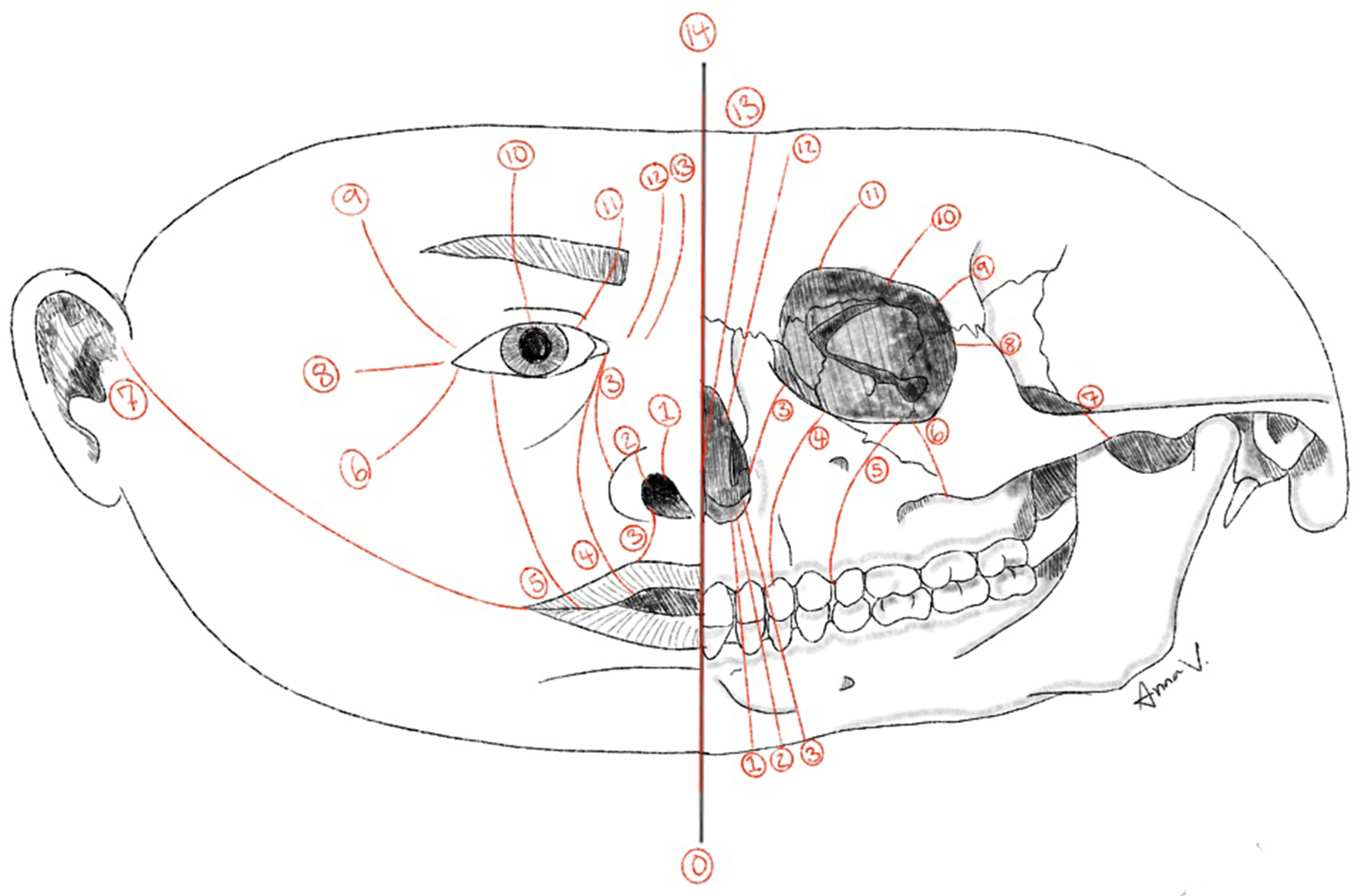
Figure 3. Diagram illustrating the soft-tissue and skeletal locations of the Tessier craniofacial clefts (numbers 0-14). Illustration designed by Anna Virginia DeMarco.
Figure 3. Diagram illustrating the soft-tissue and skeletal locations of the Tessier craniofacial clefts (numbers 0-14). Illustration designed by Anna Virginia DeMarco.


All published articles are preserved here permanently:
https://www.portico.org/publishers/oae/

