


fig2
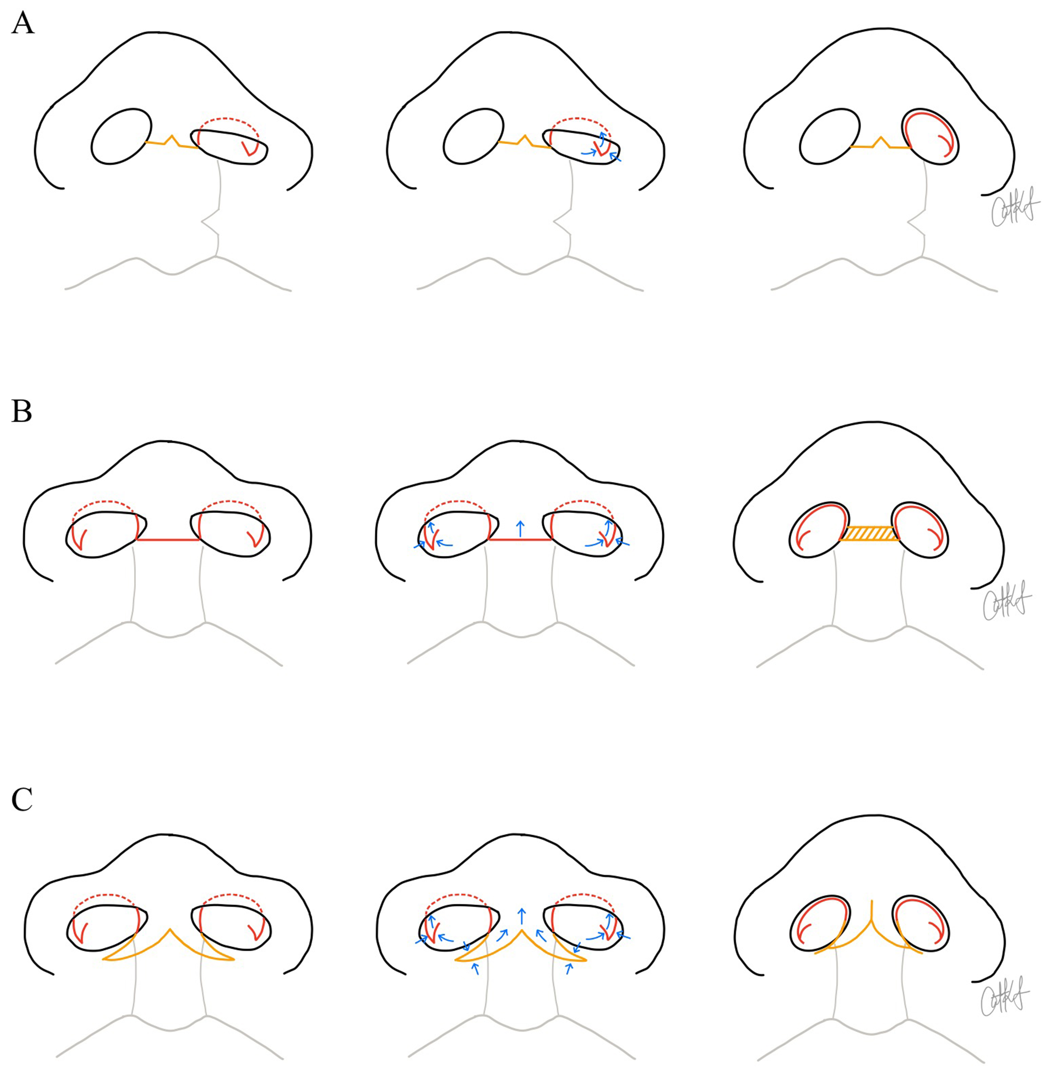
Figure 2. Schematic illustrations demonstrating surgical techniques used to increase tip projection and columellar length during primary, intermediate, or definitive rhinoplasty procedures. Yellow lines indicate trans-columellar incisions, while red lines depict additional incisional designs that may be performed with or without a trans-columellar approach. Blue arrows represent the direction of tissue advancement. The yellow hatched region denotes a composite chondrocutaneous auricular graft. (A) Left unilateral cleft lip and nasal example. From left to right, the illustrations show the planned intraoperative incisional design, including an inverted-V trans-columellar incision and a reverse-U Tajima alar incision combined with a lateral crural steal maneuver. This maneuver is performed using a composite mucosal-chondral V-to-Y advancement; (B) Bilateral cleft lip and nasal example. From left to right, the illustrations show the planned incisional design, including a horizontal columellar incision and bilateral reverse-U Tajima alar incisions, combined with lateral crural steals to increase columellar and tip projection. Columellar lengthening is achieved with insertion of a composite graft; (C) Bilateral cleft lip and nasal example. From left to right, the illustrations show the planned incision design, including a W-to-Y columellar incision and bilateral reverse-U Tajima alar incisions, also combined with lateral crural steals. The W-to-Y columellar incision allows creation of forked flaps from the nasal sill or lower columella to recruit tissue superiorly. Illustrations designed by Dr. Catharine Kappauf.


