


fig5
From: Reconstructing the lining in full-thickness nasal defects: techniques, challenges, and innovations
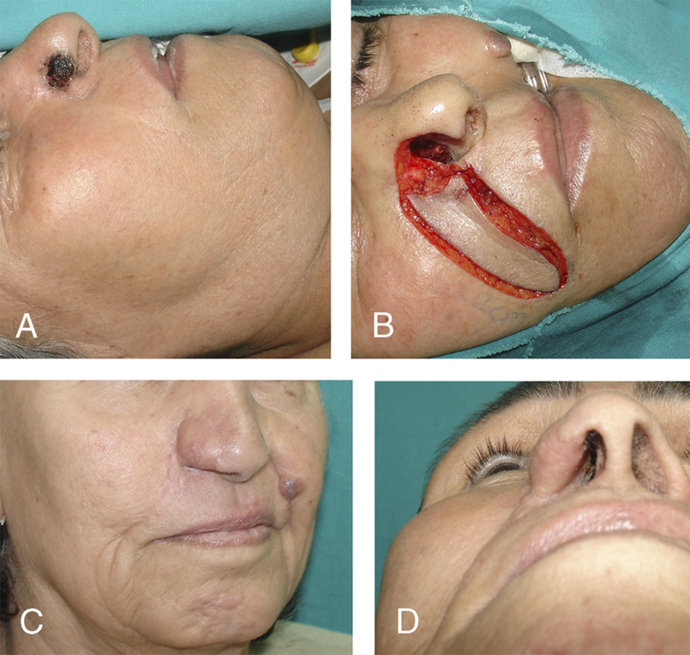
Figure 5. (A) Preoperative appearance of the skin tumor on the right ala; (B) Post-excision defect with planning of the reconstructive flap; (C) Appearance of the repair following reconstruction with a cartilage-supported nasolabial perforator flap, shown at 12 months postoperatively; (D) Inferior view of the reconstructed ala showing good contour and symmetry. Reprinted with permission from[31]. Copyright © 2025 Wolters Kluwer Health, Inc.


