


fig1
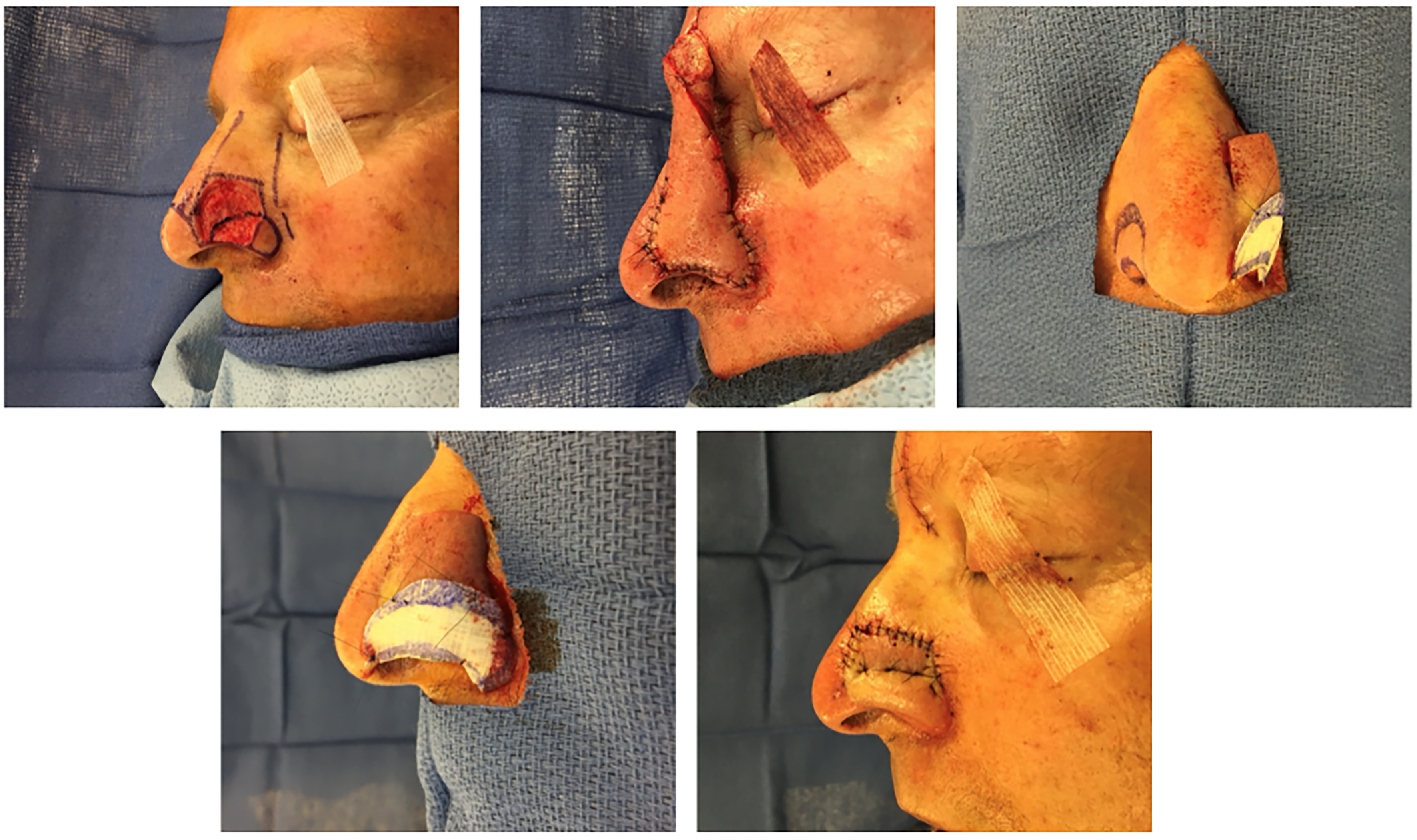
Figure 1. Partial thickness circular defect of the left ala and sidewall reconstructed with a paramedian forehead flap (top left). Note the planned subunit excision of the ala and squaring of defect edges along subunit boundaries. A template based on the non-operated side is used to facilitate symmetry. Tacking sutures are used to re-create the alar groove at the end of the second stage (pedicle division).


