


fig3
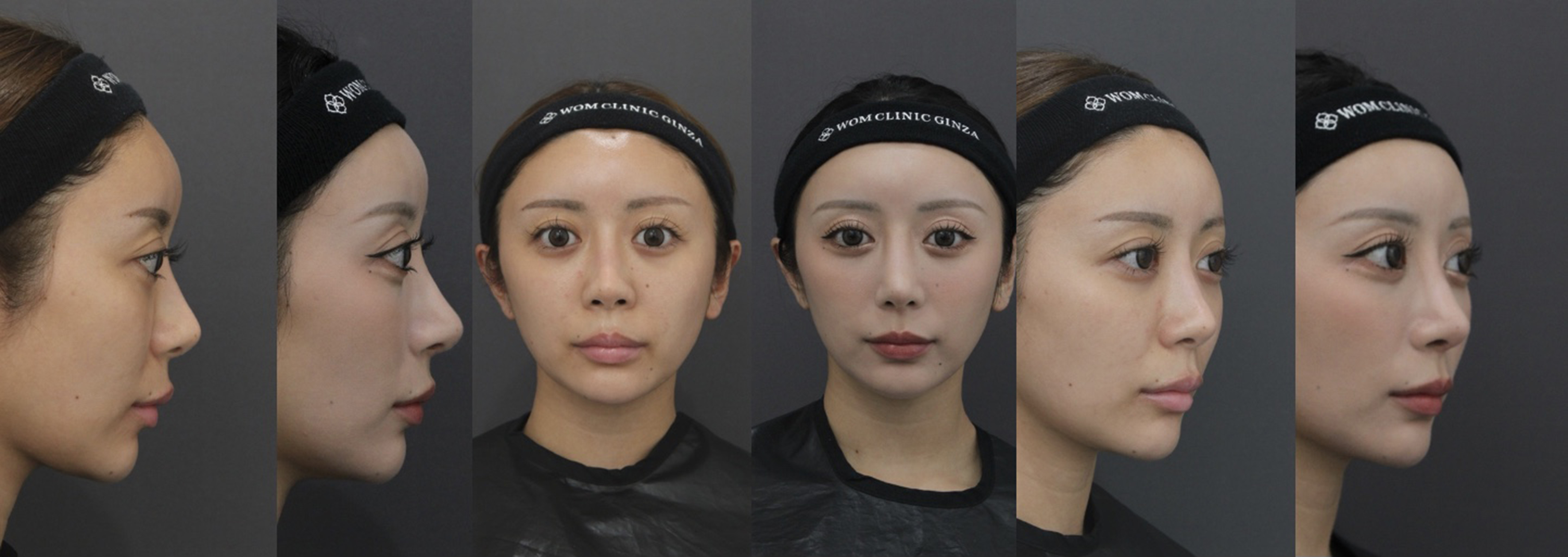
Figure 3. Primary case using posterior auricular fascia. Preoperative and postoperative (12 months) comparisons demonstrate improved tip definition and natural projection without oversharpening. (Left) Lateral view; (Middle) Frontal view; (Right) Oblique view. Postoperative images show a balanced curvature of the nasal tip and smooth contour achieved with the fascial ball graft using posterior auricular fascia.


