
fig1
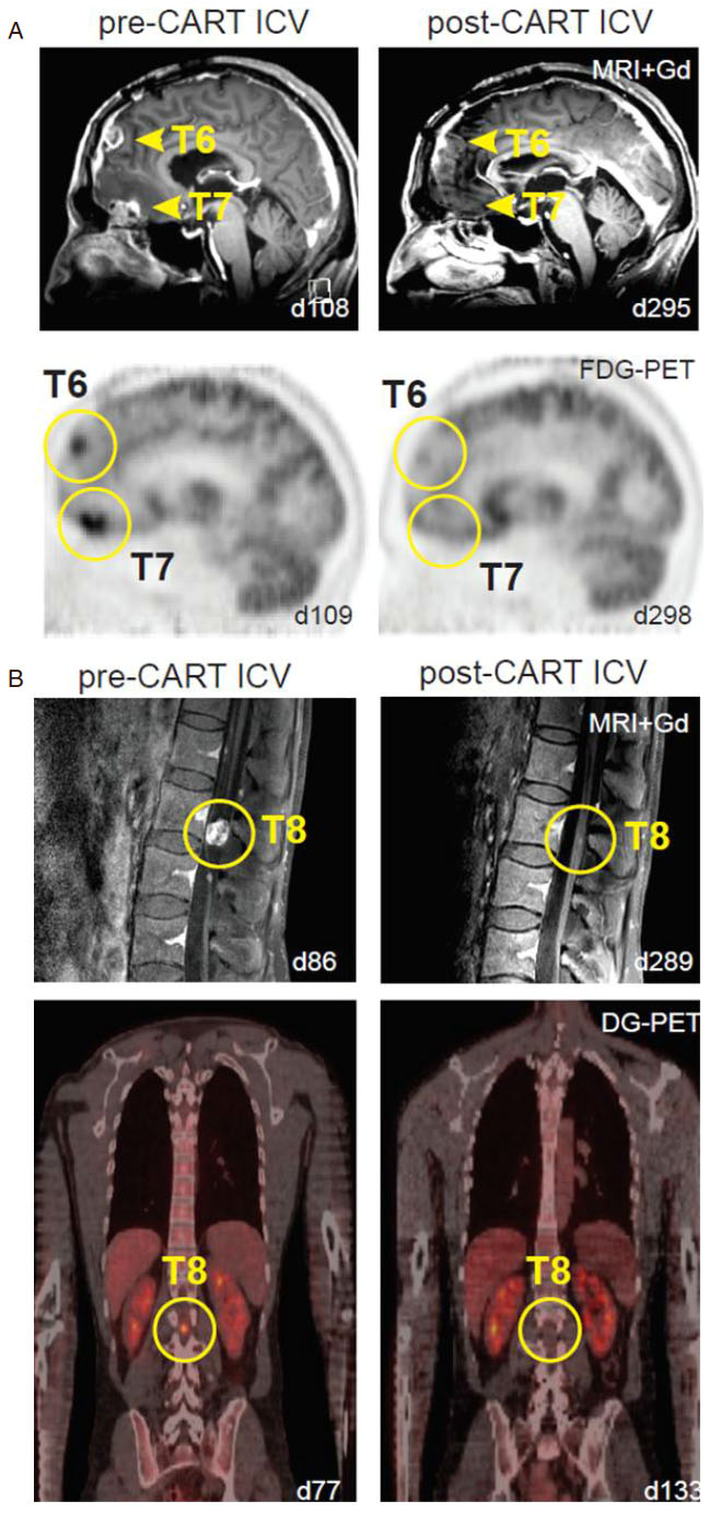
Figure 1. Multifocal glioblastoma responding to intraventricular delivery of IL13Ra2-redirected CAR T-cell therapy (modified from Brown et al.[28], permission for use granted by NEJM). A: sagittal view of gadolinium-enhanced MRI (top row) and FDG-PET (bottom row) images showing tumor regression of tumors 6 and 7 in the brain (yellow arrowheads and circles); B: sagittal gadolinium-enhanced MRI (top row) and coronal DG-PET (bottom row) images exhibiting tumor regression of tumor 8 in the spine (yellow circles)


