

Pseudo-dominant Wilson disease with an unresolved third-generation ATP7B finding
 0
Abstract
Wilson disease is an autosomal recessive copper metabolism disorder caused by pathogenic variants in the adenosine triphosphatase (ATPase) copper transporting beta gene (ATP7B). We describe a Han Chinese family from Inner Mongolia with genetically confirmed pseudo-dominant Wilson disease in two consecutive generations and an unresolved diagnosis in a third-generation relative. Whole-exome sequencing and Sanger segregation analysis showed that the proband carried c.2621C>T (p.Ala874Val) and c.2333G>T (p.Arg778Leu) in trans. Her son and daughter carried c.2621C>T (p.Ala874Val) and c.2975C>T (p.Pro992Leu); their trans configuration and paternal origin of p.Pro992Leu were inferred from the pedigree because the deceased father was unavailable for testing. The 22-year-old grandson had a previous clinical diagnosis but only monoallelic c.2621C>T, negative Kayser-Fleischer rings, and no second ATP7B variant detected by whole-exome sequencing. This family highlights the need for careful phase assignment, spouse and offspring screening, and cautious reconciliation of clinical and molecular diagnoses.
Keywords


 Download PDF
INTRODUCTION
Wilson disease (WD) is an autosomal recessive disorder of copper metabolism caused by pathogenic variants in adenosine triphosphatase (ATPase) copper transporting beta gene (ATP7B), which encodes a copper-transporting P-type ATPase required for biliary copper excretion and incorporation of copper into ceruloplasmin. Impaired ATP7B function leads to copper accumulation, most prominently in the liver and brain, and can produce hepatic, neurological, psychiatric, ocular, hematological, and renal manifestations[1-4].
Because WD is autosomal recessive, molecular confirmation usually requires pathogenic variants on both ATP7B alleles. In this report, pseudo-dominant inheritance refers to the apparent vertical transmission of an autosomal recessive disorder across consecutive generations when an affected individual has affected offspring because the reproductive partner carries a pathogenic ATP7B allele; the term does not imply a dominant molecular mechanism. Accordingly, offspring of an affected individual are obligate heterozygous carriers when the other parent has two wild-type alleles, whereas an affected individual and a heterozygous carrier have a 50% risk of an affected child in each pregnancy[3,5,6].
Here, we report a Chinese family with genetically confirmed WD in the proband and her two children and an unresolved monoallelic ATP7B finding in the grandson. The case illustrates the value of cascade screening and the need to reconcile clinical and molecular evidence when whole-exome sequencing (WES) findings are discordant.
CASE PRESENTATION
Clinical and genetic information is summarized in Tables 1-3 and Figure 1. None of the four clinically affected or suspected individuals underwent liver biopsy or hepatic copper quantification. For all copper-related measurements included in this report, the laboratory reference ranges were 0.20-0.60 g/L for serum ceruloplasmin and 15-60 μg/24 h for 24-h urinary copper. Units for the other laboratory parameters are provided in Table 1.
Clinical characteristics of family members with Wilson disease or suspected Wilson disease
| Variable | I-2 proband | II-1 son | II-2 daughter | III-1 grandson |
| Age/sex | 67 years/female | 36 years/male | 46 years/female | 22 years/male |
| Ethnicity/origin | Han; Inner Mongolia | Han; Tongliao, Inner Mongolia | Han; Inner Mongolia | Han; Inner Mongolia |
| Consanguinity | No | No | No | No |
| Age at onset/diagnosis | 54/54 years (2013) | 23/23 years (2013) | 29/29 years (2009) | 12/12 years (2016; historical clinical diagnosis) |
| Initial presentation | Abdominal distension | Asymptomatic; detected during evaluation | Upper abdominal pain, abdominal distension, liver-region pain, anorexia, hand tremor, and headache | Low ceruloplasmin, 24-h urinary copper 103 μg/24 h, and heterozygous ATP7B finding |
| Phenotype | Hepatic WD | Hepatic WD; intermittent tremor since 2024 | Predominantly hepatic WD; possible mild neurological involvement | Molecular diagnosis unresolved; previous clinical diagnosis requires re-evaluation |
| Key physical findings | Splenomegaly and K-F rings in 2013; splenomegaly, ascites, and K-F rings in 2026; transient tremor after treatment | Splenomegaly; no K-F rings; small ascites in 2026; intermittent tremor | No jaundice; no K-F rings; long-standing splenomegaly until splenectomy; subjective tremor/headache | Clinically asymptomatic; K-F rings negative; no neurological symptoms reported |
| Ceruloplasmin (reference range 0.20-0.60 g/L) | 0.03 g/L in 2013 and 2026 | 0.03 g/L in 2013 and 2026 | 0.03 g/L in 2026 | 0.13 g/L 10 years earlier; 0.143 g/L in 2026 |
| 24-h urinary copper (reference range 15-60 μg/24 h) | 186.1 μg/24 h (14 Mar 2026, before chelation therapy was restarted); 1,962.6 μg/24 h (11 May 2026, on day 5 of intravenous sodium dimercaptopropanesulfonate 500 mg/day) | 3,726.6 μg/24 h (25 Mar 2026, after 2 weeks of self-administered penicillamine 250 mg/day); 888 μg/24 h (11 May, on day 5 of intravenous sodium dimercaptopropanesulfonate 0.5 g/day); 1,304.4 μg/24 h (10 Jun, on day 5 of the second intravenous course of sodium dimercaptopropanesulfonate 0.5 g/day); 3,228.2 μg/24 h (1 Jul, after two intravenous courses and during continued penicillamine 750 mg/day) | 1,670.1 μg/24 h (21 Mar 2026, before the 2026 chelation course was started); 1,887.7 μg/24 h (1 Jul 2026, during ongoing penicillamine 750 mg/day) | 103 μg/24 h (~2016, before penicillamine); 24.6 μg/24 h (2026, during penicillamine 375 mg/day) |
| Representative liver tests | Mar 2026: ALT 13.9 U/L, AST 24.5 U/L, TBIL 21.1 μmol/L, ALB 30.7 g/L, INR 1.2 | May 2026: ALT 67.8 U/L, AST 42.1 U/L, TBIL 26.6 μmol/L, ALB 46.1 g/L, INR 1.4 | Dec 2025 pre-splenectomy: ALT 11.09 U/L, AST 14.54 U/L, TBIL 12.45 μmol/L, INR 1.14 | Jun 2026: ALT 90 U/L, AST 41 U/L, TBIL 17 μmol/L, ALB 52.2 g/L |
| Hematology | Mar 2026: WBC 3.51 × 10⁹/L, Hb 101 g/L, PLT 86 × 109/L | May 2026: WBC 3.91 × 10⁹/L, Hb 150 g/L, PLT 51 × 109/L | Dec 2025: WBC 1.85 × 10⁹/L, Hb 116 g/L, PLT 51 × 109/L; PLT increased after splenectomy | Jun 2026: WBC 9.07 × 10⁹/L, Hb 183 g/L, PLT 268 × 109/L |
| Imaging/endoscopy | Cirrhosis, splenomegaly, portal vein widening; ascites in 2026; visible esophageal varices | Cirrhosis, portal hypertension, splenomegaly, small ascites, esophageal/gastric varices, and portal hypertensive gastropathy | Cirrhosis, splenomegaly, portal hypertension, and gastric fundal varices; splenectomy and devascularization in 2025 | Ultrasound: fatty liver |
| Liver biopsy/hepatic copper quantification | Not performed | Not performed | Not performed | Not performed |
| Treatment | Initial IV sodium dimercaptopropanesulfonate; then penicillamine 750 mg/day, zinc, and dimercaptosuccinic acid; prolonged interruption; in 2026, penicillamine 500 mg/day, diuretics, and IV sodium dimercaptopropanesulfonate for two courses | Intermittent penicillamine; documented 2026 regimen 750 mg/day, with 250 mg/day for 2 weeks immediately before the March urine collection; elemental zinc 150 mg/day (as zinc gluconate); IV sodium dimercaptopropanesulfonate for two courses in 2026 | Intermittent penicillamine, generally 500 mg/day, plus elemental zinc 100 mg/day (as zinc gluconate); splenectomy plus devascularization in 2025; penicillamine intensified to 750 mg/day in 2026 | Penicillamine 375 mg/day during recent follow-up |
| Outcome | Ascites resolved by Jun 2026; transient tremor resolved | Tremor improved after second hospitalization; persistent cirrhosis/portal hypertension | Cytopenia improved after splenectomy; abdominal distension improved; tremor/headache persisted intermittently | Remained clinically asymptomatic; recent urinary copper decreased during treatment, while ALT and AST remained mildly elevated, and the molecular diagnosis remains unresolved |
Retrospective Leipzig scores based on available data
| Component | I-2 proband | II-1 son | II-2 daughter | III-1 grandson |
| K-F rings | Present = 2 | Absent = 0 | Absent = 0 | Absent = 0 |
| Neurological manifestations or typical brain MRI | No neurological signs at initial diagnosis; 2026 MRI not typical for WD = 0 | Mild tremor = 1 | Mild tremor (possible neurological involvement) = 1 | No neurological symptoms reported; typical brain MRI not assessed = 0 |
| Serum ceruloplasmin | 0.03 g/L = 2 | 0.03 g/L = 2 | 0.03 g/L = 2 | 0.13 g/L = 1 |
| Coombs-negative hemolytic anemia | Absent = 0 | Absent = 0 | Absent = 0 | Absent = 0 |
| Liver copper or rhodanine staining | Not performed; no points assigned | Not performed; no points assigned | Not performed; no points assigned | Not performed; no points assigned |
| Urinary copper | 186.1 μg/24 h before chelation; >2 × ULN = 2 | 3726.6 μg/24 h after 2 weeks of penicillamine; excluded from diagnostic score | 1,670.1 μg/24 h before chelation; >2 × ULN = 2 | 103 μg/24 h before chelation; 1.72 × ULN = 1 |
| ATP7B molecular testing | Biallelic variants = 4 | Biallelic variants = 4 | Biallelic variants = 4 | One variant = 1 |
| Total Leipzig score[2,3,7] | 10 | 7 (treatment-exposed urinary copper excluded) | 9 | 3 |
| Interpretation | Diagnosis established | Diagnosis established | Diagnosis established | Diagnosis possible; more evidence needed; molecular diagnosis unresolved |
Genetic findings, phase assignment, and inferred segregation of ATP7B variants
| Individual | Clinical status | Observed or inferred ATP7B variant(s) | Zygosity/phase interpretation | Evidence and inferred origin |
| I-2 proband | WD, genetically confirmed | c.2621C>T (p.Ala874Val); c.2333G>T (p.Arg778Leu) | Compound heterozygous; both pathogenic; trans phase demonstrated by segregation | c.2621C>T transmitted without c.2333G>T, demonstrating the two variants were in trans |
| I-1 husband of proband | Deceased; not tested | c.2975C>T (p.Pro992Leu), inferred from offspring segregation; not directly tested in I-1 | Presumed heterozygous carrier; inference only | c.2975C>T was present in both affected children but absent from the proband; paternal origin inferred because I-1 was deceased and untested |
| II-1 son | WD, genetically confirmed | c.2975C>T (p.Pro992Leu); c.2621C>T (p.Ala874Val) | Compound heterozygous; both pathogenic; trans phase inferred from pedigree segregation | c.2621C>T maternally inherited; c.2975C>T absent from mother and inferred to be paternal |
| II-2 daughter | WD, genetically confirmed | c.2621C>T (p.Ala874Val); c.2975C>T (p.Pro992Leu) | Compound heterozygous; both pathogenic; trans phase inferred from pedigree segregation | c.2621C>T maternally inherited; c.2975C>T absent from mother and inferred to be paternal |
| II-3 son-in-law | No WD-related ATP7B variant detected by WES | No pathogenic ATP7B variant detected | No pathogenic ATP7B sequence variant or phenotype-related CNV detected by WES | Overall target-region mean depth 195.60×; ≥ 20× coverage 98.41%; WES limitations remain |
| III-1 grandson | Previous clinical diagnosis; molecular diagnosis unresolved | c.2621C>T (p.Ala874Val) only | Monoallelic pathogenic ATP7B variant; no second variant detected by WES | Maternal inheritance from II-2; current findings do not establish biallelic ATP7B-related WD |
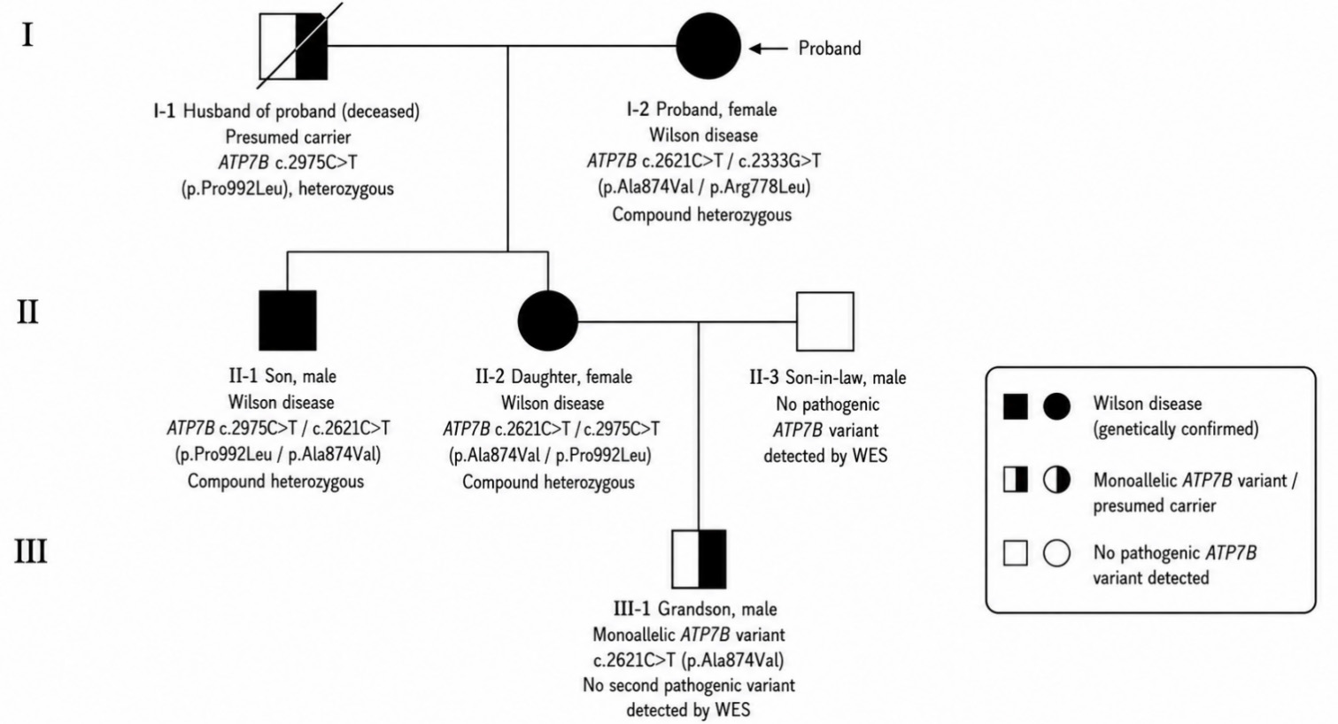
Figure 1. Pedigree of the family. Filled symbols indicate genetically confirmed Wilson disease; half-filled symbols indicate monoallelic ATP7B variant or presumed carrier status; open symbols indicate no pathogenic ATP7B variant detected by WES. The arrow indicates the proband. WES: Whole-exome sequencing; ATPase: adenosine triphosphatase; ATP7B: ATPase copper transporting beta.
Proband (I-2)
The proband (I-2) was a 67-year-old Han Chinese woman from a non-consanguineous family. She was diagnosed with hepatic WD in 2013 at 54 years of age after presenting with abdominal distension. At diagnosis, she had cirrhosis, splenomegaly, portal-vein widening, Kayser-Fleischer (K-F) rings, and a serum ceruloplasmin concentration of 0.03 g/L, without ascites or neurological signs. She improved after approximately 2 weeks of intravenous sodium dimercaptopropanesulfonate and subsequently received penicillamine 750 mg/day, zinc, and dimercaptosuccinic acid but discontinued treatment after 8 months. In March 2026, she returned with decompensated cirrhosis, ascites, esophageal varices, albumin 30.7 g/L, platelets 86 × 109/L, and a 24-h urinary copper of 186.1 μg/24 h before chelation therapy was restarted. Ascites resolved after diuretics, penicillamine 500 mg/day, and two further courses of intravenous sodium dimercaptopropanesulfonate, each consisting of 500 mg/day for 5 days. Her retrospective Leipzig score was 10[7].
Son of the proband (II-1)
The proband’s son (II-1) was a 36-year-old man who was asymptomatic when diagnosed with hepatic WD in 2013 at 23 years of age. At diagnosis, he had splenomegaly and serum ceruloplasmin of 0.03 g/L but no K-F rings or neurological signs. Cirrhosis and portal hypertension were documented in 2016, and intermittent hand tremor developed in 2024. He had previously taken penicillamine and zinc gluconate intermittently, with poor treatment adherence. Before a March 2026 urine collection, he self-administered penicillamine 250 mg/day for 2 weeks; 24-h urinary copper was 3,726.6 μg/24 h and was not used for diagnostic Leipzig scoring because it was treatment-exposed. In May 2026, he had decompensated cirrhosis with small-volume ascites, esophageal and gastric varices, hypersplenism, and portal hypertensive gastropathy. During treatment in 2026, he received penicillamine 750 mg/day, elemental zinc 150 mg/day (as zinc gluconate), and two 5-day courses of intravenous sodium dimercaptopropanesulfonate at 500 mg/day. His tremor and biochemical abnormalities partially improved. His documented retrospective Leipzig subtotal was 7 after excluding treatment-exposed urinary copper, which still supported an established diagnosis.
Daughter of the proband (II-2)
The proband’s daughter (II-2) was a 46-year-old woman diagnosed with predominantly hepatic WD in 2009 at 29 years of age after abdominal symptoms, anorexia, tremor, and headache. K-F rings were repeatedly absent, and brain magnetic resonance imaging (MRI) in 2020 was normal. Serial imaging showed cirrhosis, splenomegaly, portal hypertension, and gastric fundal varices. Progressive hypersplenism led to total splenectomy with pericardial devascularization in December 2025, after which the platelet count increased from 51 × 109/L to more than 290 × 109/L. In 2026, serum ceruloplasmin was 0.03 g/L. The 24-h urinary copper was 1,670.1 μg/24 h on March 21 before the 2026 chelation course was started and 1,887.7 μg/24 h on July 1 during ongoing penicillamine 750 mg/day. The pretreatment March value was used for retrospective Leipzig scoring, giving a total score of 9. Earlier intermittent penicillamine treatment had generally been 500 mg/day, with poor adherence. Abdominal distension improved after treatment intensification to 750 mg/day in 2026, whereas intermittent tremor and headache persisted.
Grandson of the proband (III-1)
The proband’s grandson (III-1) was a 22-year-old man who had never developed clear hepatic or neurological symptoms. Approximately 10 years earlier, before chelation therapy, serum ceruloplasmin was 0.13 g/L and 24-h urinary copper was 103 μg/24 h; together with a heterozygous ATP7B finding, these results led to a clinical diagnosis of WD and initiation of penicillamine. At recent reassessment, he remained clinically asymptomatic; K-F rings were absent, ultrasound showed fatty liver, serum ceruloplasmin was 0.143 g/L, and 24-h urinary copper had decreased to 24.6 μg/24 h during ongoing penicillamine 375 mg/day. No clinical symptomatic response could be assessed because he was asymptomatic, and the on-treatment fall in urinary copper was not considered diagnostic evidence. However, liver biochemistry was assessed on two consecutive occasions, with mild elevations of both alanine aminotransferase (ALT) (90-98 U/L) and aspartate aminotransferase (AST) (41-47 U/L). WES identified only monoallelic c.2621C>T (p.Ala874Val), with no second pathogenic ATP7B sequence variant or phenotype-related copy-number variant (CNV). Using the historical pretreatment values, his retrospective Leipzig score was 3: ceruloplasmin 0.13 g/L, 1 point; urinary copper 103 μg/24 h [1.72 × the upper limit of normal (ULN) of 60 μg/24 h], 1 point; and one pathogenic ATP7B variant, 1 point. This corresponds to a possible diagnosis requiring additional evidence; his molecular diagnosis therefore remains unresolved.
Genetic testing methods
Genomic DNA from the five tested family members was analyzed by phenotype-driven WES. Hybrid-capture libraries covered the coding exons of approximately 20,700 genes and 20-bp flanking intronic regions, together with the mitochondrial genome. Sequence reads were aligned to GRCh37/hg19 and analyzed using ISOgenetic v1.2. ATP7B variants were described using transcript NM_000053.4 and classified according to the 2015 American College of Medical Genetics and Genomics/Association for Molecular Pathology (ACMG/AMP) framework[8]. WES-based CNV analysis assessed deletions or duplications spanning at least two adjacent exons; lower-confidence findings required orthogonal confirmation by quantitative polymerase chain reaction or multiplex ligation-dependent probe amplification (MLPA) in accordance with laboratory procedures and CNV interpretation standards[9]. No phenotype-related CNV was reported in any tested individual.
Overall target-region mean sequencing depth and coverage at ≥ 20× were 167.84×/98.22% for the proband, 170.35×/98.26% for the son, 160.08×/98.17% for the daughter, 159.49×/98.31% for the grandson, and 195.60×/98.41% for the son-in-law. The capture design included all ATP7B coding exons and adjacent ±20-bp intronic sequence. Sanger sequencing was used for orthogonal confirmation and segregation analysis of c.2333G>T, c.2621C>T, and c.2975C>T [Figure 2].
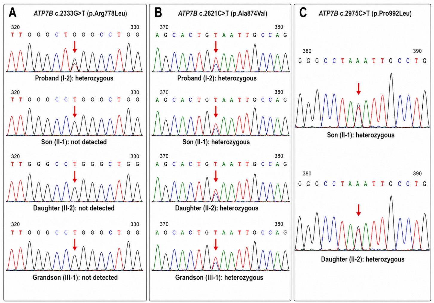
Figure 2. Sanger sequencing validation of ATP7B variants in the family. (A) ATP7B c.2333G>T (p.Arg778Leu) was heterozygous in the proband (I-2) but was not detected in her son (II-1), daughter (II-2) or grandson (III-1); (B) ATP7B c.2621C>T (p.Ala874Val) was heterozygous in the proband, son, daughter, and grandson; (C) ATP7B c.2975C>T (p.Pro992Leu) was heterozygous in the son and daughter. Red arrows indicate the variant sites. ATPase: Adenosine triphosphatase; ATP7B: ATPase copper transporting beta.
Genetic findings and segregation analysis
The proband carried ATP7B c.2621C>T (p.Ala874Val) in exon 11 and c.2333G>T (p.Arg778Leu) in exon 8. Sanger segregation analysis showed transmission of c.2621C>T without c.2333G>T to her children, thereby demonstrating that the two variants in the proband were in trans. The son and daughter each carried c.2621C>T (p.Ala874Val) and c.2975C>T (p.Pro992Leu). Their trans configuration was inferred rather than directly demonstrated: c.2621C>T was maternally transmitted, whereas c.2975C>T was absent from the proband and present in both siblings, strongly supporting inheritance from the deceased father, who was unavailable for testing and was therefore classified as a presumed heterozygous carrier.
The son-in-law had no pathogenic ATP7B sequence variant or phenotype-related CNV detected by WES. The grandson carried only the maternal c.2621C>T (p.Ala874Val) variant. Thus, current molecular findings establish biallelic ATP7B-related WD in the proband and her two children but not in the grandson.
All three reported ATP7B variants were classified as pathogenic by the diagnostic laboratory under ACMG/AMP criteria. For c.2621C>T (p.Ala874Val), the applied evidence included PM3_VeryStrong, PP4, PS3_Supporting, PM2_Supporting, and PP3; functional work has demonstrated markedly impaired copper transport[10]. For c.2333G>T (p.Arg778Leu), evidence included PM3_VeryStrong, PP4, PS3_Moderate, PM5_Strong, PM2_Supporting, and PP3. For c.2975C>T (p.Pro992Leu), evidence included PM3_VeryStrong, PS3_Moderate, PM2_Supporting, and PP3, with PP4 additionally applicable in an affected family member. Functional studies have shown that p.Arg778Leu and p.Pro992Leu impair ATP7B localization, trafficking, and/or copper excretion[11]. ClinVar was broadly concordant with these laboratory interpretations, although isolated discordant submissions remained at the time of access (28 September 2026). For c.2621C>T (p.Ala874Val), 18 contributing submissions classified the variant as pathogenic, 4 as likely pathogenic, and 1 as a variant of uncertain significance (VUS)[12]. For c.2333G>T (p.Arg778Leu), 23 classified it as pathogenic and 1 as VUS[13]. For c.2975C>T (p.Pro992Leu), 15 classified it as pathogenic, 1 as likely pathogenic, and 1 as VUS[14]. Accordingly, the current aggregate ClinVar records are formally labeled as having conflicting classifications, but the large majority of contributing submissions support pathogenic or likely pathogenic interpretations.
DISCUSSION
This family illustrates a genetically supported pseudo-dominant pattern of WD and, equally importantly, the limits of extending that label to an unresolved third-generation relative. The proband had two pathogenic variants demonstrated in trans. Both affected children inherited c.2621C>T from their mother and carried c.2975C>T, which was absent from the mother and therefore most plausibly paternal. Because the father was deceased, his carrier status and the trans phase in the children were inferred from segregation rather than directly confirmed. This distinction is important because detection of two variants alone does not establish phase.
Pseudo-dominant WD has been described previously using the same inheritance concept applied here: an autosomal recessive disease appears in successive generations because an affected individual reproduces with a heterozygous carrier[5,6,15]. Bennett et al. reported three consecutive affected generations, including a third-generation child with molecularly confirmed compound heterozygosity after carrier testing of the reproductive partner[5]. Park et al. likewise used the term pseudo-dominant inheritance for WD occurring in consecutive generations[6]. In a larger series, Dziezyc et al. identified subsequent-generation WD in nine non-consanguineous families[15]. Our definition is therefore consistent with previous reports, but we apply it only to the transmission from I-2 to the genetically confirmed affected siblings II-1 and II-2; III-1 is not counted as a third affected generation because biallelic disease has not been established.
The grandson demonstrates the need to reconcile clinical and molecular evidence and illustrates the limits of interpreting a borderline Leipzig score in isolation. A single heterozygous pathogenic ATP7B variant does not establish the molecular diagnosis of an autosomal recessive disorder[3]. Mild abnormalities in copper indices can occur in heterozygous carriers: a family-screening study showed biochemical overlap between patients with WD and carriers, with carrier median ceruloplasmin 0.18 g/L and median 24-h urinary copper 38.3 μg/24 h, and another carrier series documented mild copper-metabolism abnormalities in heterozygotes[16,17]. For III-1, ceruloplasmin 0.13 g/L and pretreatment urinary copper 103 μg/24 h (1.72 × ULN) each contribute 1 point, and the single pathogenic ATP7B variant contributes 1 point, giving a retrospective Leipzig score of 3. Under the Leipzig system, this indicates a possible diagnosis for which additional evidence is required rather than an established diagnosis. In a heterozygous carrier, modest reductions in ceruloplasmin and mild increases in urinary copper may therefore produce a borderline score, underscoring the need to integrate molecular and clinical findings. Current testing showed only the maternally inherited p.Ala874Val variant; K-F rings were absent, and his father had no pathogenic ATP7B variant detected by WES. These findings are more consistent with monoallelic carrier status than with genetically confirmed WD. Nevertheless, WES does not reliably exclude deep intronic, regulatory, or complex structural variants, and expanded testing beyond WES may be considered if clinical suspicion remains high.
The pedigree has direct genetic-counseling implications. If the carrier frequency in the reproductive population is q, an affected individual mating at random has an approximate q/2 probability that any given offspring will have WD, because an affected × carrier mating transmits the second pathogenic allele to half of the offspring. Using the traditional carrier-frequency estimate of approximately 1/90 gives an affected-offspring probability of about 1/180. Importantly, a 2026 systematic review estimated an ATP7B carrier rate of 2.51% in the Chinese Han population, which under the same simplifying assumptions corresponds to an affected-offspring probability of approximately 1.26% (about 1 in 80)[18]. These calculations are population-level approximations and do not replace partner-specific testing. Accordingly, cascade screening in WD should include reproductive partners and offspring, with targeted testing for known familial variants whenever possible[3]. In this family, the proband’s mating pattern explains the affected offspring in the second generation, whereas the grandson’s current genotype is the expected monoallelic finding from an affected mother whose partner had no detectable ATP7B pathogenic variant by WES.
The clinical histories also show the consequences of interrupted therapy, but the principal contribution of this report is diagnostic: pseudo-dominant inheritance should be assigned only where biallelic disease and phase are adequately supported, and an unresolved monoallelic relative should not be counted as a genetically confirmed third affected generation.
The main limitations are the absence of direct testing of the proband’s deceased husband, the lack of ATP7B-specific exon-level coverage metrics, and incomplete historical evaluation of the grandson. No family member underwent hepatic copper quantification. If the grandson’s diagnosis remains clinically important, expanded genetic testing beyond the WES already performed in the grandson and his father could include ATP7B-targeted deletion/duplication testing (e.g., MLPA), whole-genome sequencing, long-read sequencing, and/or RNA analysis to search for variants outside the reliable detection range of WES.
CONCLUSION
We describe a Chinese family with pseudo-dominant WD across two genetically confirmed generations. Segregation demonstrated that the proband’s two ATP7B variants were in trans, whereas the paternal origin and trans phase of p.Pro992Leu in her children were inferred because their father was deceased and untested. The grandson carried only monoallelic p.Ala874Val and had a retrospective Leipzig score of 3, corresponding to a possible diagnosis requiring additional evidence; his molecular diagnosis remains unresolved. This family emphasizes rigorous phase interpretation, partner and offspring screening, and restraint when clinical and molecular diagnoses are discordant.
DECLARATIONS
Authors’ contributions
Collected clinical and genetic data and drafted the manuscript: Mi W, Yuan Z
Conducted patient follow-up: Zhi J
Contributed to clinical interpretation and data verification: Zheng S
Conceived the report, supervised genetic interpretation, and critically revised the manuscript: Hou W
All authors read and approved the final manuscript.
Availability of data and materials
The data supporting the findings of this case report are available from the corresponding author upon reasonable request and with permission from the treating institution.
AI and AI-assisted tools statement
During the preparation of this manuscript, the AI tool ChatGPT (version GPT-5.6 Sol, released 2026-07-09) was used solely for language editing. The tool did not influence the study design, data collection, analysis, interpretation, or the scientific content of the work. All authors take full responsibility for the accuracy, integrity, and final content of the manuscript.
Financial support and sponsorship
This work was supported by the Beijing Hospitals Authority’s Ascent Plan (DFL20241701).
Conflicts of interest
All authors declared that there are no conflicts of interest.
Ethics approval and consent to participate
This case report was approved by the Ethics Committee of Beijing Youan Hospital, Capital Medical University {approval number: JYKLZ [2025] No. 140}. The study was conducted in accordance with the Declaration of Helsinki. Written informed consent was obtained from the family members included in this report. For the deceased family member, relevant family information was used with consent from next of kin.
Consent for publication
Written informed consent for publication was obtained from the family members included in this report.
Copyright
© The Author(s) 2026.
REFERENCES
1. Schilsky ML, Roberts EA, Bronstein JM, et al. A multidisciplinary approach to the diagnosis and management of Wilson disease: executive summary of the 2022 Practice Guidance on Wilson disease from the American Association for the Study of Liver Diseases. Hepatology. 2023;77:1428-55.
2. Association for the Study of the Liver. EASL-ERN clinical practice guidelines on Wilson’s disease. J Hepatol. 2025;82:690-728.
3. Weiss KH, Schilsky M. Wilson Disease. In: Adam MP, Bick S, Mirzaa GM, et al., editors. GeneReviews. Seattle (WA): University of Washington, Seattle; 1993-2026. Available from: https://www.ncbi.nlm.nih.gov/books/NBK1116/ [Last accessed on 23 Oct 2026].
4. Shribman S, Marjot T, Sharif A, et al.; British Association for the Study of the Liver Rare Diseases Special Interest Group. Investigation and management of Wilson’s disease: a practical guide from the British Association for the Study of the Liver. Lancet Gastroenterol Hepatol. 2022;7:560-75.
5. Bennett JT, Schwarz KB, Swanson PD, Hahn SH. An exceptional family with three consecutive generations affected by Wilson disease. JIMD Rep. 2013;10:1-4.
6. Park H, Park DK, Kim MS, Yoon JH. Pseudo-dominant inheritance in Wilson’s disease. Neurol Sci. 2016;37:153-5.
7. Ferenci P, Caca K, Loudianos G, et al. Diagnosis and phenotypic classification of Wilson disease. Liver Int. 2003;23:139-42.
8. Richards S, Aziz N, Bale S, et al.; ACMG Laboratory Quality Assurance Committee. Standards and guidelines for the interpretation of sequence variants: a joint consensus recommendation of the American College of Medical Genetics and Genomics and the Association for Molecular Pathology. Genet Med. 2015;17:405-24.
9. Riggs ER, Andersen EF, Cherry AM, et al. Technical standards for the interpretation and reporting of constitutional copy-number variants: a joint consensus recommendation of the American College of Medical Genetics and Genomics (ACMG) and the Clinical Genome Resource (ClinGen). Genet Med. 2020;22:245-57.
10. Schushan M, Bhattacharjee A, Ben-Tal N, Lutsenko S. A structural model of the copper ATPase ATP7B to facilitate analysis of Wilson disease-causing mutations and studies of the transport mechanism. Metallomics. 2012;4:669-78.
11. Zhu M, Dong Y, Ni W, Wu ZY. Defective roles of ATP7B missense mutations in cellular copper tolerance and copper excretion. Mol Cell Neurosci. 2015;67:31-6.
12. National Center for Biotechnology Information. ClinVar; NM_000053.4(ATP7B):c.2621C>T (p.Ala874Val), VCV000003851.82 [Internet]. Available from: https://www.ncbi.nlm.nih.gov/clinvar/variation/VCV000003851.82/ [Last accessed on 21 Oct 2026].
13. National Center for Biotechnology Information. ClinVar; NM_000053.4(ATP7B):c.2333G>T (p.Arg778Leu), VCV000003852.65 [Internet]. Available from: https://www.ncbi.nlm.nih.gov/clinvar/variation/VCV000003852.65/ [Last accessed on 21 Oct 2026].
14. National Center for Biotechnology Information. ClinVar; NM_000053.4(ATP7B):c.2975C>T (p.Pro992Leu), VCV000188831.50 [Internet]. Available from: https://www.ncbi.nlm.nih.gov/clinvar/variation/VCV000188831.50/ [Last accessed on 21 Oct 2026].
15. Dzieżyc K, Litwin T, Chabik G, Gramza K, Członkowska A. Families with Wilson’s disease in subsequent generations: clinical and genetic analysis. Mov Disord. 2014;29:1828-32.
16. Li H, Liu L, Li Y, et al. Familial screening of children with Wilson disease: necessity of screening in previous generation and screening methods. Medicine. 2018;97:e11405.
17. Zhou XX, He RX, Pu XY, et al. Clinical characteristics of the Wilson disease carrier. Natl Med J China. 2019;99:806-11.
Cite This Article
 Open Access
How to Cite
Mi W, Yuan Z, Zhi J, Zheng S, Hou W. Pseudo-dominant Wilson disease with an unresolved third-generation ATP7B finding. Rare Dis Orphan Drugs J. 2026;5:32. https://dx.doi.org/10.20517/rdodj.2026.46
Download Citation
If you have the appropriate software installed, you can download article citation data to the citation manager of your choice. Simply select your manager software from the list below and click on download.
Export Citation File
Type of Import
Tips on Downloading Citation
Citation Manager File Format
Type of Import
Direct Import: When the Direct Import option is selected (the default state), a dialogue box will give you the option to Save or Open the downloaded citation data. Choosing Open will either launch your citation manager or give you a choice of applications with which to use the metadata. The Save option saves the file locally for later use.
Indirect Import: When the Indirect Import option is selected, the metadata is displayed and may be copied and pasted as needed.
About This Article
Copyright

Data & Comments
Data
0









Comments
Comments must be written in English. Spam, offensive content, impersonation, and private information will not be permitted. If any comment is reported and identified as inappropriate content by OAE staff, the comment will be removed without notice. If you have any queries or need any help, please contact us at [email protected].