

Microglia replacement in CSF1R-associated microgliopathy: from preclinical rationale to emerging clinical evidence
 0


 Download PDF
Colony-stimulating factor 1 receptor (CSF1R)-associated microgliopathy (CAMP), a major genetic cause of adult-onset leukoencephalopathy with axonal spheroids and pigmented glia (ALSP), represents one of the most devastating yet underrecognized entities among neurodegenerative leukodystrophies. In the literature, genetically confirmed disease has also been described as hereditary diffuse leukoencephalopathy with spheroids (HDLS), CSF1R-related leukoencephalopathy, or CSF1R-ALSP; these terms partly reflect historical and clinicopathological nomenclature. Patients typically present in the fourth or fifth decade of life with progressive cognitive decline, neuropsychiatric disturbances, motor dysfunction, and seizures. The discovery that heterozygous mutations in CSF1R - encoding the tyrosine kinase receptor essential for microglial proliferation, survival, and differentiation - cause HDLS established a direct link between CSF1R dysfunction and a primary microglial disorder[1]. This genetic insight supported framing ALSP/HDLS as a microgliopathy and shifted attention toward microglia as a therapeutic target. Despite this mechanistic rationale, no disease-modifying therapy has yet been established in controlled clinical studies, and clinical management remains largely symptomatic and supportive. The concept of microglia replacement - exchanging dysfunctional, genetically defective microglia for healthy counterparts - has long been proposed as a logical strategy, given that the disease is cell-autonomous and the causal gene is expressed predominantly in microglia. However, this approach has been constrained by a biological barrier: the adult central nervous system (CNS) has historically been considered relatively refractory to sustained engraftment of exogenous microglia-like cells. Early preclinical work established efficient microglia replacement protocols, including microglia replacement by bone marrow transplantation (Mr BMT), yet clinical translation remained uncertain[2]. A key question has therefore been whether modifying the microglial compartment can meaningfully alter disease progression in patients. The study by Wu et al.[3] addresses this question through a translational trajectory from engineered mouse models to an eight-patient clinical cohort.
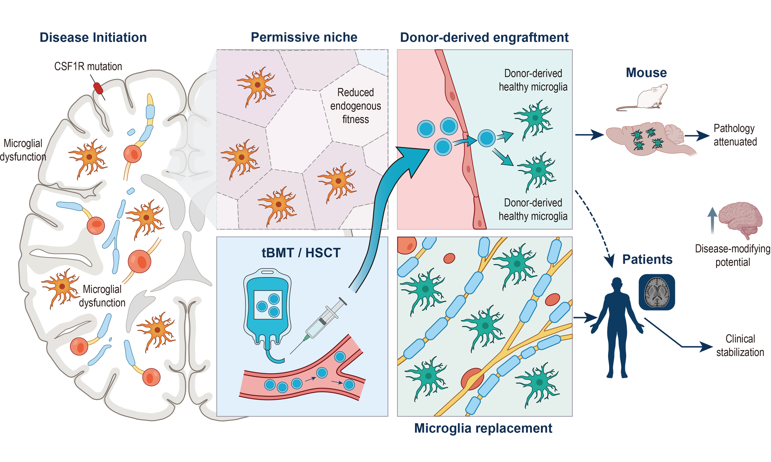
Wu et al.[3] designed a stepwise translational study. They first generated knock-in mouse models harboring human CAMP hotspot mutations in Csf1r (the mouse ortholog of human CSF1R p.I794T), which recapitulated key disease features including ventricular enlargement, axonal spheroids, demyelination, microglial loss, and motor deficits. Using these models, the authors demonstrated that Mr BMT - replacing CSF1R-deficient resident microglia with CSF1R-normal donor-derived cells in mice - significantly attenuated neuropathological hallmarks and functional decline. A key finding emerged from a direct comparison between the optimized Mr BMT protocol and traditional bone marrow transplantation (tBMT). tBMT is generally inefficient at repopulating the adult CNS parenchyma with donor-derived microglia-like cells when the endogenous niche is intact. In the Csf1r-mutant mouse models, however, Wu et al.[3] reported that 84.08% of microglia were replaced by green fluorescent protein (GFP)-positive donor-derived cells after tBMT, a level comparable to the approximately 91.15% replacement achieved with Mr BMT in the reported experiments, with similar therapeutic effects. These animal data support the interpretation that CSF1R deficiency reduces the competitiveness of endogenous microglia and creates a more permissive niche for donor-derived myeloid-cell engraftment. Building on this preclinical insight, the authors evaluated tBMT in eight patients with ALSP. Over 24 months, treatment was associated with stabilization of clinical and neuroimaging measures[3]. These observations extend a growing clinical literature in which hematopoietic stem cell transplantation (HSCT)/tBMT had already been explored in individual cases and small cohorts of CSF1R-related disease[4,5]. Importantly, Wu et al.[3] did not directly demonstrate lineage-defined microglial replacement in the human brain: the reported clinical, magnetic resonance imaging (MRI), and positron emission tomography (PET) outcomes are not microglia-specific, and the precise identity of donor-derived CNS-engrafting cells in patients remains inferential. Thus, the human findings are consistent with a microglia-replacement mechanism supported by the mouse experiments, but they should not be described as direct proof of microglial replacement in patients. The convergence of murine mechanistic data and human clinical stabilization therefore strengthens the rationale for tBMT as a potential disease-modifying strategy while leaving the cellular mechanism in humans to be established.
The significance of these findings extends beyond the immediate context of CAMP/ALSP, although the implications should be framed cautiously. At its core, this study provides a clinically relevant proof of concept: correcting a genetically defective microglial compartment may stabilize a progressive neurodegenerative disease. This differs from many prevailing therapeutic approaches in neurodegeneration, which primarily aim to slow progression or modulate downstream pathology rather than address a primary cellular defect. The finding in the mouse models that Csf1r deficiency creates a permissive niche for donor-derived microglia-like engraftment carries implications for neuroimmunology. It supports a view of the brain’s immune niche as a dynamic, disease-responsive environment rather than a uniformly static barrier to cellular replacement. The traditional view held that the CNS parenchyma resists exogenous microglial colonization; the present mouse data suggest that this resistance is influenced by microglial fitness and abundance, and that CSF1R deficiency can open a therapeutic window. This observation suggests that the efficiency of cell replacement therapy in the CNS may depend not only on the delivery method but also on the receptivity of the recipient niche - a principle that may be relevant to other disease contexts characterized by a primary microglial defect. However, the findings do not imply that all disorders with secondary microglial activation should be reclassified as microgliopathies. Recent studies have demonstrated other microglia replacement strategies as promising therapeutic approaches for CAMP. Chadarevian et al.[6] transplanted human induced pluripotent stem cell (iPSC)-derived microglia, including clustered regularly interspaced short palindromic repeats (CRISPR)-corrected autologous cells, into xenotolerant mice lacking the fms-intronic regulatory element (FIRE) enhancer, preventing and even reversing established ALSP-like pathology, although intracranial delivery limits clinical translation. Li et al.[7] developed duplicate-cyclic microglial depletion for transplantation (DCMDT), ameliorating microgliopathy in Csf1rI792T/+ mice, an independently generated knock-in model carrying the same hotspot mutation as Wu et al. Compared with these preclinical strategies, which differ in cell source, delivery route, and conditioning requirements, the distinctive contribution of Wu et al.[3] is clinical validation: the integration of disease-relevant knock-in models, direct comparison of Mr BMT with tBMT, and longitudinal observation of an eight-patient cohort within one translational framework. Furthermore, because tBMT is a well-established clinical procedure, it leverages existing transplant infrastructure and may facilitate clinical evaluation relative to entirely novel engineered cell products[3]. However, conditioning toxicity, infection, graft-versus-host disease, early neurological worsening, and patient selection remain important limitations. This practical advantage, combined with the biological insight that disease pathology may facilitate engraftment, positions tBMT as a candidate disease-modifying strategy that warrants further evaluation in CAMP and related primary microglial disorders.
Despite the importance of these findings, two critical issues warrant focused investigation. First, the long-term durability of the clinical benefit beyond 24 months remains uncertain. While clinical stabilization at 24 months is encouraging, the persistence of benefit and the long-term fate of donor-derived CNS-engrafting cells require longer observation. A complementary international cohort study of HSCT in CSF1R-related disorder reported an initial clinical and radiological deterioration in the first six months post-transplantation before subsequent stabilization, and observed that reduced-intensity conditioning regimens yielded neurological outcomes comparable to myeloablative protocols[8]. Together with earlier case reports and small series, these findings indicate that outcomes can be heterogeneous and that transplant-related morbidity must be weighed against the possibility of stabilization. Longitudinal studies with multi-year follow-up incorporating serial neurofilament light chain measurements, quantitative diffusion tensor imaging, standardized cognitive and motor assessments, and systematic recording of transplant-related adverse events are essential to define durability and safety. Future prospective multicenter studies should enroll genetically confirmed patients before advanced irreversible disability when feasible, stratify participants by baseline disease burden and rate of progression, standardize conditioning and graft parameters where possible, and use natural-history or matched comparator data when randomization is not practical. Earlier intervention may be biologically advantageous because established axonal loss is unlikely to be reversed, but the optimal treatment window remains to be defined prospectively. Second, the molecular mechanism by which CSF1R-deficient microglia create a permissive niche for donor engraftment remains incompletely understood. The niche may be generated by active microglial degeneration and physical niche emptying, altered chemokine or trophic signaling that recruits donor cells, changes in blood-brain barrier integrity, or a combination of these processes. Elucidating this mechanism through single-cell transcriptomic profiling of the niche microenvironment at serial post-transplantation time points, combined with targeted perturbation of candidate signaling pathways, could advance biological understanding and identify biomarkers predictive of engraftment efficiency. In human studies, direct lineage tracing is not feasible; therefore, convergent measures such as donor chimerism in accessible compartments, serial biomarkers, advanced imaging, and, where ethically available, neuropathological or single-cell analyses may help distinguish cellular engraftment from nonspecific transplant effects. Notably, emerging alternative approaches - such as peripheral delivery of CSF1R inhibitor-resistant hematopoietic cells carrying the G795A variant - offer complementary strategies designed to improve CNS myeloid replacement while potentially reducing conditioning-related toxicity[9]. Overall, the work of Wu et al.[3] provides a mechanistically informative step toward clinical translation, but larger cohorts, longer follow-up, clearer cellular biomarkers, and prospective evaluation of treatment timing are still required before tBMT-mediated microglial replacement can be considered an established clinical therapy.
DECLARATIONS
Authors’ contributions
Made substantial contributions to the conception and design of the manuscript and performed data analysis and interpretation: Yang Q, Wang Y
Contributed to the writing of the manuscript: Wang Y, Yang Q, Tong J
All authors contributed to the review and editing of the manuscript.
Availability of data and materials
Not applicable.
AI and AI-assisted tools statement
During the preparation of this manuscript, the AI tool ChatGPT (version GPT-5.4, released 2026-03-05) was used solely for language editing. The tool did not influence the study design, data collection, analysis, interpretation, or the scientific content of the work. All authors take full responsibility for the accuracy, integrity, and final content of the manuscript.
Financial support and sponsorship
This work was supported by the National Natural Science Foundation of China (No. 82272925) and Shanghai Jiao Tong University School of Medicine Research Physician Program (No. 20240815).
Conflicts of interest
Yang Q is a Junior Editorial Board Member of Rare Disease and Orphan Drugs Journal. Yang Q was not involved in any steps of editorial processing, notably including reviewers’ selection, manuscript handling, or decision-making. The other authors declare that there are no conflicts of interest.
Ethical approval and consent to participate
Not applicable.
Consent for publication
Not applicable.
Copyright
© The Author(s) 2026.
REFERENCES
1. Rademakers R, Baker M, Nicholson AM, et al. Mutations in the colony stimulating factor 1 receptor (CSF1R) gene cause hereditary diffuse leukoencephalopathy with spheroids. Nat Genet. 2012;44:200-5.
2. Xu Z, Rao Y, Huang Y, et al. Efficient strategies for microglia replacement in the central nervous system. Cell Rep. 2020;32:108041.
3. Wu J, Wang Y, Li X, et al. Microglia replacement halts the progression of microgliopathy in mice and humans. Science. 2025;389:eadr1015.
4. Bergner CG, Schäfer L, Vucinic V, et al. Case report: treatment of advanced CSF1-receptor associated leukoencephalopathy with hematopoietic stem cell transplant. Front Neurol. 2023;14:1163107.
5. Dulski J, Heckman MG, White LJ, Żur-Wyrozumska K, Lund TC, Wszolek ZK. Hematopoietic stem cell transplantation in CSF1R-related leukoencephalopathy: retrospective study on predictors of outcomes. Pharmaceutics. 2022;14:2778.
6. Chadarevian JP, Hasselmann J, Lahian A, et al. Therapeutic potential of human microglia transplantation in a chimeric model of CSF1R-related leukoencephalopathy. Neuron. 2024;112:2686-2707.e8.
7. Li X, Hu B, Wu C, et al. A strategy of microglia replacement alleviates microgliopathy in a CSF1R I794T hotspot mutation mouse model of CSF1R-related disorder. Cell Rep Med. 2026;7:102641.
8. Yska HAF, Golse M, Beerepoot S, et al.; and the International CSF1R-RD Working Group. Hematopoietic stem cell transplantation in an international cohort of colony stimulating factor-1 receptor (CSF1R)-related disorder. Mov Disord. 2025;40:1826-35.
Cite This Article
 Open Access
How to Cite
Wang Y, Tong J, Yang Q. Microglia replacement in CSF1R-associated microgliopathy: from preclinical rationale to emerging clinical evidence. Rare Dis Orphan Drugs J. 2026;5:31. https://dx.doi.org/10.20517/rdodj.2026.42
Download Citation
If you have the appropriate software installed, you can download article citation data to the citation manager of your choice. Simply select your manager software from the list below and click on download.
Export Citation File
Type of Import
Tips on Downloading Citation
Citation Manager File Format
Type of Import
Direct Import: When the Direct Import option is selected (the default state), a dialogue box will give you the option to Save or Open the downloaded citation data. Choosing Open will either launch your citation manager or give you a choice of applications with which to use the metadata. The Save option saves the file locally for later use.
Indirect Import: When the Indirect Import option is selected, the metadata is displayed and may be copied and pasted as needed.
About This Article
Copyright

Data & Comments
Data
0










Comments
Comments must be written in English. Spam, offensive content, impersonation, and private information will not be permitted. If any comment is reported and identified as inappropriate content by OAE staff, the comment will be removed without notice. If you have any queries or need any help, please contact us at [email protected].