

Genetic defects in programmed meiotic DNA double-strand break formation and repair in human infertility
 0
Graphical Abstract
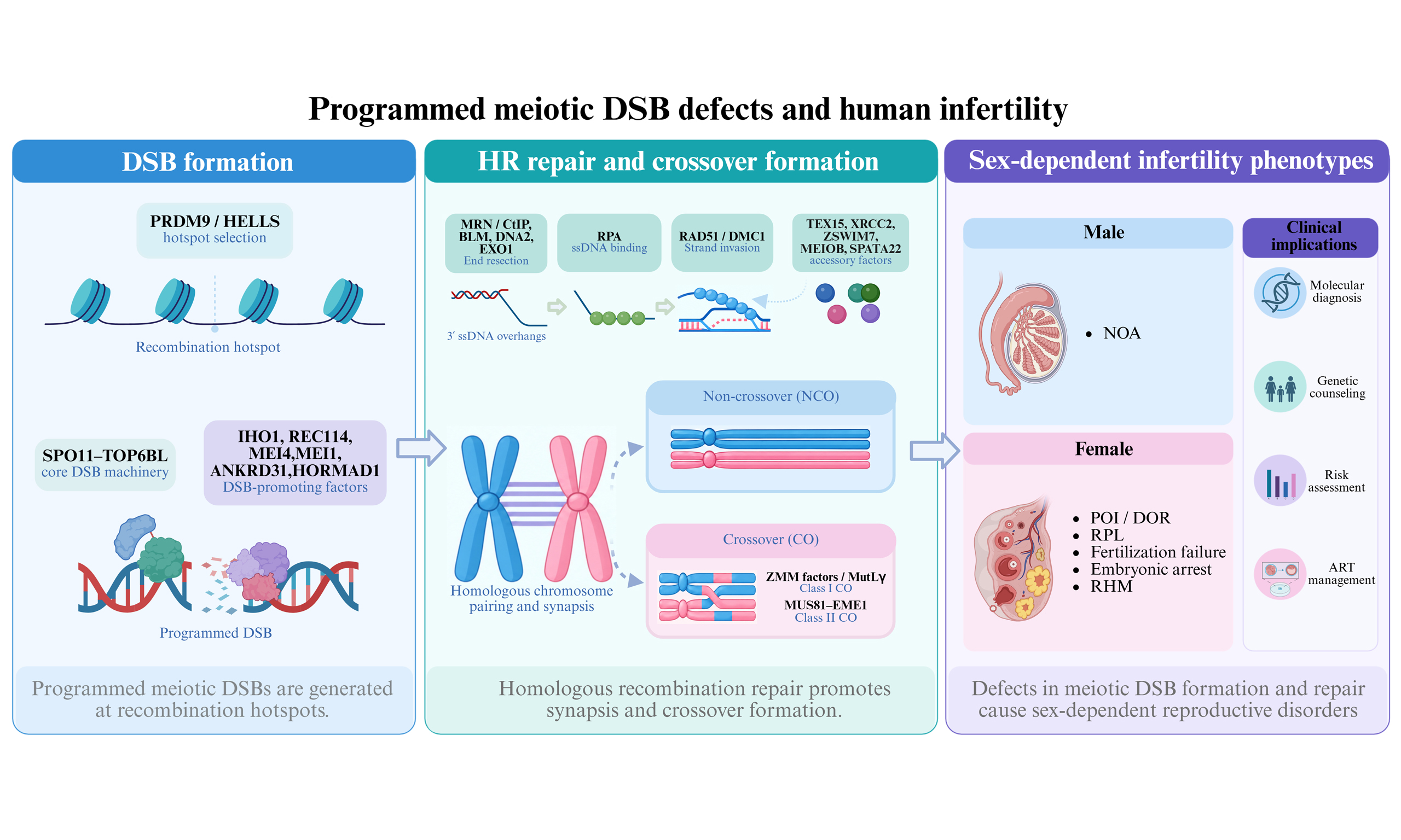
Abstract
In meiosis, the occurrence of programmed DNA double-strand breaks (DSBs) and their repair via homologous recombination are indispensable for homologous chromosome synapsis, crossover formation, and faithful chromosome segregation during gametogenesis. Over the past decade, advances in next-generation sequencing have expanded the spectrum of infertility-associated genes involved in meiotic DSB formation and repair, establishing this pathway as a major genetic determinant of human reproductive disorders. The disruption of these processes impairs homologous recombination and meiotic progression, leading to defective gametogenesis and a wide range of reproductive phenotypes. Clinically, genetic defects affecting meiotic DSB formation and repair have been associated with non-obstructive azoospermia in males, and a broader spectrum of reproductive phenotypes in females, such as premature ovarian insufficiency, recurrent pregnancy loss, fertilization failure, embryonic developmental arrest, recurrent hydatidiform moles, and poor outcomes following assisted reproductive technologies. This review summarizes current advances in genetic defects affecting programmed meiotic DSB formation and repair, with an emphasis on the molecular functions of disease-associated genes, evidence from animal models and genetic studies in humans, sex-dependent genotype-phenotype relationships, and their clinical relevance. Together, these advances provide a foundation for improving molecular diagnosis, genetic counseling, and precision reproductive medicine in patients with unexplained infertility.
Keywords


 Download PDF
INTRODUCTION
Infertility has emerged as a major global public health challenge, affecting approximately 17.5% of reproductive-aged couples worldwide[1]. Its etiology is highly heterogeneous, encompassing endocrine dysfunction, anatomical abnormalities, immunological disorders, environmental exposures, lifestyle factors, and genetic defects[2]. With the rapid advancement of next-generation sequencing (NGS) and other molecular diagnostic technologies, the contribution of genetic factors to infertility has become increasingly evident, with genetic abnormalities estimated to account for approximately 30%-50% of the cases[2,3].
Meiosis is a specialized cell division process that generates haploid gametes through a single round of DNA replication followed by two consecutive cell divisions. This process ensures the reduction of chromosome number by half and the faithful transmission of genetic information across generations[4]. A central molecular event during meiosis is homologous recombination (HR), which is initiated by programmed DNA double-strand breaks (DSBs)[5]. The formation, localization, and repair of meiotic DSBs are tightly regulated by a highly conserved network of proteins. Defects in these processes can disrupt meiotic progression, impair gametogenesis, and increase the risk of chromosomal abnormalities, ultimately leading to infertility, recurrent pregnancy loss, and embryonic developmental arrest[6].
Over the past decade, advances in human genetics and functional genomics have markedly expanded the landscape of infertility-associated genes involved in meiotic DSB formation and repair. Previous reviews have addressed important aspects of meiotic genetics, including general mechanisms of meiotic recombination or specific infertility phenotypes, whereas the focus on genes involved in programmed meiotic DSB formation and repair in human infertility has remained limited[7-9]. In this review, we summarize the current knowledge on infertility-associated genes involved in programmed meiotic DSB formation and repair, with particular emphasis on their molecular functions, pathogenic mechanisms, genotype-phenotype correlations, and translational implications for molecular diagnosis, genetic counseling, and precision reproductive medicine.
FORMATION AND REPAIR OF MEIOTIC PROGRAMMED DNA DOUBLE-STRAND BREAKS
Programmed DNA DSBs and their subsequent repair via homologous recombination are central events in meiosis that enable homologous chromosome pairing, synapsis, crossover formation, and accurate chromosome segregation [Figure 1][10].
Figure 1. Mechanistic Overview of Programmed Meiotic Double-Strand Breaks (DSBs) Formation, Repair, and Crossover Pathway Choice. Programmed meiotic DSBs are initiated at PRDM9-defined recombination hotspots through recruitment of the SPO11-TOP6BL complex by the pre-DSB recombinosome. Following DSB formation, SPO11-bound DNA ends are processed by the MRE11-RAD50-NBS1 (MRN) complex together with CtIP, generating 3’ single-stranded DNA (ssDNA) overhangs. The exposed ssDNA is initially bound by replication protein A (RPA), while the MEIOB-SPATA22 complex contributes to the stabilization and processing of recombination intermediates. RAD51 and DMC1 are subsequently loaded onto ssDNA and promote homology search and strand invasion into the homologous chromosome. Recombination intermediates may be repaired by synthesis-dependent strand annealing (SDSA), resulting in non-crossover products, or proceed through crossover pathways. Class I crossovers are promoted by ZMM-associated factors, including MSH4-MSH5, HFM1, RNF212, HEI10, and the SHOC1-TEX11-SPO16 complex, which stabilize recombination intermediates and facilitate their maturation into double Holliday junction (dHJ) structures. These intermediates are subsequently resolved primarily by the MutLγ complex composed of MLH1 and MLH3 to generate interference-sensitive class I crossover products. Class II crossovers arise through an alternative, interference-independent pathway involving MUS81-EME1, whereas GEN1 may also contribute to the resolution of meiotic recombination intermediates. The blue and red lines represent chromatids from homologous chromosomes, whereas colored symbols represent key meiotic proteins or protein complexes involved in each step. Solid arrows indicate the major progression of meiotic recombination, and dashed arrows indicate intermediate processing steps. The pathways shown are based largely on studies of mammalian meiosis, particularly in mouse models; direct experimental evidence in humans remains limited for some steps. ZMM: Zip–Mer–Msh proteins; ZZS: Zip2–Zip4–Spo16 complex; CtIP: CtBP-interacting protein. Created in BioRender. yi, S. (2026) https://BioRender.com/48i53yr
Formation of programmed DSBs
Programmed DSBs are generated during meiotic prophase I and serve as initiating events for homologous recombination[8]. Unlike accidental DNA damage, meiotic DSBs occur in defined genomic regions known as recombination hotspots and are tightly regulated both spatially and temporally. Hotspot selection is primarily determined by the coordinated activity of the meiosis-specific DNA-binding protein PRDM9 and chromatin remodeler helicase, lymphoid specific (HELLS)[11-13]. PRDM9 recognizes specific DNA motifs through its zinc-finger domain and catalyzes the trimethylation of histone H3 lysine 4 (H3K4me3) and lysine 36 (H3K36me3) in nearby nucleosomes. These epigenetic modifications designate potential recombination hotspots and create a chromatin environment conducive to DSB formation. Subsequently, HELLS and other chromatin-remodeling factors promote local chromatin opening, thereby increasing DNA accessibility and facilitating the recruitment of DSB-forming proteins[14].
DSB formation is directly catalyzed by the SPO11-TOP6BL complex, an evolutionarily conserved topoisomerase VI-like heterotetramer[15]. Precise targeting of this complex to recombination hotspots requires a specialized chromosome axis-associated pre-DSB recombinosome[16]. During meiotic prophase I, IHO1, REC114, and MEI4 assemble into a ternary complex with a 4:4:2 stoichiometry, which subsequently associates with MEI1 and ANKRD31 to form a functional pre-DSB complex. Through interactions between IHO1 and the chromosome-axis protein HORMAD1, this complex anchors the DSB-forming machinery to unsynapsed chromosome axes and promotes the recruitment of SPO11-TOP6BL to PRDM9-defined hotspots, thereby ensuring efficient and accurately positioned formation of DSBs[17,18]. Following recruitment, SPO11-TOP6BL catalyzes the site-specific cleavage of both DNA strands through a Mg2+-dependent, adenosine triphosphate (ATP)-independent transesterification reaction. During this process, SPO11 becomes covalently attached to the 5’ end of the cleaved DNA, generating protein-bound DSB intermediates that trigger meiotic homologous recombination[19,20].
Repair of programmed DSBs
Following DSB formation, high-fidelity repair pathways are rapidly activated to preserve genome integrity and stability. During meiosis prophase I, programmed DSBs generated by SPO11 are predominantly repaired through HR, whereas non-homologous end joining is largely suppressed[21].
DSB repair is initiated by the removal of SPO11-linked DNA ends and resection of DSB termini by the MRE11-RAD50-NBS1 (MRN) complex together with CtBP-interacting protein (CtIP), generating 3’ single-stranded DNA (ssDNA) overhangs. Long-range resection is subsequently extended by bloom syndrome helicase (BLM) helicase along with DNA2 and EXO1[22]. The resulting ssDNA is rapidly coated by replication protein A (RPA) and subsequently replaced by RAD51 and the meiosis-specific recombinase DMC1, with the assistance of mediator proteins including BRCA2, TEX15, MEILB2-BRME1, XRCC2, and ZSWIM7[23,24].
RAD51-DMC1 nucleoprotein filaments promote homology search and strand invasion into homologous chromosomes, generating displacement loop (D-loop) intermediates that serve as the central structures of meiotic recombination. Following repair DNA synthesis, these intermediates are processed via crossover (CO) or non-crossover (NCO) pathways[25]. Most recombination events are resolved as NCOs through synthesis-dependent strand annealing (SDSA) or dissolution by the bloom syndrome helicase-topoisomerase IIIα-RMI1–RMI2 complex (BTR) complex. In contrast, a subset of intermediates is stabilized by Zip-Mer-Msh (ZMM) proteins, including MSH4-MSH5, HFM1, RNF212, and HEI10, and matures into double Holliday junctions (dHJs). These crossover precursors are subsequently resolved by the MutLγ (MLH1-MLH3) complex to generate class I COs, whereas a smaller proportion of class II COs are generated through the MUS81-EME1-dependent pathway. GEN1 may also contribute to the resolution of meiotic recombination intermediates[26-28].
Molecular mechanisms linking aberrant DSB formation and repair to infertility
Defective DSB formation or repair can profoundly disrupt meiotic progression, leading to abnormalities in homologous chromosome synapsis, recombination, and segregation. These defects may result in aneuploid gametes or meiotic arrest, ultimately resulting in infertility. The underlying molecular mechanisms mainly include the following:
(1) Insufficient DSB formation: reduced SPO11 activity or dysfunction of key DSB- forming factors such as IHO1, REC114, and MEI1 may lead to a decreased number of DSBs. Consequently, homologous chromosomes fail to generate sufficient COs and chiasmata to maintain the proper bivalent structure and tension required for accurate chromosome segregation, resulting in chromosome mis-segregation and increased risk of aneuploidy[29].
(2) Aberrant DSB positioning: defects in PRDM9, components of the pre-DSB complex, or chromatin regulatory pathways can impair the spatial distribution of DSB hotspots. This may misdirect the SPO11-TOP6BL complex to inappropriate genomic loci, thereby increasing the risk of aberrant recombination and chromosomal structural abnormalities, such as translocations and insertions[15].
(3) Impaired DSB repair: defects in key recombination factors such as RAD51 and DMC1 compromise homologous recombination repair, thereby leading to persistent DNA breaks, defective synapses, and meiotic arrest. In addition, escape from meiotic checkpoint control may result in chromosome fragmentation or structural aberrations in gametes[30].
GENES INVOLVED IN DSB FORMATION AND REPAIR THAT ARE ASSOCIATED WITH HUMAN INFERTILITY
An increasing number of genes involved in DSB formation and repair have been implicated in human infertility[31]. Genetic variants in these genes may disrupt distinct stages of meiotic recombination, giving rise to a broad spectrum of reproductive phenotypes. Below, we summarize the genes implicated in human infertility according to their functions in meiotic DSB formation and repair. To reflect differences in the strength of available human genetic evidence, genes are further stratified according to the number of independent human families reported to date, with evidence levels assessed separately for male and female infertility [Table 1 and Supplementary Table 1].
Summary of genotype-phenotype associations for genes involved in programmed meiotic DSB formation and repair
| Gene | Molecular function | Mouse knockout phenotype | Males | Females | References | ||||
| Phenotype | Mode of inheritance | Evidence level & | Phenotype | Mode of inheritance | Evidence level & | ||||
| SPO11 | Catalyzes programmed meiotic DSB formation | Both-sex infertility; meiotic arrest/synapsis failure in males; postnatal oocyte loss in females | NOA | AR | Limited | Recurrent blastulation failure | AR | Limited | [35,36] |
| TOP6BL | SPO11 partner required for meiotic programmed DSB formation | Both-sex infertility; meiotic arrest/synapsis failure in males; oocyte maturation failure in females | NOA | AR | Emerging | Hydatidiform mole, recurrent, 4 (OMIM: #618432); Primary infertility | AR | Emerging | [37-40] |
| MEI1 | Pre-DSB factor required for SPO11-dependent meiotic DSB formation | Both-sex infertility; meiotic DSB/synapsis defects with male meiotic arrest; female oocyte loss and segregation defects | Spermatogenic failure 103 (OMIM: #621619) | AR | Confirmed | Hydatidiform mole, recurrent, 3 (OMIM: #618431); Oocyte/zygote/embryo maturation arrest 26 (OMIM: #621620); Embryonic chromosomal abnormalities | AR | Confirmed | [36,37,42-48,50-52] |
| REC114 | REC114-MEI4-IHO1 pre-DSB factor required for meiotic DSB formation | Both-sex infertility; male zygotene-like arrest/synapsis failure with absent DSBs; impaired oocyte development in females | NOA | AR | Emerging | Oocyte/zygote/embryo maturation arrest 10 (OMIM: #619176); POI | AR | Confirmed | [36,37,55-59] |
| MEI4 | Pre-DSB factor regulating meiotic DSB formation and positioning | Both-sex infertility; male zygotene-like arrest/synapsis failure with absent DSBs; female oocyte loss/follicle depletion, POI-like | / | / | / | Embryonic developmental arrest; POI$ | AR; AD$ | Confirmed; Emerging$ | [60-62] |
| IHO1 | Axis-associated pre-DSB factor required for meiotic DSB formation | Both-sex infertility; male pachytene arrest/synapsis failure with absent or severely defective DSBs; female oocyte loss/follicle depletion by 6 weeks | / | / | / | / | / | / | / |
| ANKRD31 | REC114-interacting scaffold-like pre-DSB factor regulating meiotic DSB number, timing and positioning, especially in the PAR | Male infertility; PAR DSB defects with X-Y synapsis failure; female oocyte loss and age-dependent fertility decline | / | / | / | POI | AD | Emerging | [66] |
| HORMAD1 | Unsynapsed-axis HORMA protein regulating meiotic DSB formation, synapsis and prophase I checkpoint signaling | Both-sex infertility; male mid-pachytene arrest/synapsis failure with apoptosis; female synapsis and spindle defects with aneuploid embryos | NOA | AR | Limited | / | / | / | [69] |
| DMC1 | Meiosis-specific recombinase mediating inter-homolog DSB repair | Both-sex infertility; male zygotene arrest/synapsis failure with unrepaired DSBs; female oogenesis arrest and oocyte loss | NOA | AR | Confirmed | POI; DOR | AR | Emerging | [24,36,73-76,78] |
| TEX15 | Recombination mediator required for RAD51/DMC1 loading and meiotic DSB repair | Male infertility with female fertility; male pre-pachytene arrest with defective DSB repair, impaired RAD51/DMC1 loading and synapsis failure | Spermatogenic failure 25 (OMIM: #617960) | AR | Confirmed | / | / | / | [80,84-87] |
| XRCC2 | RAD51 paralog/BCDX2 component required for RAD51/DMC1-mediated meiotic DSB repair | Null mice are embryonic lethal; Xrcc2 p.Leu14Pro mice show male meiotic arrest/azoospermia and female follicle depletion/POI-like ovarian failure | Spermatogenic failure 50 (OMIM: #619145) | AR | Emerging | Premature ovarian failure 17 (OMIM: #619146) | AR | Limited | [82,88,89] |
| ZSWIM7 | SWSAP1-ZSWIM7/Shu complex component supporting RAD51/DMC1 assembly during meiotic DSB repair | Both-sex infertility; male pachytene arrest with defective HR, synapsis failure and apoptosis; female oocyte/follicle loss with POI-like phenotype | Spermatogenic failure 71 (OMIM: #619831) | AR | Confirmed | Ovarian dysgenesis 10 (OMIM: #619834) | AR | Confirmed | [89-95] |
| MEIOB | MEIOB-SPATA22 ssDNA-binding factor required for meiotic HR-mediated DSB repair | Both-sex infertility; male zygotene/pachytene-like arrest with unrepaired DSBs, synapsis failure and absent crossovers; female rapid follicle depletion/POI-like ovarian failure | Spermatogenic failure 22 (OMIM: #617706) | AR | Confirmed | Premature ovarian failure 23 (OMIM: #620686) | AR | Emerging | [50,76,97-103] |
| SPATA22 | MEIOB-interacting ssDNA-binding factor required for meiotic recombination intermediate stability | Both-sex infertility; male zygotene arrest with defective DSB repair, synapsis failure and apoptosis; female follicle depletion/POI-like ovarian failure | Spermatogenic failure 96 (OMIM: #621001) | AR | Limited | Premature ovarian failure 25 (OMIM: #621002) | AR | Limited | [100,104,105] |
| MSH4 | MutSγ component stabilizing crossover-fated recombination intermediates for class I crossover formation | Both-sex infertility; male zygotene-like arrest with synapsis/crossover failure and absent mature sperm; female postnatal oocyte depletion/ovarian atrophy | Spermatogenic failure 2 (OMIM: #108420) | AR | Confirmed | Premature ovarian failure 20 (OMIM: #619938) | AR | Confirmed | [50,59,111-120,126,128] |
| MSH5 | MutSγ subunit that stabilizes crossover-designated meiotic recombination intermediates and promotes class I crossover formation | Both-sex infertility; male zygotene-like arrest with synapsis/crossover failure and azoospermia; female oocyte depletion and ovarian atrophy | Spermatogenic failure 74 (OMIM: #619937) | AR | Confirmed | Premature ovarian failure 13 (OMIM: #617442) | AR | Confirmed | [57,59,76,113,120-124,127] |
| MLH3# | MLH1-MLH3/MutLγ endonuclease required for class I crossover maturation and chiasma formation | Both-sex infertility; male MI arrest/crossover failure; female oocyte aneuploidy and post-fertilization arrest | NOA | AR | Emerging | POI; DOR; RPL; Embryonic arrest | AR | Confirmed | [36,74,129-132] |
| HFM1 | Meiosis-specific ZMM helicase promoting class I crossover formation and complete homologous chromosome synapsis | Both-sex infertility; male meiotic arrest with reduced crossovers and azoospermia; female follicle depletion and ovarian atrophy | NOA, severe oligozoospermia | AR | Confirmed | Premature ovarian failure 9 (OMIM: 615724); Recurrent pregnancy loss; Recurrent androgenetic hydatidiform moles; Preimplantation embryonic arrest | AR | Confirmed | [116,126,135-148] |
| RNF212 | SUMO E3 ligase stabilizing MutSγ-bound recombination intermediates for crossover designation | Both-sex infertility; male metaphase I arrest with crossover failure and azoospermia; female infertility despite normal ovaries and oocyte numbers | Spermatogenic failure 62 (OMIM: #619673) | AR | Limited | / | / | / | [151] |
| SHOC1 | ZZS complex component that binds meiotic recombination intermediates and promotes class I crossover formation | Conventional knockout: embryonic lethal; Germ cell-specific knockout: both-sex infertility with male synapsis/crossover failure and azoospermia, and female follicle depletion | Spermatogenic failure 75 (OMIM: #619949) | AR | Confirmed | / | / | / | [50,154,156-159] |
| TEX11 | Meiosis-specific ZMM factor promoting recombination intermediate processing, class I crossover formation, and homologous chromosome synapsis | Male infertility with pachytene arrest, asynapsis, reduced crossovers, and azoospermia; females largely fertile | Spermatogenic failure, X-linked 2 (OMIM: #309120) | XLR | Confirmed | / | / | / | [50,155,157,160-169] |
| SPO16 | Component of the evolutionarily conserved ZZS complex with SHOC1 and TEX11; recruited by SHOC1 to stabilize early recombination intermediates and facilitate their progression toward double Holliday junction (dHJ) formation and class I crossover maturation | Spo16-deficient mice exhibit severe defects in homologous chromosome synapsis, failure of crossover formation, and meiotic arrest at a zygotene-like or early pachytene-like stage, resulting in infertility | NOA | AR | Limited | POI | AR | Limited | [157,170] |
Genes involved in meiotic DSB formation
SPO11 and TOP6BL
SPO11 and TOP6BL (TOPOVIBL) constitute the catalytic core responsible for meiotic DSB formation[15,32]. Knockout mouse models of either gene exhibit infertility in both sexes, which is characterized by defective DSB formation, failed homologous chromosome synapsis, meiotic arrest, and extensive germ-cell depletion[33,34]. However, evidence differs between these two genes in humans. A single homozygous missense variant, c.556G>A (p.Glu186Lys), was reported in two brothers with non-obstructive azoospermia (NOA) and maturation arrest[35]. More recently, biallelic SPO11 variants were reported in female patients with recurrent blastulation failure, thereby expanding the reproductive phenotypic spectrum associated with SPO11 deficiency[36]. In contrast, biallelic TOP6BL variants have been identified in both sexes and are associated with NOA in men and recurrent hydatidiform mole (RHM) in women, establishing TOP6BL as a gene responsible for infertility in both sexes[37-40].
MEI1
MEI1 is an essential component of the meiosis-specific pre-DSB recombinosome and is required for efficient meiotic DSB formation and homologous chromosome synapsis[7]. Mei1-/- mice closely phenocopy Spo11-/- mice, exhibiting complete infertility caused by defective DSB formation and meiotic arrest[41]. Studies in humans have shown that biallelic MEI1 variants are a cause of infertility in both sexes. Male patients consistently present with NOA resulting from spermatocyte-stage meiotic arrest, whereas affected females exhibit a broader reproductive spectrum, including RHM, recurrent implantation failure (RIF), preimplantation embryonic arrest (PREMBA), and embryonic chromosomal abnormalities[37,42-52].
REC114, MEI4, and IHO1
REC114, MEI4, and IHO1 form a core trimeric subcomplex of the pre-DSB recombinosome, which is required for recruitment and stabilization of the SPO11-TOP6BL catalytic machinery on chromosome axes. Homozygous knockout mouse models of Rec114, Mei4, and Iho1 are infertile in both sexes[17,53,54]. All three mutants share a core meiotic phenotype characterized by defective programmed DSB formation and impaired homologous chromosome synapsis. In males, these defects result in early spermatocyte arrest accompanied by extensive apoptosis, whereas females exhibit severe oocyte depletion and near-complete loss of primordial and primary follicles.
Among these genes, REC114 is the best-characterized infertility-associated gene in humans. Biallelic pathogenic variants have been repeatedly identified in women with recurrent androgenetic complete hydatidiform moles, abnormal fertilization with multiple pronuclei, and early embryonic developmental arrest[37,55]. In males, REC114 deficiency has been consistently associated with NOA characterized by defective meiotic recombination and zygotene-stage arrest[36,37,55-58]. More recently, biallelic pathogenic REC114 variants have also been associated with premature ovarian insufficiency (POI), further expanding the phenotypic spectrum of REC114-related female infertility[59].
Recent studies established that MEI4 is a female infertility gene associated with a spectrum of reproductive phenotypes. The first MEI4 variants were identified in four unrelated families with female infertility that was predominantly characterized by preimplantation embryonic arrest. Six homozygous or compound heterozygous missense variants were shown to impair the DNA-binding capacity of MEI4, and knock-in mouse models have recapitulated the human phenotype, demonstrating defective oogenesis and female infertility[60,61]. More recently, Wang et al. identified three heterozygous loss-of-function variants of MEI4 in 1,530 women with POI. Functional analyses demonstrated that these variants impaired meiotic DSB formation by disrupting stabilization of the MEI4-REC114 subcomplex. The corresponding mouse models exhibited oocyte depletion, follicular loss, and early embryonic arrest, supporting a haploinsufficiency mechanism underlying MEI4-associated reproductive disorders[62].
To date, no pathogenic IHO1 variants have been reported in humans. Complete loss of Iho1 causes infertility in both male and female mice, whereas disruption of the C-terminal HORMAD1-interacting motif reduces DSB formation and results in subfertility[63]. These findings suggest IHO1 is a strong candidate gene for infertility in humans.
ANKRD31
ANKRD31 is a key regulator of the number, timing, and genomic distribution of meiotic DSBs, particularly in the pseudoautosomal region[64,65]. Ankrd31-/- male mice are sterile, whereas females exhibit reduced fertility accompanied by oocyte depletion and POI-like phenotypes[65]. However, genetic evidence in humans is limited. Heterozygous loss-of-function variants were identified in women with POI[66]. Functional studies have shown that these variants impair the interaction between ANKRD31 and REC114, supporting a haploinsufficiency mechanism and implicating ANKRD31 insufficiency in human ovarian dysfunction[66].
HORMAD1
HORMAD1 is a meiosis-specific HORMA-domain protein that localizes to unsynapsed chromosome axes and plays a critical role in meiotic DSB formation, homologous chromosome synapsis, and meiotic checkpoint surveillance via its interaction with IHO1 and other chromosome-axis components[67,68]. Hormad1 deficiency in mice causes infertility in both sexes, which is characterized by defective homologous chromosome synapsis and impaired meiotic progression[68].
Evidence in humans is currently limited to a single consanguineous family carrying a homozygous loss-of-function variant, in which affected males presented with NOA caused by spermatocyte meiotic arrest[69]. Functional studies have demonstrated that this variant abolishes the nuclear localization of HORMAD1, resulting in cytoplasmic retention of the mutant protein and meiotic arrest of primary spermatocytes[69]. Although clinical evidence remains limited, these findings support a conserved role of HORMAD1 in human male fertility.
Genes involved in meiotic DSB repair
DMC1
DMC1 is a meiosis-specific RecA-family recombinase that catalyzes homologous strand invasion during meiotic homologous recombination and is therefore indispensable for meiotic DSB repair[70,71]. Dmc1-/- mice are infertile in both sexes owing to defective homologous chromosome synapsis, meiotic arrest, and extensive germ-cell loss. Specifically, male mice exhibit spermatocyte arrest at the zygotene-to-pachytene transition, whereas female mice undergo severe oocyte depletion as a result of extensive apoptosis during meiotic prophase I[72]. In humans, clinical genetic evidence has firmly established DMC1 as an autosomal recessive infertility gene affecting both males and females. Biallelic pathogenic variants have been identified in men with NOA and in women with POI or diminished ovarian reserve (DOR)[73-77]. Functional studies have consistently demonstrated that these variants impair DMC1 recombinase activity, resulting in defective homologous recombination and meiotic DSB repair. Interestingly, female patients carrying loss-of-function variants may retain residual ovarian function and are capable of producing mature oocytes, suggesting that humans and mice differ in terms of the requirement for DMC1 during oogenesis[75]. Subsequent studies have further expanded the mutational spectrum of DMC1-associated infertility[24,36,78]. Collectively, these findings establish DMC1 as the central recombinase mediating meiotic DSB repair and underscore its essential role in human fertility.
TEX15, XRCC2, and ZSWIM7
TEX15, XRCC2, and ZSWIM7 function as key accessory factors that promote RAD51-DMC1 recombinase loading, nucleoprotein filament assembly and stabilization, homologous chromosome synapsis, and efficient meiotic DSB repair. Studies involving mouse models have provided evidence of essential roles for all three genes in meiotic recombination, although the phenotypic severity differs among them. Tex15 deficiency in mice results in defective meiotic DSB repair, impaired homologous chromosome synapsis, and spermatogenic arrest, leading to complete male infertility, whereas female fertility is largely preserved[79,80]. In contrast, conventional Xrcc2 knockout mice are embryonically lethal. Pathogenic knock-in models and Zswim7-null mice exhibit defective homologous recombination, impaired chromosome synapsis, meiotic arrest, and infertility in both sexes[81-83].
Genetic studies in humans have provided evidence supporting the critical roles of these genes in fertility. Biallelic TEX15 variants have been recurrently identified in men with phenotypes ranging from severe oligozoospermia to NOA, establishing TEX15 as one of the most frequently implicated meiosis-specific genes underlying spermatogenic failure[80,84-87]. XRCC2 has been implicated in infertility affecting both sexes, with a homozygous c.41T>C (p.Leu14Pro) variant identified in patients with NOA and POI[82,88]. Functional analyses, including studies in knock-in mouse models, demonstrated that this variant disrupts meiotic DSB repair and homologous chromosome synapsis, confirming the pathogenic role of XRCC2 deficiency. Likewise, biallelic ZSWIM7 variants have been implicated in both male and female infertility. Affected males present with NOA characterized by spermatocyte maturation arrest and defective meiotic recombination, whereas affected females develop POI associated with primary amenorrhea, absent pubertal development, and prepubertal ovaries and uterus[89-95].
MEIOB and SPATA22
MEIOB and SPATA22 form a meiosis-specific heterodimer that cooperates with the RPA complex to stabilize resected ssDNA, thereby protecting recombination intermediates and promoting homologous chromosome synapsis, crossover formation, and efficient meiotic DSB repair[25]. Meiob-/- and Spata22-/- mice display highly similar phenotypes, including defective homologous recombination and chromosome
MSH4, MSH5, MLH3, HFM1, RNF212, SHOC1, and TEX11: the ZMM pathway and core factors in class I crossover formation
The ZMM pathway comprises a functionally coordinated network of pro-crossover factors that promote the maturation of recombination intermediates into interference-sensitive class I crossovers.
Originally identified in Saccharomyces cerevisiae, canonical ZMM proteins include Zip, Mer, and Msh, whose mammalian functional counterparts comprise SYCP1, SHOC1, TEX11, SPO16, HFM1, MSH4, MSH5, RNF212, and HEI10. Acting downstream of these proteins, the MutLγ complex (MLH1-MLH3) resolves designated double Holliday junctions into mature class I crossovers. Disruption of the ZMM pathway impairs crossover maturation and redirects most DSB repair events to the non-crossover pathway, leaving only a small proportion of class II crossovers generated through the MUS81-dependent pathway. This significantly reduces crossover numbers and compromises chromosome segregation[106]. Accordingly, biallelic variants of ZMM genes have been associated with a broad spectrum of reproductive disorders, including NOA, POI, recurrent pregnancy loss, and recurrent hydatidiform moles.
MSH4, MSH5, and MLH3
MSH4 and MSH5 form the meiosis-specific MutSγ heterodimer, which stabilizes recombination intermediates and designates crossover sites, whereas the MLH1-MLH3 (MutLγ) complex acts downstream as the endonuclease that mediates crossover resolution[107,108]. Msh4-/-, Msh5-/-, and Mlh3-/- mice are infertile in both sexes. Msh4/Msh5 deficiency causes defective homologous chromosome synapsis and early meiotic arrest, whereas Mlh3 deficiency primarily disrupts class I crossover formation, leading to chromosome missegregation, metaphase I arrest in males, and defective oocyte maturation in females[109,110].
In humans, biallelic MSH4 and MSH5 variants have already been established as causes of NOA in men and POI in women[50,57,59,76,111-127]. MSH4 variants have also been associated with DOR, suggesting that they impair both oocyte quantity and quality[128]. Interestingly, a heterozygous MSH4 missense variant has been linked to moderate oligozoospermia in a male carrier, whereas female heterozygotes remained fertile, raising the possibility of dosage-dependent effects on male fertility[114]. Recent genetic studies in humans have implicated the biallelic loss-of-function variants of MLH3 in male infertility. Homozygous frameshift variants have been identified in men with spermatogenic failure or NOA, providing further evidence that the disruption of MLH3 impairs meiotic crossover formation in humans[74,129,130]. In females, biallelic MLH3 variants have been associated with a broader spectrum of reproductive phenotypes, including POI, DOR, recurrent pregnancy loss (RPL), and recurrent embryonic arrest[36,131,132].
HFM1
HFM1 encodes a meiosis-specific SF2 DNA helicase that is predominantly expressed in germ cells and promotes class I crossover formation by stabilizing early recombination intermediates, facilitating heteroduplex extension, and supporting homologous chromosome synapsis[133]. Hfm1-/- mice are infertile in both sexes, characterized by gonadal hypoplasia, incomplete homologous chromosome synapsis, and markedly reduced crossover formation. In males, spermatocytes are arrested at diakinesis and undergo apoptosis, resulting in azoospermia. Females have smaller ovaries with reduced numbers of follicles and corpora lutea and only infrequent ovulation, consistent with a POI-like phenotype[133,134]. Genetic studies in humans have established that biallelic HFM1 variants cause diverse reproductive disorders. In men, biallelic pathogenic variants are associated with phenotypes ranging from severe oligozoospermia to NOA. In
RNF212
RNF212 is a dosage-sensitive SUMO E3 ligase that cooperates antagonistically with the ubiquitin ligase HEI10 to regulate crossover designation and maturation by stabilizing the MutSγ complex at selected recombination sites[149,150]. Rnf212-/- mice are infertile in both sexes despite largely normal homologous chromosome synapsis. Male mutants exhibit > 90% reduction in crossovers, metaphase I arrest, and azoospermia, whereas females retain morphologically normal ovaries but are infertile because of severe crossover failure[149]. However, genetic evidence in humans is limited. A homozygous truncating RNF212 variant was identified in two brothers with NOA characterized by normal meiotic entry but markedly reduced crossover formation, increased univalent chromosomes, and complete metaphase I arrest, emerging as the first evidence that RNF212 deficiency causes human male infertility[151].
SHOC1, TEX11, and SPO16
SHOC1, TEX11, and SPO16 form the evolutionarily conserved mammalian Zip2–Zip4–Spo16 (ZZS) complex that stabilizes early recombination intermediates following strand invasion and promotes their maturation into interference-sensitive class I crossovers[152]. Mouse models have supported an essential role of the ZZS complex in meiotic recombination. Although conventional Shoc1-null mice are embryonic lethal, hypomorphic and germ cell-specific knockout models demonstrate severe defects in homologous chromosome synapsis, crossover formation, and meiotic progression[153,154]. Similarly, Spo16-null mice exhibit severe defects in homologous chromosome synapsis and meiotic progression, which lead to meiotic arrest at the zygotene-like or early pachytene-like stage, impaired crossover formation, and reproductive failure in both males and females[152]. In mice, Tex11 deficiency in males (Tex11-/Y) results in reduced crossover formation, metaphase I arrest, and infertility, whereas Tex11-/- females remain fertile but exhibit reduced litter size and impaired crossover formation[155].
Genetic evidence in humans implicates SHOC1 and TEX11 in male infertility. Multiple biallelic loss-of-function SHOC1 variants, including homozygous and compound heterozygous truncating mutations, have been identified in men with spermatogenic maturation arrest and NOA, establishing SHOC1 deficiency as a cause of human spermatogenic failure[50,154,156-159]. In contrast, TEX11 is an X-linked meiosis-specific gene and is one of the best-established monogenic causes of idiopathic NOA. Numerous hemizygous pathogenic TEX11 variants have been reported in men with spermatogenic failure. Functional studies have demonstrated that these variants impair meiotic DSB repair, homologous chromosome synapsis, and crossover maturation, leading to spermatocyte arrest and azoospermia[50,155,157,160-169]. Human evidence for SPO16-associated infertility remains limited. A homozygous splice-altering variant (c.160+8A>G) was identified in a woman with POI and functionally shown to disrupt splicing, while a homozygous frameshift variant (c.266del, p.Leu89Trpfs*15) was subsequently reported in an infertile man[157,170]. To date, there have been no reports establishing a convincing association between SHOC1 or TEX11 variants and female infertility. The discrepancy between mouse models and genetic evidence in humans may reflect species-specific differences in meiotic regulation, sex-dependent tolerance to meiotic defects, and limited ascertainment of patients with reproductive disorders.
SEX-DEPENDENT GENOTYPE-PHENOTYPE RELATIONSHIPS
Although most genes involved in programmed meiotic DSB formation and repair are expressed in both male and female germ cells, pathogenic variants often produce strikingly different clinical manifestations in the two sexes. In males, these variants predominantly cause NOA, particularly meiotic arrest-associated NOA (MA-NOA)[171,172]. In contrast, females exhibit a much broader phenotypic spectrum, including POI, DOR, oocyte maturation defects, fertilization failure, embryonic developmental arrest, embryonic aneuploidy, RHM, RPL, and poor outcomes following assisted reproductive technologies (ART)[173,174].
This sexual dimorphism reflects fundamental differences in meiotic timing, checkpoint surveillance, germ-cell developmental dynamics, and reproductive physiology[4]. During spermatogenesis, meiosis begins after puberty and proceeds continuously with highly stringent quality-control mechanisms. Defects in DSB formation, homologous recombination, or crossover formation readily activate the pachytene checkpoint, leading to the apoptotic elimination of defective spermatocytes[175]. In addition, male meiosis requires an accurate pairing and recombination of X and Y chromosomes within pseudoautosomal regions, imposing an additional constraint on meiotic chromosome organization[176]. Consequently, pathogenic variants that affect early meiotic recombination frequently cause arrest of spermatogenic maturation and complete infertility[8]. In contrast, female meiosis begins during fetal development, with oocytes entering meiosis and subsequently being arrested at the dictyate stage around birth. Compared with spermatogenesis, oogenesis exhibits greater tolerance to certain recombination defects. Although severe impairment of the DSB pathway similarly triggers meiotic arrest and accelerated oocyte depletion, partial defects may still permit oocytes to progress through meiosis despite reduced crossover numbers or increased chromosome segregation errors[177]. Moreover, as human oocytes subsequently remain arrested at the dictyate stage for years or even decades before ovulation, prolonged meiotic arrest may increase their vulnerability to chromosome instability, and unrepaired or improperly repaired meiotic defects can further compromise chromosome stability and developmental competence[178]. Beyond sex-dependent biological differences, genotype-phenotype relationships are also influenced by variant-specific functional consequences, inheritance pattern, and the specific stage of meiotic recombination at which gene function is disrupted[8]. Hypomorphic variants with residual activity may permit partial meiotic progression, whereas null variants or defects affecting critical early meiotic processes are more likely to cause severe gametogenic failure. However, substantial phenotypic variability remains, reflecting the complex interplay among genetic dosage, residual protein function, and biological context[7,8].
GENETIC TESTING STRATEGIES AND CLINICAL IMPLICATIONS
Genetic testing should be considered in individuals with phenotypes indicative of meiotic dysfunction, including NOA, POI, RPL, recurrent ART failure, and unexplained embryonic developmental abnormalities. Disease-associated gene panels provide cost-effective screening of established infertility genes, whereas whole-exome sequencing (WES) or whole-genome sequencing enables broader variant detection and facilitates future data reanalysis when initial testing is inconclusive.
Importantly, defects in meiotic DSB formation and repair may result in distinct reproductive consequences between males and females. Therefore, the interpretation of pathogenic variants in meiotic DSB genes should incorporate sex-dependent genotype-phenotype relationships to provide more accurate prognostic information for reproductive counseling. In addition, the molecular function of the affected gene, the stage at which meiosis was disrupted, and variant severity, including residual protein activity, should be integrated into individualized risk assessment. For males with NOA, pathogenic variants in programmed meiotic DSB genes may provide prognostic information for testicular sperm retrieval. Recent multicenter studies suggest that patients with NOA carrying pathogenic variants in early meiotic genes, including SPO11, MEI1, TEX11, and MSH4, have a very low likelihood of sperm retrieval via micro-testicular sperm extraction. In contrast, individuals with variants in some genes, such as MLH3, may retain residual spermatogenesis and occasionally achieve sperm retrieval[179]. These findings highlight the potential value of gene-specific molecular diagnosis in guiding treatment selection and reproductive counseling, although larger cohorts are needed to validate these genotype-phenotype associations. For females, genetic diagnosis may provide important information for reproductive planning and risk anticipation[180]. Identification of pathogenic variants in meiotic DSB-related genes associated with ovarian dysfunction or impaired oocyte competence may prompt earlier assessment of the ovarian reserve and consideration of fertility preservation options before a substantial decline in reproductive potential. For individuals undergoing ART, genetic findings may also facilitate counseling regarding expected reproductive outcomes and the potential risks of impaired oocyte quality, fertilization failure, or early embryonic developmental abnormalities. This information may support individualized reproductive strategies, including the consideration of embryo genetic evaluation when clinically appropriate.
CONCLUSION AND PERSPECTIVES
Programmed meiotic DNA DSB formation and repair constitute the molecular foundation of homologous recombination, faithful chromosome segregation, and genome integrity during meiosis. The widespread application of NGS has considerably expanded the repertoire of DSB-associated genes implicated in human infertility, enabling more accurate molecular diagnosis while providing new insights into the mechanisms underlying meiotic failure[181].
Despite these advances, the genetic basis of infertility remains unclear in a substantial proportion of patients. Although this review focuses on genes directly involved in programmed meiotic DSB formation and repair, other infertility-associated genes involved in meiotic checkpoint activation, chromosome segregation, and oocyte maturation also contribute substantially to reproductive failure, but are not comprehensively covered here. Recent studies have shown that the diagnostic yield of genetic testing in patients with oocyte and early embryo competence defects is only approximately 13.2%-23.1%, indicating that many disease-causing genes and molecular mechanisms remain unidentified[132,182]. Moreover, current genetic studies have largely relied on WES, which may fail to detect clinically relevant variants in non-coding regions, mosaic variants, structural variants, polygenic contributions, or epigenetic alterations. Additionally, the interpretation of newly identified variants remains challenging, particularly for newly proposed mechanisms or genes with limited genetic evidence in human studies. Accurate variant classification requires the integration of multiple lines of evidence, including population data, computational prediction, functional validation, segregation analysis, and phenotype-genotype correlation, following established American College of Medical Genetics and Genomics (ACMG)/ Association for Molecular Pathology (AMP) guidelines. Furthermore, the mechanisms underlying variable expressivity and sex-specific phenotypes remain poorly understood[2]. Addressing these challenges may require the integration of larger and ethnically diverse patient cohorts, whole-genome and long-read sequencing, artificial intelligence -assisted gene discovery, and high-throughput functional validation using clustered regularly interspaced short palindromic repeats -based models[183]. Together, these complementary approaches will accelerate the discovery of novel infertility genes, improve the interpretation of variants of uncertain significance, refine genotype-phenotype correlations, and deepen our understanding of the molecular mechanisms governing human meiosis.
Nevertheless, translating these discoveries into effective therapies remains challenging. Unlike postnatal genetic disorders, defects in meiotic DSB formation and repair arise during prophase I of meiosis. In females, this developmental program is completed during fetal life, leaving little opportunity for therapeutic intervention after diagnosis[178]. In the foreseeable future, the greatest clinical impact of identifying DSB-associated defects will lie in early molecular diagnosis, genetic counseling, reproductive risk assessment, and individualized reproductive management, thereby laying the foundation for precision reproductive medicine. As our understanding of meiotic DSB biology continues to evolve, integration of genomic, functional, and clinical evidence will be critical for bridging the gap between gene discovery and clinical practice, ultimately enabling precision diagnosis and personalized reproductive care.
DECLARATIONS
Acknowledgments
The Graphical Abstract was created using BioRender.com (Publication License No. YC2ADR0XH4 https://BioRender.com/nxww604).
Authors’ contributions
Collected relevant papers and drafted the manuscript: Sui Y
Participated in revising the review, provided financial support for this review: Li S
Involved in the initiation and revision of this review: Sui Y, Yin Y, Ma T, Chen Y, Li S
Finalized the manuscript: Li S, Chen Y
All authors read and approved the final manuscript.
Availability of data and materials
Not applicable.
AI and AI-assisted tools statement
During the preparation of this manuscript, the AI tool ChatGPT (version GPT-5.5, released 2026-04-23) was used solely for language editing. The tool did not influence the study design, data collection, analysis, interpretation, or the scientific content of the work. All authors take full responsibility for the accuracy, integrity, and final content of the manuscript.
Financial support and sponsorship
This work was supported by the National Natural Science Foundation of China (82471687) and the Shanghai Hospital Development Center Foundation (SHDC12026127).
Conflicts of interest
All authors declared that there are no conflicts of interest.
Ethical approval and consent to participate
Not applicable.
Consent for publication
Not applicable.
Copyright
© The Author(s) 2026.
Supplementary Materials
REFERENCES
1. Cox CM, Thoma ME, Tchangalova N, et al. Infertility prevalence and the methods of estimation from 1990 to 2021: a systematic review and meta-analysis. Hum Reprod Open. 2022;2022:hoac051.
2. Ioannou D, Tempest HG. The genetic basis of male and female infertility. Syst Biol Reprod Med. 2025;71:143-69.
3. Sudhakar DVS, Shah R, Gajbhiye RK. Genetics of male infertility - present and future: a narrative review. J Hum Reprod Sci. 2021;14:217-27.
5. Hunter N. Meiotic recombination: the essence of heredity. Cold Spring Harb Perspect Biol. 2015;7:a016618.
6. Brick K, Pratto F, Camerini-Otero RD. After the break: DSB end processing in mouse meiosis. Genes Dev. 2020;34:731-2.
7. Huang C, Guo T, Qin Y. Meiotic recombination defects and premature ovarian insufficiency. Front Cell Dev Biol. 2021;9:652407.
8. Xie C, Wang W, Tu C, et al. Meiotic recombination: insights into its mechanisms and its role in human reproduction with a special focus on non-obstructive azoospermia. Hum Reprod Update. 2022;28:763-97.
9. Huang Y, Roig I. Genetic control of meiosis surveillance mechanisms in mammals. Front Cell Dev Biol. 2023;11:1127440.
10. Cooper TJ, Garcia V, Neale MJ. Meiotic DSB patterning: a multifaceted process. Cell Cycle. 2016;15:13-21.
11. Baudat F, Buard J, Grey C, et al. PRDM9 is a major determinant of meiotic recombination hotspots in humans and mice. Science. 2010;327:836-40.
12. Chen Y, Lyu R, Rong B, et al. Refined spatial temporal epigenomic profiling reveals intrinsic connection between PRDM9-mediated H3K4me3 and the fate of double-stranded breaks. Cell Res. 2020;30:256-68.
13. Li J, Lu J, Chen J, et al. Multi-omics analysis reveals single-cell meiotic hotspot dynamics and epigenomic regulations in female mammals. Cell Rep. 2025;44:116082.
14. Guckelberger P, Haut L, Tornisiello R, Kretzmer H, Meissner A. HELLS is required for maintaining proper DNA modification at human satellite repeats. Genome Biol. 2025;26:211.
15. Zheng Z, Zheng L, Arter M, et al. Reconstitution of SPO11-dependent double-strand break formation. Nature. 2025;639:784-91.
16. Guan Y, Lin H, Leu NA, et al. SCF ubiquitin E3 ligase regulates DNA double-strand breaks in early meiotic recombination. Nucleic Acids Res. 2022;50:5129-44.
17. Stanzione M, Baumann M, Papanikos F, et al. Meiotic DNA break formation requires the unsynapsed chromosome axis-binding protein IHO1 (CCDC36) in mice. Nat Cell Biol. 2016;18:1208-20.
18. Nore A, Juarez-Martinez AB, Clément J, et al. TOPOVIBL-REC114 interaction regulates meiotic DNA double-strand breaks. Nat Commun. 2022;13:7048.
19. Oger C, Claeys Bouuaert C. SPO11 dimers are sufficient to catalyse DNA double-strand breaks in vitro. Nature. 2025;639:792-9.
20. Tang X, Hu Z, Ding J, et al. In vitro reconstitution of meiotic DNA double-strand-break formation. Nature. 2025;639:800-7.
21. Lopez BS. RAD51-mediated homologous recombination is a pro-tumour driver pathway. Oncogene. 2025;44:4006-16.
22. DeFoer MB, Mostafa AM, Hartlerode AJ, et al. Differential expression of a disease-associated MRE11 variant reveals distinct phenotypic outcomes. Hum Mol Genet. 2025;34:1964-78.
23. Shin Y, Kim SY, Greene EC. ATP hydrolysis-driven structural transitions within the Saccharomyces cerevisiae Rad51 and Dmc1 nucleoprotein filaments. J Biol Chem. 2025;301:110528.
24. Wang Y, Wang R, Bao Z, et al. Compound heterozygous DMC1 variants cause non-obstructive azoospermia by defective replication protein A replacement. Reprod Biomed Online. 2025;51:105021.
25. Cheng R, Jiang H, Qin W, et al. ssDNA dynamics during mammalian meiotic recombination†. Biol Reprod. 2026:ioag102.
26. Holloway JK, Booth J, Edelmann W, McGowan CH, Cohen PE. MUS81 generates a subset of MLH1-MLH3-independent crossovers in mammalian meiosis. PLoS Genet. 2008;4:e1000186.
27. Wang X, Wang H, Guo B, et al. Gen1 and eme1 play redundant roles in DNA Repair and meiotic recombination in mice. DNA Cell Biol. 2016;35:585-90.
28. Ip SC, Rass U, Blanco MG, Flynn HR, Skehel JM, West SC. Identification of Holliday junction resolvases from humans and yeast. Nature. 2008;456:357-61.
29. Verdyck P, Altarescu G, Santos-Ribeiro S, et al. Aneuploidy in oocytes from women of advanced maternal age: analysis of the causal meiotic errors and impact on embryo development. Hum Reprod. 2023;38:2526-35.
30. Ines O, Bazile J, Gallego ME, White CI. DMC1 attenuates RAD51-mediated recombination in Arabidopsis. PLoS Genet. 2022;18:e1010322.
31. Yuan L, Liu JG, Hoja MR, Wilbertz J, Nordqvist K, Höög C. Female germ cell aneuploidy and embryo death in mice lacking the meiosis-specific protein SCP3. Science. 2002;296:1115-8.
32. Diagouraga B, Tambones I, Carivenc C, et al. The TOPOVIBL meiotic DSB formation protein: new insights from its biochemical and structural characterization. Nucleic Acids Res. 2024;52:8930-46.
33. Baudat F, Manova K, Yuen JP, Jasin M, Keeney S. Chromosome synapsis defects and sexually dimorphic meiotic progression in mice lacking Spo11. Mol Cell. 2000;6:989-98.
34. Robert T, Nore A, Brun C, et al. The TopoVIB-Like protein family is required for meiotic DNA double-strand break formation. Science. 2016;351:943-9.
35. Fakhro KA, Elbardisi H, Arafa M, et al. Point-of-care whole-exome sequencing of idiopathic male infertility. Genet Med. 2018;20:1365-73.
36. Chen X, Leng L, He W, et al. Meiotic gene variants contribute to recurrent blastulation failure. Hum Reprod Open. 2026;2026:hoag039.
37. Nguyen NMP, Ge ZJ, Reddy R, et al. Causative mutations and mechanism of androgenetic hydatidiform moles. Am J Hum Genet. 2018;103:740-51.
38. Jiao Y, Fan S, Jabeen N, et al. A TOP6BL mutation abolishes meiotic DNA double-strand break formation and causes human infertility. Sci Bull. 2020;65:2120-9.
39. Wang C, Sha Y, Ji Y, Zhou P, Wang S, Zhou H. Mechanisms of non-obstructive azoospermia caused by TOP6BL variants. Reproduction. 2025;170:e250298.
40. Okutman O, Sassi A, Salvarci A, et al. Disruption of meiotic double-strand break dynamics provokes germline human infertility in both sexes. J Assist Reprod Genet. 2026;43:1209-18.
41. Libby BJ, Reinholdt LG, Schimenti JC. Positional cloning and characterization of Mei1, a vertebrate-specific gene required for normal meiotic chromosome synapsis in mice. Proc Natl Acad Sci U S A. 2003;100:15706-11.
42. Ben Khelifa M, Ghieh F, Boudjenah R, et al. A MEI1 homozygous missense mutation associated with meiotic arrest in a consanguineous family. Hum Reprod. 2018;33:1034-7.
43. Jiao X, Zhu Y, Liu W, et al. Novel biallelic MEI1 variants cause female infertility characterized by multiple pronuclei formation and aberrant embryonic development. J Ovarian Res. 2025;18:300.
44. Chen B, Chai M, Zhu Y, et al. Novel variants in MEI1 cause female infertility characterized by early embryonic arrest and implantation failure. J Assist Reprod Genet. 2026;43:457-68.
45. Dong J, Zhang H, Mao X, et al. Novel biallelic mutations in MEI1: expanding the phenotypic spectrum to human embryonic arrest and recurrent implantation failure. Hum Reprod. 2021;36:2371-81.
46. Chen J, Li FH, Zhang XB, et al. Novel MEI1 compound heterozygous mutation in a sporadic family associated with spermatogenetic failure. Asian J Androl. 2023;25:544-6.
47. Wu X, Tian Y, Yu Y, et al. Novel MEI1 mutations cause chromosomal and DNA methylation abnormalities leading to embryonic arrest and implantation failure. Mol Genet Genomics. 2024;299:18.
48. Liu B, Tong K, Gao Y, et al. Identification of the novel homozygous whole exon deletion in MEI1 underlying azoospermia and embryonic arrest in one consanguineous family. Reprod Sci. 2025;32:1968-78.
49. Tan C, Wang T, Tu C, et al. A common cause of non-obstructive azoospermia: biallelic MEI1 variants and implications for infertility diagnostics. J Assist Reprod Genet. 2026;43:1243-59.
50. Krausz C, Riera-Escamilla A, Moreno-Mendoza D, et al. Genetic dissection of spermatogenic arrest through exome analysis: clinical implications for the management of azoospermic men. Genet Med. 2020;22:1956-66.
51. Zhang Y, Li N, Ji Z, et al. Bi-allelic MEI1 variants cause meiosis arrest and non-obstructive azoospermia. J Hum Genet. 2023;68:383-92.
52. Huang X, Zhang C, Zhang C, et al. Novel biallelic mutation c.131G>T in MEI1 causes meiotic arrest and nonobstructive azoospermia. Andrologia. 2024;2024:6683212.
53. Kumar R, Oliver C, Brun C, et al. Mouse REC114 is essential for meiotic DNA double-strand break formation and forms a complex with MEI4. Life Sci Alliance. 2018;1:e201800259.
54. Kumar R, Ghyselinck N, Ishiguro K, et al. MEI4 - a central player in the regulation of meiotic DNA double-strand break formation in the mouse. J Cell Sci. 2015;128:1800-11.
55. Wang W, Dong J, Chen B, et al. Homozygous mutations in REC114 cause female infertility characterised by multiple pronuclei formation and early embryonic arrest. J Med Genet. 2020;57:187-94.
56. Xu S, Zhao J, Gao F, et al. A bi-allelic REC114 loss-of-function variant causes meiotic arrest and nonobstructive azoospermia. Clin Genet. 2024;105:440-5.
57. Liu Y, Li J, Li L, et al. Genetic and functional analysis reveals novel mutations in meiotic genes underlying non-obstructive azoospermia. J Assist Reprod Genet. 2025;42:2247-61.
58. Ni W, Zhang C, Xu S, et al. Compound heterozygous REC114 variants in dizygotic twins causes meiotic arrest and non-obstructive azoospermia. Basic Clin Androl. 2025;35:41.
59. Xu Q, Ding H, Liu Y, et al. Genetic variants in diminished ovarian reserve and premature ovarian insufficiency: implications for assisted reproductive outcomes. J Assist Reprod Genet. 2025;42:3791-807.
60. Pan Z, Wang W, Wu L, et al. Bi-allelic missense variants in MEI4 cause preimplantation embryonic arrest and female infertility. Hum Genet. 2024;143:1049-60.
61. Kong F, Yin Z, Zhou H, Liu Z, Xie W. A genetic landscape of euploid miscarriages from couples with recurrent pregnancy loss through whole exome sequencing. Mol Genet Genomic Med. 2026;14:e70220.
62. Wang Y, Qi Y, Xu K, et al. MEI4 variations drive female reproductive disorders via impaired oocyte abundance and developmental potential. J Genet Genomics. 2026;53:615-29.
63. Dereli I, Telychko V, Papanikos F, et al. Seeding the meiotic DNA break machinery and initiating recombination on chromosome axes. Nat Commun. 2024;15:2941.
64. Papanikos F, Clément JAJ, Testa E, et al. Mouse ANKRD31 regulates spatiotemporal patterning of meiotic recombination initiation and ensures recombination between X and Y sex chromosomes. Mol Cell. 2019;74:1069-1085.e11.
65. Boekhout M, Karasu ME, Wang J, et al. REC114 partner ANKRD31 controls number, timing, and location of meiotic DNA breaks. Mol Cell. 2019;74:1053-1068.e8.
66. Wang Y, Guo T, Ke H, et al. Pathogenic variants of meiotic double strand break (DSB) formation genes PRDM9 and ANKRD31 in premature ovarian insufficiency. Genet Med. 2021;23:2309-15.
67. Koornneef L, Slotman JA, Sleddens-Linkels E, et al. Multi-color dSTORM microscopy in Hormad1-/- spermatocytes reveals alterations in meiotic recombination intermediates and synaptonemal complex structure. PLoS Genet. 2022;18:e1010046.
68. Shin YH, Choi Y, Erdin SU, et al. Hormad1 mutation disrupts synaptonemal complex formation, recombination, and chromosome segregation in mammalian meiosis. PLoS Genet. 2010;6:e1001190.
69. Okutman O, Boivin M, Muller J, Charlet-Berguerand N, Viville S. A biallelic loss of function variant in HORMAD1 within a large consanguineous Turkish family is associated with spermatogenic arrest. Hum Reprod. 2023;38:306-14.
70. Li Z, Golub EI, Gupta R, Radding CM. Recombination activities of HsDmc1 protein, the meiotic human homolog of RecA protein. Proc Natl Acad Sci U S A. 1997;94:11221-6.
71. Sehorn MG, Sigurdsson S, Bussen W, Unger VM, Sung P. Human meiotic recombinase Dmc1 promotes ATP-dependent homologous DNA strand exchange. Nature. 2004;429:433-7.
72. Pittman DL, Cobb J, Schimenti KJ, et al. Meiotic prophase arrest with failure of chromosome synapsis in mice deficient for Dmc1, a germline-specific RecA homolog. Mol Cell. 1998;1:697-705.
73. He WB, Tu CF, Liu Q, et al. DMC1 mutation that causes human non-obstructive azoospermia and premature ovarian insufficiency identified by whole-exome sequencing. J Med Genet. 2018;55:198-204.
74. Chen S, Wang G, Zheng X, et al. Whole-exome sequencing of a large Chinese azoospermia and severe oligospermia cohort identifies novel infertility causative variants and genes. Hum Mol Genet. 2020;29:2451-9.
75. Cao D, Shi F, Guo C, et al. A pathogenic DMC1 frameshift mutation causes nonobstructive azoospermia but not primary ovarian insufficiency in humans. Mol Hum Reprod. 2021;27:gaab058.
76. Kherraf ZE, Cazin C, Bouker A, et al. Whole-exome sequencing improves the diagnosis and care of men with non-obstructive azoospermia. Am J Hum Genet. 2022;109:508-17.
77. Witham M, Hengel SR. The role of RAD51 regulators and variants in primary ovarian insufficiency, endometriosis, and polycystic ovary syndrome. NAR Mol Med. 2024;1:ugae010.
78. Ullah N, Pombar C, Hvasta-Gloria R, et al. Identification of missense DMC1 variants in males with non-obstructive azoospermia. J Assist Reprod Genet. 2025;42:3723-35.
79. Yang F, Eckardt S, Leu NA, McLaughlin KJ, Wang PJ. Mouse TEX15 is essential for DNA double-strand break repair and chromosomal synapsis during male meiosis. J Cell Biol. 2008;180:673-9.
80. Podgrajsek R, Hodzic A, Maver A, et al. Genetic testing for monogenic forms of male infertility contributes to the clinical diagnosis of men with severe idiopathic male infertility. World J Mens Health. 2025;43:908-17.
81. Deans B, Griffin CS, Maconochie M, Thacker J. Xrcc2 is required for genetic stability, embryonic neurogenesis and viability in mice. EMBO J. 2000;19:6675-85.
82. Yang Y, Guo J, Dai L, et al. XRCC2 mutation causes meiotic arrest, azoospermia and infertility. J Med Genet. 2018;55:628-36.
83. Abreu CM, Prakash R, Romanienko PJ, Roig I, Keeney S, Jasin M. Shu complex SWS1-SWSAP1 promotes early steps in mouse meiotic recombination. Nat Commun. 2018;9:3961.
84. Okutman O, Muller J, Baert Y, et al. Exome sequencing reveals a nonsense mutation in TEX15 causing spermatogenic failure in a Turkish family. Hum Mol Genet. 2015;24:5581-8.
85. Qureshi S, Hardy JJ, Pombar C, et al. Genomic study of TEX15 variants: prevalence and allelic heterogeneity in men with spermatogenic failure. Front Genet. 2023;14:1134849.
86. Colombo R, Pontoglio A, Bini M. Two novel TEX15 mutations in a family with nonobstructive azoospermia. Gynecol Obstet Invest. 2017;82:283-6.
87. Wyrwoll MJ, Köckerling N, Vockel M, et al. Genetic architecture of azoospermia-time to advance the standard of care. Eur Urol. 2023;83:452-62.
88. Zhang YX, Li HY, He WB, et al. XRCC2 mutation causes premature ovarian insufficiency as well as non-obstructive azoospermia in humans. Clin Genet. 2019;95:442-3.
89. Alhathal N, Maddirevula S, Coskun S, et al. A genomics approach to male infertility. Genet Med. 2020;22:1967-75.
90. Li Y, Wu Y, Zhou J, et al. A recurrent ZSWIM7 mutation causes male infertility resulting from decreased meiotic recombination. Hum Reprod. 2021;36:1436-45.
91. McGlacken-Byrne SM, Le Quesne Stabej P, Del Valle I, et al. ZSWIM7 is associated with human female meiosis and familial primary ovarian insufficiency. J Clin Endocrinol Metab. 2022;107:e254-63.
92. Yatsenko SA, Gurbuz F, Topaloglu AK, et al. Pathogenic variants in ZSWIM7 cause primary ovarian insufficiency. J Clin Endocrinol Metab. 2022;107:e2359-64.
93. Christofolini DM, Pinn G, Vilella T, et al.; Rare Genomes Project Consortium. Compound heterozygous variants in ZSWIM7 gene linked to infertility and its role in gonadal development. Am J Med Genet A. 2025;197:e64078.
94. Lokchine A, Zhang F, Cluzeau L, et al. SWS1-complex in premature ovarian insufficiency: SWSAP1 as a new POI gene. Hum Reprod. 2025;40:2189-201.
95. Hussain S, Nawaz S, Khan I, et al. A novel homozygous variant in homologous recombination repair gene ZSWIM7 causes azoospermia in males and primary ovarian insufficiency in females. Eur J Med Genet. 2022;65:104629.
96. Hays E, Majchrzak N, Daniel V, et al. Spermatogenesis associated 22 is required for DNA repair and synapsis of homologous chromosomes in mouse germ cells. Andrology. 2017;5:299-312.
97. Gershoni M, Hauser R, Yogev L, et al. A familial study of azoospermic men identifies three novel causative mutations in three new human azoospermia genes. Genet Med. 2017;19:998-1006.
98. Gershoni M, Hauser R, Barda S, et al. A new MEIOB mutation is a recurrent cause for azoospermia and testicular meiotic arrest. Hum Reprod. 2019;34:666-71.
99. Caburet S, Todeschini AL, Petrillo C, et al. A truncating MEIOB mutation responsible for familial primary ovarian insufficiency abolishes its interaction with its partner SPATA22 and their recruitment to DNA double-strand breaks. EBioMedicine. 2019;42:524-31.
100. Wu Y, Li Y, Murtaza G, et al. Whole-exome sequencing of consanguineous families with infertile men and women identifies homologous mutations in SPATA22 and MEIOB. Hum Reprod. 2021;36:2793-804.
101. Wang Y, Liu L, Tan C, et al. Novel MEIOB variants cause primary ovarian insufficiency and non-obstructive azoospermia. Front Genet. 2022;13:936264.
102. Zhu X, Hu K, Cheng H, et al. Novel MEIOB pathogenic variants including a homozygous non-canonical splicing variant, cause meiotic arrest and human non-obstructive azoospermia. Clin Genet. 2024;105:99-105.
103. Sharifi S, Dursun M, Şahin A, et al. Genetic insights into non-obstructive azoospermia: implications for diagnosis and TESE outcomes. J Assist Reprod Genet. 2025;42:1223-37.
104. Yao C, Hou D, Ji Z, et al. Bi-allelic SPATA22 variants cause premature ovarian insufficiency and nonobstructive azoospermia due to meiotic arrest. Clin Genet. 2022;101:507-16.
105. Xia L, Zhang W, Liu M, et al. SPATA22 heterozygous variants drive premature ovarian insufficiency: mechanism of granulosa cells dysfunction and therapeutic potential of N-acetylcysteine (NAC) treatment. Reprod Biol Endocrinol. 2026:24.
106. Pyatnitskaya A, Borde V, De Muyt A. Crossing and zipping: molecular duties of the ZMM proteins in meiosis. Chromosoma. 2019;128:181-98.
107. Cannavo E, Sanchez A, Anand R, et al. Regulation of the MLH1-MLH3 endonuclease in meiosis. Nature. 2020;586:618-22.
108. Santucci-Darmanin S, Neyton S, Lespinasse F, Saunières A, Gaudray P, Paquis-Flucklinger V. The DNA mismatch-repair MLH3 protein interacts with MSH4 in meiotic cells, supporting a role for this MutL homolog in mammalian meiotic recombination. Hum Mol Genet. 2002;11:1697-706.
109. Kneitz B, Cohen PE, Avdievich E, et al. MutS homolog 4 localization to meiotic chromosomes is required for chromosome pairing during meiosis in male and female mice. Genes Dev. 2000;14:1085-97.
110. Lipkin SM, Moens PB, Wang V, et al. Meiotic arrest and aneuploidy in MLH3-deficient mice. Nat Genet. 2002;31:385-90.
111. Carlosama C, Elzaiat M, Patiño LC, Mateus HE, Veitia RA, Laissue P. A homozygous donor splice-site mutation in the meiotic gene MSH4 causes primary ovarian insufficiency. Hum Mol Genet. 2017;26:3161-6.
112. Tang D, Xu C, Geng H, et al. A novel homozygous mutation in the meiotic gene MSH4 leading to male infertility due to non-obstructive azoospermia. Am J Transl Res. 2020;12:8185-91.
113. Wyrwoll MJ, van Walree ES, Hamer G, et al. Bi-allelic variants in DNA mismatch repair proteins MutS homolog MSH4 and MSH5 cause infertility in both sexes. Hum Reprod. 2021;37:178-89.
114. Akbari A, Padidar K, Salehi N, et al. Rare missense variant in MSH4 associated with primary gonadal failure in both 46, XX and 46, XY individuals. Hum Reprod. 2021;36:1134-45.
115. Li P, Ji Z, Zhi E, et al. Novel bi-allelic MSH4 variants causes meiotic arrest and non-obstructive azoospermia. Reprod Biol Endocrinol. 2022;20:21.
116. Luo W, Ke H, Tang S, et al. Next-generation sequencing of 500 POI patients identified novel responsible monogenic and oligogenic variants. J Ovarian Res. 2023;16:39.
117. Zeng Y, Li L, Li Q, et al. Genetic screening in patients with ovarian dysfunction. Clin Genet. 2023;103:352-7.
118. Sheikhshabani S, Ghafouri-Fard S, Hosseini E, Omrani MD. A novel homozygote nonsense variant of MSH4 leads to primary ovarian insufficiency and non-obstructive azoospermia. Mol Biol Rep. 2024;51:68.
119. Zhou H, Yin Z, Ni B, Lin J, Luo S, Xie W. Whole exome sequencing analysis of 167 men with primary infertility. BMC Med Genomics. 2024;17:230.
120. Podgrajsek R, Hodzic A, Maver A, et al. The role of DNA mismatch repair mutS/mutL homolog genes in spermatogenesis and male infertility: a systematic review and cohort study. Reprod Biol Endocrinol. 2025;23:149.
121. Guo T, Zhao S, Zhao S, et al. Mutations in MSH5 in primary ovarian insufficiency. Hum Mol Genet. 2017;26:1452-7.
122. Shen J, Qu D, Gao Y, et al. Genetic etiologic analysis in 74 Chinese han women with idiopathic premature ovarian insufficiency by combined molecular genetic testing. J Assist Reprod Genet. 2021;38:965-78.
123. Gong C, Abbas T, Muhammad Z, et al. A homozygous loss-of-function mutation in MSH5 abolishes MutSγ axial loading and causes meiotic arrest in NOA-affected individuals. Int J Mol Sci. 2022;23:6522.
124. Chen M, Yao C, Qin Y, et al. Mutations of MSH5 in nonobstructive azoospermia (NOA) and rescued via in vivo gene editing. Signal Transduct Target Ther. 2022;7:1.
125. Fergus-Mackie J, Kline BL, Robevska G, et al. MSH4 and MSH5 variants in premature ovarian insufficiency: a literature review and case study. Maturitas. 2026;211:109034.
126. Ke H, Tang S, Guo T, et al. Landscape of pathogenic mutations in premature ovarian insufficiency. Nat Med. 2023;29:483-92.
127. Mao B, Zhang L, Zhao X, et al. Mutation in MSH5 causes primary ovarian insufficiency and successful therapeutic intervention by in vitro fertilisation. J Cell Mol Med. 2026;30:e70745.
128. Wan Y, Hong Z, Ma B, et al. Identification of compound heterozygous variants in MSH4 as a novel genetic cause of diminished ovarian reserve. Reprod Biol Endocrinol. 2023;21:76.
129. Nawaz S, Ullah MI, Hamid BS, et al. A loss-of-function variant in DNA mismatch repair gene MLH3 underlies severe oligozoospermia. J Hum Genet. 2021;66:725-30.
130. Gu J, Wang J, Zhou D, Jin H. A case of homozygous germline mutation at the MLH3 splicing site causing infertility, colon polyposis with synchronous multiple colon cancers, and osteoma. Gastroenterol Rep. 2026;14:goag082.
131. Wu Y, Liang T, Liu Y, et al. Maternal MLH3 dysfunction drives unexplained recurrent pregnancy loss via impaired oocyte maturation and defective decidualization. Cell Mol Life Sci. 2026:83.
132. Zhang C, Zheng W, Zhang H, et al. Genetic architecture and phenotypic diversity of oocyte and early embryo competence defects in female infertility. Nat Med. 2026;32:72-81.
133. Tanaka K, Miyamoto N, Shouguchi-Miyata J, Ikeda JE. HFM1, the human homologue of yeast Mer3, encodes a putative DNA helicase expressed specifically in germ-line cells. DNA Seq. 2006;17:242-6.
134. Guiraldelli MF, Eyster C, Wilkerson JL, Dresser ME, Pezza RJ. Mouse HFM1/Mer3 is required for crossover formation and complete synapsis of homologous chromosomes during meiosis. PLoS Genet. 2013;9:e1003383.
135. Wang J, Zhang W, Jiang H, Wu BL; Primary ovarian insufficiency collaboration. Mutations in HFM1 in recessive primary ovarian insufficiency. N Engl J Med. 2014;370:972-4.
136. Liu H, Wei X, Sha Y, et al. Whole-exome sequencing in patients with premature ovarian insufficiency: early detection and early intervention. J Ovarian Res. 2020;13:114.
137. Okutman O, Tarabeux J, Muller J, Viville S. Evaluation of a custom design gene panel as a diagnostic tool for human non-syndromic infertility. Genes. 2021;12:410.
138. Tang D, Lv M, Gao Y, et al. Novel variants in helicase for meiosis 1 lead to male infertility due to non-obstructive azoospermia. Reprod Biol Endocrinol. 2021;19:129.
139. Xie X, Murtaza G, Li Y, et al. Biallelic HFM1 variants cause non-obstructive azoospermia with meiotic arrest in humans by impairing crossover formation to varying degrees. Hum Reprod. 2022;37:1664-77.
140. Saebnia N, Ebrahimzadeh-Vesal R, Haddad-Mashhadrizeh A, et al. Identification of a new splice-acceptor mutation in HFM1 and functional analysis through molecular docking in nonobstructive azoospermia. J Assist Reprod Genet. 2022;39:1195-203.
141. Tucker EJ, Bell KM, Robevska G, et al. Meiotic genes in premature ovarian insufficiency: variants in HROB and REC8 as likely genetic causes. Eur J Hum Genet. 2022;30:219-28.
142. Yu L, Li M, Zhang H, et al. Novel pathogenic splicing variants in helicase for meiosis 1 (HFM1) are associated with diminished ovarian reserve and poor pregnancy outcomes. J Assist Reprod Genet. 2022;39:2135-41.
143. Yao L, Ge Y, Du T, Chen T, Ma J, Song N. A novel splicing mutation in helicase for meiosis 1 leads to non-obstructive azoospermia. J Assist Reprod Genet. 2023;40:2493-8.
144. Tang F, Gao Y, Li K, et al. Novel deleterious splicing variant in HFM1 causes gametogenesis defect and recurrent implantation failure: concerning the risk of chromosomal abnormalities in embryos. J Assist Reprod Genet. 2023;40:1689-702.
145. Rezaei M, Liang M, Yalcin Z, et al. Defects in meiosis I contribute to the genesis of androgenetic hydatidiform moles. J Clin Invest. 2024:134.
146. Liu L, Zhou YL, Tian WD, et al. Novel biallelic HFM1 variants cause severe oligozoospermia with favorable intracytoplasmic sperm injection outcome. Asian J Androl. 2025;27:751-6.
147. Yuan Z, Cheng M, Meng X, Cao B, Gong C. Case report and literature review: a 46,XX infant with atypical genitalia diagnosed with primary ovarian insufficiency caused by HFM1 gene variants. Horm Res Paediatr. 2026;99:541-8.
148. Zhang YW, Zhang XG, Li PY, et al. A homozygous variant in HFM1 causes preimplantation embryo developmental arrest by disrupting zygotic genome activation. Hum Reprod. 2026;41:296-309.
149. Reynolds A, Qiao H, Yang Y, et al. RNF212 is a dosage-sensitive regulator of crossing-over during mammalian meiosis. Nat Genet. 2013;45:269-78.
150. Ahuja JS, Sandhu R, Mainpal R, et al. Control of meiotic pairing and recombination by chromosomally tethered 26S proteasome. Science. 2017;355:408-11.
151. Riera-Escamilla A, Enguita-Marruedo A, Moreno-Mendoza D, et al. Sequencing of a 'mouse azoospermia' gene panel in azoospermic men: identification of RNF212 and STAG3 mutations as novel genetic causes of meiotic arrest. Hum Reprod. 2019;34:978-88.
152. Zhang Q, Ji SY, Busayavalasa K, Yu C. SPO16 binds SHOC1 to promote homologous recombination and crossing-over in meiotic prophase I. Sci Adv. 2019;5:eaau9780.
153. Guiraldelli MF, Felberg A, Almeida LP, Parikh A, de Castro RO, Pezza RJ. SHOC1 is a ERCC4-(HhH)2-like protein, integral to the formation of crossover recombination intermediates during mammalian meiosis. PLoS Genet. 2018;14:e1007381.
154. Wang W, Meng L, He J, et al. Bi-allelic variants in SHOC1 cause non-obstructive azoospermia with meiosis arrest in humans and mice. Mol Hum Reprod. 2022;28:gaac015.
155. Yang F, Silber S, Leu NA, et al. TEX11 is mutated in infertile men with azoospermia and regulates genome-wide recombination rates in mouse. EMBO Mol Med. 2015;7:1198-210.
156. Yao C, Yang C, Zhao L, et al. Bi-allelic SHOC1 loss-of-function mutations cause meiotic arrest and non-obstructive azoospermia. J Med Genet. 2021;58:679-86.
157. Rotte N, Dunleavy JEM, Runkel MD, et al. Genotype-specific differences in infertile men due to loss-of-function variants in M1AP or ZZS genes. EMBO Mol Med. 2025;17:1417-51.
158. Zhang Y, Sun Q, Ji Z, et al. XPF-like domain in human SHOC1 is required for crossover formation and protecting autosome from MSUC. Nucleic Acids Res. 2026:54.
159. Nagirnaja L, Lopes AM, Charng WL, et al. Diverse monogenic subforms of human spermatogenic failure. Nat Commun. 2022;13:7953.
160. Yatsenko AN, Georgiadis AP, Röpke A, et al. X-linked TEX11 mutations, meiotic arrest, and azoospermia in infertile men. N Engl J Med. 2015;372:2097-107.
161. Sha Y, Zheng L, Ji Z, et al. A novel TEX11 mutation induces azoospermia: a case report of infertile brothers and literature review. BMC Med Genet. 2018;19:63.
162. An M, Liu Y, Zhang M, et al. Targeted next-generation sequencing panel screening of 668 Chinese patients with non-obstructive azoospermia. J Assist Reprod Genet. 2021;38:1997-2005.
163. Yu XC, Li MJ, Cai FF, Yang SJ, Liu HB, Zhang HB. A new TEX11 mutation causes azoospermia and testicular meiotic arrest. Asian J Androl. 2021;23:510-5.
164. Ji Z, Yao C, Yang C, et al. Novel hemizygous mutations of TEX11 cause meiotic arrest and non-obstructive azoospermia in Chinese han population. Front Genet. 2021;12:741355.
165. Tang D, Li K, Geng H, et al. Identification of deleterious variants in patients with male infertility due to idiopathic non-obstructive azoospermia. Reprod Biol Endocrinol. 2022;20:63.
166. Song J, Sha Y, Liu X, Zeng X, Zhao X. Novel mutations of TEX11 are associated with non-obstructive azoospermia. Front Endocrinol. 2023;14:1159723.
167. Ozturk S. Genetic variants underlying spermatogenic arrests in men with non-obstructive azoospermia. Cell Cycle. 2023;22:1021-61.
168. Muranishi Y, Kobori Y, Katoh-Fukui Y, et al. Systematic molecular analyses for 115 karyotypically normal men with isolated non-obstructive azoospermia. Hum Reprod. 2024;39:1131-40.
169. Ma A, Wang H, Ke S, et al. Variants in ZZS complex-associated genes TEX11 and M1AP are responsible for male infertility and nonobstructive azoospermia. Andrology. 2026;14:1847-58.
170. Qi Y, Wang Y, Li W, et al. Pathogenic bi-allelic variants of meiotic ZMM complex gene SPO16 in premature ovarian insufficiency. Clin Genet. 2023;104:486-90.
171. Houston BJ, Riera-Escamilla A, Wyrwoll MJ, et al. A systematic review of the validated monogenic causes of human male infertility: 2020 update and a discussion of emerging gene-disease relationships. Hum Reprod Update. 2021;28:15-29.
172. Jiao SY, Yang YH, Chen SR. Molecular genetics of infertility: loss-of-function mutations in humans and corresponding knockout/mutated mice. Hum Reprod Update. 2021;27:154-89.
173. Fragouli E, Alfarawati S, Spath K, et al. The origin and impact of embryonic aneuploidy. Hum Genet. 2013;132:1001-13.
174. Gruhn JR, Hoffmann ER. Errors of the Egg: The establishment and progression of human aneuploidy research in the maternal germline. Annu Rev Genet. 2022;56:369-90.
175. MacQueen AJ, Hochwagen A. Checkpoint mechanisms: the puppet masters of meiotic prophase. Trends Cell Biol. 2011;21:393-400.
176. Lampitto M, Barchi M. Recent advances in mechanisms ensuring the pairing, synapsis and segregation of XY chromosomes in mice and humans. Cell Mol Life Sci. 2024;81:194.
177. Tyc KM, McCoy RC, Schindler K, Xing J. Mathematical modeling of human oocyte aneuploidy. Proc Natl Acad Sci U S A. 2020;117:10455-64.
178. Charalambous C, Webster A, Schuh M. Aneuploidy in mammalian oocytes and the impact of maternal ageing. Nat Rev Mol Cell Biol. 2023;24:27-44.
179. Riera-Escamilla A, Arafa MM, Farnetani G, et al. Genetic determinants of testicular sperm extraction outcomes: insights from a large multicentre study of men with non-obstructive azoospermia. Hum Reprod Open. 2025;2025:hoaf049.
180. Biswas L, Tyc K, El Yakoubi W, Morgan K, Xing J, Schindler K. Meiosis interrupted: the genetics of female infertility via meiotic failure. Reproduction. 2021;161:R13-35.
182. Chen B, Wang W, Shi J, et al. Genetic landscape of human oocyte/embryo defects. Cell Genom. 2026;6:101012.
Cite This Article
 Open Access
How to Cite
Sui Y, Yin Y, Ma T, Chen Y, Li S. Genetic defects in programmed meiotic DNA double-strand break formation and repair in human infertility. J Transl Genet Genom. 2026;10:604-25. https://dx.doi.org/10.20517/jtgg.2026.97
Download Citation
If you have the appropriate software installed, you can download article citation data to the citation manager of your choice. Simply select your manager software from the list below and click on download.
Export Citation File
Type of Import
Tips on Downloading Citation
Citation Manager File Format
Type of Import
Direct Import: When the Direct Import option is selected (the default state), a dialogue box will give you the option to Save or Open the downloaded citation data. Choosing Open will either launch your citation manager or give you a choice of applications with which to use the metadata. The Save option saves the file locally for later use.
Indirect Import: When the Indirect Import option is selected, the metadata is displayed and may be copied and pasted as needed.
About This Article
Copyright

Data & Comments
Data
0









Comments
Comments must be written in English. Spam, offensive content, impersonation, and private information will not be permitted. If any comment is reported and identified as inappropriate content by OAE staff, the comment will be removed without notice. If you have any queries or need any help, please contact us at [email protected].