

Current and emerging therapeutic strategies for mitochondrial disorders
 0
0 Graphical Abstract
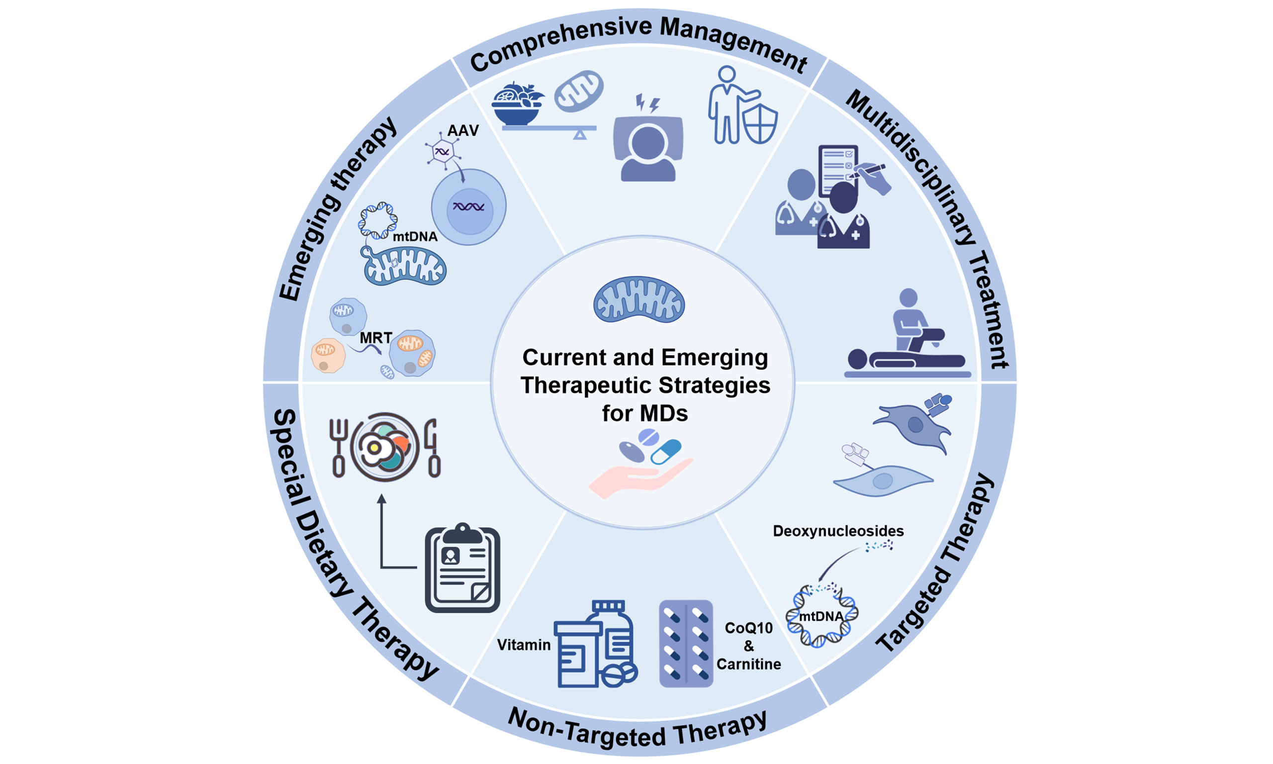
Abstract
Mitochondrial diseases are a group of inherited metabolic disorders caused by defects in mitochondrial DNA (mtDNA) or nuclear DNA leading to oxidative phosphorylation dysfunction. Clinical manifestations are highly heterogeneous, typically involving high-energy-demand organs (brain, muscle, heart, etc.), and curative treatments remain largely unavailable. This review examines the literature over the past decades on mitochondrial disease treatments, including traditional Cocktail therapy, target therapy, multidisciplinary management, gene therapy, mtDNA base editing, and mitochondrial replacement therapy. Although most current treatments remain symptomatic, breakthrough advances in gene therapy and base editing over recent years have brought new hope for precision medicine in mitochondrial diseases.
Keywords


 Download PDF
INTRODUCTION
Primary mitochondrial diseases (PMDs) represent one of the most common inherited genetic disorders. The estimated prevalence is approximately 1 in 4,300 adults, with mitochondrial DNA (mtDNA) mutation carriers occurring in about 1 in 5,000 individuals and nuclear DNA (nDNA)-related cases affecting around 2.9 per 100,000 adults[1]. PMDs are characterized by impaired oxidative phosphorylation of the mitochondrial respiratory chain, resulting from defects in either mtDNA or nDNA[2]. These disorders exhibit significant clinical and genetic heterogeneity, predominantly impacting tissues and organs with high energy demands. Clinically, they are categorized based on the involved systems into mitochondrial encephalopathy, encephalomyopathy, myopathy, and peripheral neuropathy. Notable examples include MELAS syndrome (mitochondrial encephalomyopathy, lactic acidosis, and stroke-like episodes) and Leigh syndrome. Definitive diagnosis relies on molecular and/or pathological confirmation. With recent advancements in molecular techniques and a deeper understanding of pathogenesis, the recognition, diagnosis and treatment including emerging gene therapies of PMDs are evolving rapidly[3,4]. This article offers a review of the significant advancements referring to treatment that have shaped this field in recent decades.
MECHANISM OF PMDS
Dual genomic control by nuclear and mitochondrial genomes
Mitochondria represent the only organelles within human cells that possess their own DNA (mtDNA). mtDNA is a circular double-stranded molecule approximately 16.6 kb in length that encodes 37 genes: 13 core subunits of the OXPHOS complexes, 22 tRNAs and 2 rRNAs that are necessary for protein translation within mitochondria[5]. However, the vast majority (> 99%) of the ~1,500 mitochondrial proteins are encoded by the nuclear genome (nDNA); these proteins are synthesized in the cytosol and are then transported into mitochondria via dedicated translocation mechanisms.
This dual genetic control dictates the diverse inheritance patterns observed in PMDs. mtDNA mutations are transmitted maternally and may occur in coding regions (directly affecting subunit structure) or in regulatory regions (impairing transcription and translation). As every cell carries hundreds to thousands of copies of mtDNA, the coexistence of mutant and wild-type mtDNA, termed heteroplasmy, is a hallmark. When the proportion of mutant mtDNA exceeds a tissue-specific bioenergetic threshold, dysfunction ensues—a phenomenon known as the threshold effect, which accounts for much of the clinical variability[6,7]. To date, over 100 confirmed pathogenic point mutations and rearrangements (mainly deletions) of mtDNA have been identified, among which the most frequent are m.3243A>G, m.8344A>G, and m.8993T>G[7]. Mutations in nDNA follow the Mendelian inheritance pattern, including autosomal dominant, autosomal recessive or X-linked inheritance, and exert broader effects, not only involving structural subunits of OXPHOS but also genes critical for mitochondrial biogenesis, dynamics (e.g., OPA1), protein import (e.g., TOMM40), respiratory supercomplex assembly (e.g., COX10), mtDNA replication and repair (e.g., POLG), and coenzyme Q10 biosynthesis[7-9]. Currently, over 1,000 nuclear genes encoding proteins related to mitochondrial function have been identified, among which approximately > 400 are clearly associated with pathogenic mutations. Epidemiological data indicate that approximately 80% of childhood-onset PMDs are attributable to nDNA defects, whereas about 60% of adult-onset cases are linked to mtDNA mutations[7].
Heteroplasmy in mtDNA-related PMDs
The ultimate clinical manifestations of PMDs are remarkably tissue-specific, stemming from differential tissue reliance on adenosine triphosphate (ATP) and variable susceptibility to mitochondrial dysfunction. The brain, cardiac muscle, skeletal muscle, and retinal photoreceptors—owing to their high energy demand, low reserves, and limited regenerative capacity—are the most frequently affected systems[10]. Furthermore, the variable heteroplasmic burden across various tissues within the same individual accounts for the striking phenotypic heterogeneity observed with identical gene mutations, ranging from Leigh syndrome to MELAS and MERRF (myoclonic epilepsy with ragged red fibers)[11].
Disrupted mitochondrial biogenesis, dynamics and mitophagy
Mitochondrial quality control (MQC) constitutes a sophisticated interconnected network that surveils mitochondrial integrity, counteracts damage or stress, and sustains mitochondrial homeostasis. This system coordinates multiple cellular processes, such as mt-ISR, mitochondrial biogenesis, mitochondrial dynamics, mitophagy, and intercellular mitochondrial transfer[12]. MQC serves a critical function in a large variety of human disorders, encompassing cancer, cardiovascular diseases and neurodegenerative diseases[13].
Oxidative phosphorylation dysfunction and dissipation of the proton motive force
The core pathological hallmark of PMDs is impaired oxidative phosphorylation (OXPHOS) within the mitochondrial respiratory chain, resulting in insufficient ATP production[4]. OXPHOS is the primary aerobic energy-producing process, executed by the electron transport chain (ETC) complexes I to IV, which are embedded in the inner mitochondrial membrane, working in concert with ATP synthase (Complex V). In PMDs, a defect in any single complex disrupts this finely tuned system. For instance, Complex I deficiency impairs nicotinamide adenine dinucleotide (NADH) oxidation, reducing electron flux and proton pumping, thereby lowering Δp[14]. Complex III or IV deficiencies directly block electron transfer to the terminal acceptor, similarly dissipating Δp. Notably, although Complex V dysfunction does not directly affect electron transfer, it reduces proton utilization, leading to a relative increase in Δp, which in turn exerts negative feedback on upstream electron transport, further impairing energy output[15]. The dissipation of Δp not only cripples ATP production but also compromises other Δp-dependent processes, including mitochondrial protein import (requiring membrane potential), calcium uptake, and metabolite transport, thereby triggering a global cellular energetic failure[4].
Electron leakage and the vicious cycle of oxidative stress
Bioenergetic impairment initiates a self-perpetuating cascade wherein ETC blockage causes upstream hyper-reduction[16], prolonging electron-transfer intermediate lifetime at Complex I (FMN) and Complex III (Q0) and promoting electron leakage to O2•-)[17]. Subsequent dismutation to H2O2 and Fenton-mediated conversion to •OH[18]. Collectively, constitute reactive oxygen species (ROS).
This ROS burst inflicts spatially targeted damage. Owing to its histone-depleted state and proximity to the ETC, mtDNA is highly vulnerable to hydroxyl radical attack, generating 8-oxoguanine (8-oxoG) and oxidative lesions that cause base mispairings, point mutations, and strand breaks during replication. This impairs respiratory chain subunit synthesis, establishing a self-amplifying cycle: ETC deficiency → electron leakage → ROS → mtDNA damage → aggravated ETC deficiency[19]. The inner membrane, rich in cardiolipin and polyunsaturated fatty acids, undergoes lipid peroxidation upon ROS attack, altering membrane fluidity and permeability to cause proton leakage and Δp dissipation, while disrupting cardiolipin binding to ETC complexes and destabilizing respirasome assembly[20]. Oxidative modifications of fusion proteins OPA1 and MFN2 further drive mitochondrial fragmentation and metabolic failure[21].
Mitochondrial apoptosis and ferroptosis in PMDs
When increased oxidative stress and depleted ATP accumulate beyond a critical threshold, cells execute downstream injury programs. When permeability of the outer mitochondrial membrane is increased, cytochrome c and apoptosis-inducing factor (AIF) are released into the cytosol, activating both caspase-dependent and caspase-independent apoptotic pathways[22,23]. As an iron-dependent, lipid-peroxidation-driven form of regulated cell death, ferroptosis has been recently implicated in neuronal loss in specific PMD subtypes characterized by glutathione depletion and extensive lipid peroxidation[24,25]. Grasping the crosstalk between apoptosis and ferroptosis is critical for developing targeted treatments for mitochondrial disorders. A combination of mitochondria-targeted antioxidants, GPX4 modulators, and mPTP inhibitors, paired with personalized diagnostic frameworks, constitutes the most promising therapeutic approach to date. Further in-depth exploration of the apoptosis-ferroptosis interaction is key to driving progress in the clinical management of mitochondrial diseases.
Mitochondrial inflammation
Upon stress or damage, mitochondria release damage-associated molecular patterns (DAMPs) including mtDNA and cardiolipin. They are recognized by microglial Toll-like receptor 9 (TLR9) and the NLRP3 inflammasome, activating innate immune responses and sustaining chronic neuroinflammation. This inflammatory state not only directly harms adjacent neurons but also exacerbates the local microenvironment through reactive astrogliosis, driving disease progression independently of the primary energetic defect. mtDNA-mediated inflammation is primarily driven by the cGAS (cyclic GMP-AMP synthase)-STING (stimulator of interferon genes) signaling pathway[26]. In addition to the cGAS-STING pathway, the mtROS and mtDNA can activate NLRP3 inflammasome to promote IL-1β and IL-18 cleavage. A link between inflammation and mitochondrial bioenergetics has already been confirmed. Studies indicate that mitochondrial dysfunction can exacerbate inflammation, and the exacerbated inflammation in turn impairs OXPHOS and disrupts MQC[27]. Histopathological analyses of postmortem human brain tissue and mouse models of PMDs indicate that neuroinflammation is not merely a consequence of neurodegeneration but a potential pathogenic driver of disease progression warranting further investigation. This insight may pave the way for novel therapeutic strategies for these devastating disorders, which currently lack effective treatments[28].
In summary, the pathogenesis of PMDs is a multi-dimensional process initiated by genetic defects, centered on OXPHOS dysfunction, amplified by oxidative stress and impaired mitochondrial dynamics, and culminating in cell death and inflammatory injury. A thorough elucidation of these mechanisms not only enhances our understanding of disease heterogeneity but also provides a solid theoretical foundation for developing novel therapeutic strategies targeting mitochondrial biogenesis, ROS scavenging, inflammation modulation, and gene editing.
TREATMENT STRATEGIES IN PMDs
Clinical care standards for PMDs are evolving. Currently, as specific and curative treatments remain largely unavailable, therapeutic strategies focus on several key objectives: stabilizing and enhancing the function of residual enzymes and cells; alleviating symptoms; preventing or slowing clinical progression; supporting cellular demands during metabolic stress to avert acute decompensation; and avoiding catabolic triggers as well as medications with known mitochondrial toxicity[29].
Pharmacological treatment strategies
The mitochondrial medicine society (MMS) initially published its guidelines for the management of mitochondrial medicine therapies in 2009. In 2015, the MMS released a consensus statement that reviewed the existing literature on mitochondrial diseases and provided recommendations for the optimal diagnosis and treatment of PMDs[30]. Subsequently, a comprehensive update in 2020 offered detailed dosage protocols for the clinical treatment of these diseases[29], alongside an international Delphi-based consensus on drug safety in patients with PMDs[31]. Most recently, an Australian adaptation of the MMS recommendations, titled Patient Care Standards for PMDs in Australia, was published[32]. While multimodal therapeutic strategies (Cocktail therapy) are currently prevalent in the management of PMDs, the rapid evolution of targeted medication offers a promising new direction for future treatment paradigms.
Classical non-target pharmacological treatment strategies
In both published research and clinical practice, mitochondrial medicine regimens (colloquially called "mitochondrial cocktails") include a wide range of nutrients [Figure 1], with considerable variation in their combinations and dosages[29]. Despite their widespread use, the dosages and formulations of these agents remain non-uniform, and the majority of patients are treated on an empirical basis [Table 1]. Critically, this practice is not underpinned by large-scale randomized controlled trials (RCTs) or high-grade evidence-based medicine. A Cochrane systematic review concluded that evidence remains insufficient to endorse any pharmacological intervention for mitochondrial diseases, underscoring how clinical practice in this domain remains constrained by small case series, open-label studies, and expert opinion rather than randomized controlled data[33]. This evidence gap is compounded by the extreme clinical and genetic heterogeneity of mitochondrial disorders, which makes conventional RCT designs challenging to execute in adequately powered cohorts. In this review we comprehensively summarized the agents used in PMDs, including the mechanism and clinical evidence levels [Table 2].
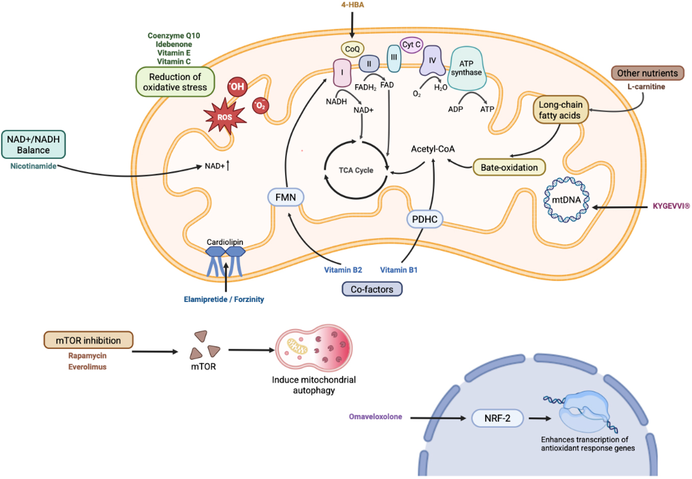
Figure 1. Pharmacological treatment strategies in PMDs and their mechanistic targets. Created in BioRender.
Classical pharmacological treatment strategies for PMDs
| Agent | Doses | Clinical evidence | Adverse event | |
| Agents enhancing electron transport chain (ETC) function | ||||
| Thiamine (Vitamin B1) C12H17N4OS+ | adults 100-1,000 mg/day children 5 mg/kg/day | • Case reports: Thiamine improved myopathy and lactic acidosis in MELAS patients with A3243G mutation[97] • Expert consensus | Occasional tremors and herpes | |
| Riboflavin (Vitamin B2) C17H20N4O6 | 10 mg/kg/d | • Complex I deficiency: case reports of clinical improvement with riboflavin[98] • Systematic review (23 studies): riboflavin promotes neuroprotection via redox modulation and enhanced complex activity | Anorexia, nausea | |
| Coenzyme Q10 (CoQ10, Ubiquinone, Ubiquinol) C59H90O4 | 10 mg/kg/d | • 2015 PMD consensus: should be offered to most PMD patients, not only primary CoQ10 deficiency[30] • RCT (30 patients, 1,200 mg/day, 60 days): minor effects on post-exercise lactate and VO2/lean mass; no effect on strength or resting lactate[99] • Phase 3 trial in children (NCT00432744): no statistically significant difference in primary outcomes • Most supportive data from anecdotal reports, uncontrolled open-label investigations, or combination-treatment regimens; minimal benefit reported with CoQ10 alone | Insomnia, lowering warfarin blood concentration | |
| Antioxidant actions and scavenging of oxygen free radicals | ||||
| Vitamin E (α‐tocopherol) C29H50O2 | 10 mg/kg/d | Global prescribing patterns of vitamins and cofactor supplements in the management of PMDs[100] | Occasionally associated with cardiac adverse events | |
| Vitamin C (Ascorbic acid) C6H8O6 | 25 mg/kg/d | Case reports: Improvement was reported in 9 patients with combined treatment with Vitamin C, CoQ10, thiamin and riboflavin, of whom 4 demonstrated acquisition of additional developmental milestones, but this was transient in 6 patients four[101] | Diarrhea, stones in some cases | |
| Substrates supplementation & energy buffers | ||||
| L-carnitine (Levocarnitine) C7H15NO3 | 50 mg/kg/d | • 2015 PMD consensus: administer when documented deficiency exists; monitor levels during therapy[30] • RCT crossover (12 CPEO patients, 3 g/day, 2 months): significantly improved constant work rate exercise tolerance[102] | Gastrointestinal discomfort, fishy odor | |
| Nitric oxide (NO) precursors - arginine and citrulline | ||||
| Arginine (L-Arginine) C6H14N4O2 | Acute stroke-like episodes: IV arginine (0.5 g/kg, maximum 20-30 g); Chronic oral prophylaxis: 0.15-0.3 g/kg/day | • Acute stroke-like episodes: open-label trials show symptom improvement[103] • Multicenter prospective trial (13 patients, 0.3-0.5 g/kg/day, 2 years): MELAS stroke scale trend toward improvement[104] • 7-year follow-up: systematic arginine significantly improved survival vs natural history. Maintaining plasma arginine ≥ 168 umol/L may prevent seizures. • MMS 2017 consensus: IV arginine recommended for acute MELAS stroke-like episodes | Occasional hypotension, hypoglycemia, diarrhea | |
Target therapy in PMDs
| Strategy and pathway | Disease | Specific therapy | Situation/evidence | Dose |
| Activating endogenous defense pathways | Friedreich’s ataxia | Omaveloxolone | Approved in 2023 | 150 mg qd |
| Stabilizing mitochondrial architecture | Barth syndrome | Elamipretide | Approved in 2025 | 40 mg subcutaneous qd |
| Replacing depleted nucleotide pools | Thymidine kinase 2 (TK2) deficiency | Deoxycytidine and deoxythymidine | Approved in 2025 | Up to 800 mg/kg/day |
| Cofactor restoration | ACAD9 mutations | Riboflavin | Case series[51] | 20 mg/kg/day, maximum 200 mg/day |
| NAXE/NAXD deficiency | Nicotinamide | Case series[53] | 100 mg/day titrated weekly to 500 mg/day | |
| TPK1, SLC25A19 and SLC19A3 mutations | Thiamine | Case series[105] | 20-40 mg/kg/day | |
| Primary coenzyme Q10 (CoQ10) biosynthesis defects (PDSS1, COQ2, COQ6) | CoQ 10 | 2015 PMD consensus[30] | 30-50 mg/kg/day | |
| LHON | Idebenone | EMA-approved for LHON (≥ 12 years) | 300 mg tid | |
| Biotinidase deficiency (BTD mutations) | Biotin administration | Case series[106] | 5-10 mg/day | |
| Metabolic bypass and dietary modification | Pyruvate dehydrogenase deficiency | L-glutamine and the ketone body β-hydroxybutyrate | Global clinical practice from literature and survey data[72] | / |
| Valine catabolism: ECHS1 and HIBCH deficiencies | Restricting dietary valine and fat | Case report[60] | / |
Emerging/target pharmacological therapy
The therapeutic landscape for mitochondrial disorders has undergone a paradigm shift with the advent of precision medicine. Rather than relying solely on supportive care, clinicians now increasingly tailor treatment to the specific molecular lesion-whether it involves correcting a depleted cofactor, bypassing a blocked metabolic step, or activating endogenous cytoprotective pathways. This section reviews the emerging targeted strategies that have moved from mechanistic insight to clinical application, grouped by their underlying therapeutic rationale: activation of cellular defense pathways, direct supplementation of deficient substrates or cofactors, and metabolic reprogramming through dietary intervention.
Activating endogenous defense pathways
In Friedreich’s ataxia, the most common autosomal recessive ataxia, frataxin deficiency leads to impaired iron-sulfur cluster biogenesis and increased oxidative stress[34]. Omaveloxolone, approved by the U.S. Food and Drug Administration in February 2023 for patients aged ≥ 16 years[35], addresses this pathophysiology not by replacing the missing protein but by activating the nuclear factor (erythroid-derived 2)-like 2 (NRF2) pathway. By inhibiting the ubiquitination and proteasomal degradation of NRF2, omaveloxolone enhances the transcription of antioxidant response genes. In Part 2 of the phase II MOXIe trial (NCT02255435), treatment with 150 mg once daily resulted in a significant improvement in the modified Friedreich Ataxia Rating Scale (mFARS) score at week 48 compared with placebo[36], establishing a disease-modifying precedent for disorders driven by oxidative imbalance.
Stabilizing mitochondrial architecture
Barth syndrome, characterized by cardiolipin remodeling defects and mitochondrial membrane instability, illustrates a second mechanistic approach: organelle-targeted pharmacotherapy. Elamipretide (Forzinity) is an aromatic-cationic tetrapeptide that easily crosses cell membranes and temporarily accumulates in the inner mitochondrial membrane, where it attaches to cardiolipin and maintains the structural integrity of the inner mitochondrial membrane. A phase 2/3 randomized, double-blind, placebo-controlled clinical trial demonstrated that daily subcutaneous administration of 40 mg elamipretide showed significant improvements in 6-minute walk (6MWT) with a mean increase of 95.9 m (P = 0.024) as well as in the Barth Syndrome Symptom Assessment (BTHS-SA) score with a mean reduction of 2.1 points (P = 0.031)[37]. Subsequent 168-week open-label extension study[38] and a natural history comparative study[39] further validated the long-term efficacy and safety of elamipretide in patients with Barth syndrome. This drug was granted accelerated approval in the USA for use to improve muscle strength in adult and pediatric patients with Barth syndrome weighing more than 30 kg[40].
It is also potentially effective for other mitochondrial disorders. Initial clinical evidence from a randomized dose-escalation trial in adults with primary mitochondrial myopathy (PMM) indicated that 5 days of elamipretide treatment improved exercise performance with a favorable safety profile[41]. Subsequently, a randomized crossover trial evaluated four weeks of daily subcutaneous elamipretide in adults with PMM. Although the primary endpoint-a clinically meaningful change in the 6MWT-did not achieve statistical significance, secondary endpoints suggested a potential treatment effect compared with placebo[42]. However, a later Phase 3 study failed to demonstrate improvements in either the 6MWT or the PMM symptom assessment total fatigue score (TFS)[43]. Notably, post hoc analyses revealed beneficial effects in patients with mtDNA replisome disorders, underscoring the critical importance of stratifying by genetic subtype in clinical trials. These findings provide the rationale for the ongoing Phase 3 NuPOWER trial, which is specifically designed to evaluate elamipretide efficacy in patients with mtDNA maintenance-related disorders[44].
Nucleotide bypass therapy
A fundamentally different strategy is substrate replacement. In thymidine kinase 2 (TK2) deficiency, impaired mtDNA synthesis leads to progressive myopathy due to inadequate deoxypyrimidine supply. Deoxycytidine and deoxythymidine (marketed as KYGEVVI®) was approved in the United States in November 2025 for the treatment of TK2d in adult and pediatric patients with an age of symptom onset on or before 12 years[45], bypassing the enzymatic block by providing the nucleoside precursors required for mtDNA replication. Clinical data showed significant efficacy compared with natural history[46,47], and the regimen—up to 800 mg/kg/day of deoxycytidine and deoxythymidine (or their monophosphates)—may also hold promise for other mtDNA depletion syndromes[48].
mTOR inhibitors
Rapamycin and its analog everolimus, as mTOR inhibitors, have shown potential therapeutic value in some mitochondrial diseases. Studies have shown that rapamycin delays symptoms and extends lifespan in the NDUFS4-/- mouse model of Leigh disease[49]. A clinical study treated two pediatric patients with everolimus: a child with Leigh syndrome (nuclear NDUFS4 mutation) showed significantly improved motor function, reduced MRI brain lesions, and progressive weaning from ventilator and gastrostomy after 19 months of treatment; however, a MELAS child carrying the m.3243A>G mutation showed no response[50], suggesting that efficacy may depend on disease stage, mutation type, and heteroplasmy levels, requiring individualized assessment.
Cofactor restoration for special genotypes in PMDs
A substantial proportion of treatable mitochondrial disorders respond to high-dose vitamin or cofactor supplementation, reflecting the fact that many genetic lesions impair the binding, transport, or recycling of essential metabolites rather than causing absolute protein loss. Riboflavin (vitamin B2) at 20 mg/kg/day (maximum 200 mg/day) dramatically improves survival and alleviates symptoms in patients with ACAD9 mutations, particularly when treatment is initiated before 1 year of age[51,52]. Similarly, high-dose nicotinamide (100 mg/day titrated weekly to 500 mg/day) has emerged as an effective therapy for NAXE/NAXD deficiency, enhancing clinical outcomes and quality of life by restoring cellular NAD homeostasis[53]. In thiamine-responsive disorders—encompassing TPK1[54], SLC25A19, and SLC19A3 mutations—high-dose thiamine remains the cornerstone of therapy, although whether biotin co-administration is necessary in SLC19A3-related disease remains unresolved.
High-dose CoQ supplementation is effective for both primary and secondary CoQ deficiencies[55]. For biosynthesis defects (e.g., PDSS1, COQ2, COQ6), ubiquinone or its reduced form ubiquinol at
Metabolic bypass and dietary modification
When enzymatic blocks disrupt catabolic flux, alternative fuel sources or precursor restriction can mitigate toxic metabolite accumulation. Mitochondrial pyruvate carrier (MPC) deficiency caused by MPC1 mutations may be amenable to anaplerotic therapy with L-glutamine and the ketone body β-hydroxybutyrate, although rigorous clinical trials are still needed to confirm efficacy[59]. In contrast, disorders of valine catabolism—specifically ECHS1 and HIBCH deficiencies—are managed by restricting dietary valine and fat (particularly dairy fat), a strategy that limits disease progression when implemented early[60-63].
Other agents
Additionally, several other agents are in preclinical or early clinical evaluation, including resveratrol/SRT2104 (SIRT1 activators), cysteine donors (N-acetylcysteine, whey protein), among others[1]. The table below summarizes the clinical trial status of major therapeutic agents.
Viewed thematically, the targeted therapies for mitochondrial disease cluster into three broad categories. First, cofactor pathway restoration—exemplified by riboflavin, nicotinamide, thiamine, CoQ10, and biotin—represents the most immediately translatable strategy, capitalizing on the high plasticity of vitamin-responsive enzymology. Second, gene-product-independent compensation seeks to bypass the genetic defect by supplying downstream substrates (deoxycytidine/deoxythymidine in TK2 deficiency), providing alternative fuels (L-glutamine/βHB in MPC deficiency), or activating parallel protective pathways (NRF2 activation by omaveloxolone). Third, organelle-directed biophysical stabilization (elamipretide) and dietary metabolic reprogramming (valine/fat restriction) offer complementary avenues for selected disorders. While cofactor replacement and dietary modification currently provide the broadest applicability, the approval of mechanism-based agents such as omaveloxolone, elamipretide, and deoxycytidine/deoxythymidine signals a maturing field in which therapy is increasingly dictated by molecular diagnosis rather than phenomenology alone.
Symptomatic treatment and supportive care
Since mitochondrial diseases often involve multiple systems, clinical practice requires collaboration among multidisciplinary teams including neurology, endocrinology, cardiology, nephrology, hepatology, respiratory medicine, and gastroenterology[32,64]. Current symptomatic management of mitochondrial diseases primarily involves targeted pharmacological or interventional therapies for affected systems or organs.
Neurological symptoms
Stroke-like episodes
Arginine is commonly used for acute-phase management and prophylaxis in MELAS patients or those with stroke-like seizures with m.3243A>G mutation or other genetic mutations[65]. Intravenous arginine (0.5 g/kg, maximum dose 20-30 g; for higher-weight patients, start with a low dose of 5 g) should be administered as early as possible during the acute phase of a stroke, followed by re-evaluation after 3-5 days. For MELAS patients in the interictal period, oral arginine (0.15-0.3 g/kg) is recommended to prevent stroke-like seizures[65]. There is currently no consensus on the monitoring of plasma arginine and citrulline levels or the efficacy of oral citrulline.
Seizures
A 2024 consensus statement on seizure management in patients with PMD has been published[66], which recommends selecting appropriate antiepileptic drugs according to specific seizure types. Most anti-seizure medications are not contraindicated for use in adults and children with PMDs. For patients with MELAS syndrome, levetiracetam, lamotrigine, lacosamide and benzodiazepines are the preferred therapeutic options, with particular emphasis on optimized seizure control during the stroke-like episode phase[67]. Patients with early-onset disease frequently present with refractory seizures, which may necessitate dual antiepileptic drug therapy. Propofol should be closely monitored for prolonged use in the context of refractory status epilepticus due to the risk of propofol infusion syndrome. The consensus also explicitly outlines clinically notable exceptions, including the use of valproic acid in POLG-related disorders, vigabatrin in patients with gamma-aminobutyric acid transaminase deficiency, and topiramate in patients with pre-existing risk for renal tubular acidosis[66]. In case a new treatment is initiated in a PMD patient, careful monitoring is suggested for potential side effects, including measurement of blood lactate and other laboratory parameters.
Dyskinesia & dystonia
PMDs may present with a mixed movement and tone disorder that encompasses hyperkinetic, hypokinetic, or cerebellar movement patterns, alongside hypotonia, spasticity, rigidity, and dystonia[64]. The management of movement and tone abnormalities in PMDs follows a comparable framework to that applied to patients with movement disorders of alternative etiologies. Multidisciplinary physical medicine and rehabilitation assessments are strongly recommended to optimize functional mobility, prevent the development of joint contractures, and mitigate associated discomfort and pain[64]. However, agents that modify muscle tone should be administered with extreme caution, as these medications may selectively impair cognitive performance, reduce muscle strength and secondarily compromise respiratory effort, and adversely affect both gastrointestinal motility and urinary function[32]. While botulinum toxin has demonstrated therapeutic benefits for alleviating spasticity and dystonia, its use requires careful risk-benefit stratification due to potential adverse effects, most notably the exacerbation of pre-existing muscle weakness.
Cardiac management
The prevalence of cardiomyopathy among PMDs has been estimated at 20%-40% in earlier studies, although not all enrolled cases received genetic confirmation for the disease[64]. While hypertrophic cardiomyopathy appears to be the most prevalent phenotype, dilated cardiomyopathy, restrictive cardiomyopathy, left ventricular noncompaction, and histiocytoid cardiomyopathy have all been well documented in published cohorts. Cardiac conduction disorders with unpredictable clinical trajectories are also a highly prevalent disease manifestation, and arrhythmias are a leading cause of death, particularly in KSS patients and those carrying the m.3243A>G pathogenic variant. Regular annual monitoring of electrocardiogram (ECG) and echocardiography is recommended, with 24-h Holter monitoring and cardiac MRI performed when necessary[32,64]. For patients with supraventricular tachycardia, ablation therapy may be considered. For cardiomyopathy patients at risk of sudden cardiac death, such as KSS patients, implantable cardioverter-defibrillator (ICD) or pacemaker implantation is advised.
Endocrinology
Endocrine dysfunction represents a common clinical manifestation of PMDs, with incidence rates differing markedly by specific underlying mitochondrial disorder and the involved endocrine organ. Diabetes is the most frequent symptom, but other endocrine phenotypes, including growth hormone deficiency, adrenal insufficiency, hypogonadism and parathyroid gland dysfunction, represent well-recognized but less frequently reported manifestations across the PMD clinical spectrum[68]. The endocrine component of the multidisciplinary management pathway is generally straightforward: it relies on formal biochemical verification of hormone deficiency, followed by physiological hormone replacement and careful dose titration tailored to individual clinical outcomes and serial biochemical monitoring results. Regularly measure height, weight, body mass index (BMI), and growth rate (including nutritional status assessment), monitor blood glucose and glycated hemoglobin (HbA1c), and test thyroid function, parathyroid function, and electrolyte levels when necessary. For patients with elevated blood glucose, use metformin with caution and consider alternative hypoglycemic agents. It is recommended to consider combined therapy with oral hypoglycemic agents and insulin, as some patients may exhibit both insulin deficiency and insulin resistance. Sodium-glucose cotransporter-2 inhibitors and glucagon-like peptide-1 receptor agonists exhibit multifaceted beneficial effects that directly counteract oxidative stress and ameliorate mitochondrial dysfunction, making them exceptionally well-suited first-line pharmacological agents for the targeted management of maternally inherited diabetes and deafness, a monogenic diabetes subtype driven by m.3243A>G mtDNA mutation[69]. For patients with confirmed growth hormone deficiency, growth hormone replacement has been reported to have varying degrees of success without adverse effects. However, long-term efficacy of growth hormone therapy is inconclusive[68].
Hearing
50% of mitochondrial patients exhibit hearing impairment especially in m.3243A>G mutations, requiring close monitoring of auditory function (including otoacoustic emissions and auditory brainstem evoked responses)[70]. All patients should receive a comprehensive baseline audiological workup at the time of diagnosis, and undergo scheduled surveillance audiology assessments on an annual to biennial basis[32]. Avoid exposure to adverse environments and excessive noise, and refrain from using ototoxic medications. For patients with symptomatic moderate to severe hearing loss, appropriately programmed hearing aids represent the established first-line intervention to ameliorate auditory impairment and reduce associated communication difficulties in daily life. For severe hearing loss (> 90 dB), cochlear implantation should be considered, particularly in pediatric patients, where early intervention can enhance speech function[32,64].
Vision
Patients who may be affected should undergo evaluation and close monitoring of ision, visual field, ocular motility (including ptosis), and fundus examination[64]. Visual evoked potentials and electroretinography should be performed when necessary. Surgical interventions may be considered for strabismus, ptosis, or cataracts. Oral idebenone is strongly recommended for Leber Hereditary Optic Neuropathy (LHON) patients[64]. Patients with visual impairment should be closely monitored for potential cardiac and neurological involvement.
Kidneys
Renal involvement in mitochondrial diseases includes: asymptomatic kidney disease, reduced glomerular filtration rate (GFR), proteinuria and/or hematuria (with or without hypertension), metabolic acidosis, renal tubular acidosis, focal segmental glomerulosclerosis, progressive kidney disease, and rapid progression to end-stage renal failure[71]. Therefore, all patients with mitochondrial diseases require evaluation of blood pressure, serum urea, creatinine, sodium, potassium, chloride, calcium, magnesium, phosphate, and anion gap[64]. For patients with renal involvement, close monitoring of urinalysis, urinary microprotein, cystatin C (particularly in pediatric patients with muscle involvement), renal ultrasound, and renal function parameters (urinary albumin/creatinine ratio, serum creatinine, urinary electrolytes and bicarbonate, and other metabolic parameters) is recommended[32]. Patients with significant abnormalities should be referred to nephrology specialists with experience in managing mitochondrial diseases. For end-stage renal disease, the decision to proceed with dialysis or renal transplantation should be based on quality of life, other comorbidities, and survival prospects.
Dietary interventions
The ketogenic diet (KD) is the first-line treatment for patients with pyruvate dehydrogenase deficiency[72]. KD may be effective for some refractory epilepsy patients with MELAS[73]. However, individuals with a confirmed diagnosis of fatty acid oxidation disorders or pyruvate carboxylase deficiency should be categorically excluded from KD interventions, as this dietary pattern may precipitate life-threatening metabolic decompensation. For patients with certain fatty acid metabolic disorders, special dietary interventions are required, and consultation with a clinical nutrition specialist is necessary. For patients with significantly elevated lactate levels, it is recommended to restrict the intake of sugars and carbohydrates.
Other dietary strategies
Individualized diets are recommended based on the specific metabolic defect—high-fat, low-carbohydrate for pyruvate dehydrogenase deficiency; fat restriction for carnitine deficiency; and high-protein, high-carbohydrate, low-fat for pyruvate carboxylase deficiency[20]. General principles include avoiding fasting, maintaining adequate caloric intake, and avoiding high-fat, low-carbohydrate diets (except for specific indications)[20].
Rehabilitation training
Patients should receive annual assessments for potential musculoskeletal complications, including kyphoscoliosis, joint contractures, dislocations and limb deformities, with particular focus on individuals with pre-existing abnormalities in muscle tone, muscle strength or neurological function[32]. Exercise helps improve mitochondrial function and reduce abnormal mtDNA load. Children with mitochondrial disorders are prone to exercise intolerance due to their lower maximal oxygen uptake. Therefore, a personalized rehabilitation training program should be developed under the guidance of a professional rehabilitation therapist, starting with low-intensity and short-duration sessions, and progressing gradually. However, this is not recommended during acute disease phases or when fasting.
GENE THERAPY
Mitochondrial gene therapy is rapidly evolving into a highly promising etiological intervention that directly targets either nuclear genes or the mitochondrial genome to rectify the primary genetic mutations and downstream functional impairments that drive mitochondrial pathologies. The design and optimization of bespoke mtDNA delivery platforms capable of specific organelle targeting and efficient mammalian mitochondrial transfection has emerged as a dynamic and rapidly advancing research area, enabling transformative breakthroughs in the historically intractable goal of rescuing impaired mitochondrial function.
Gene replacement in PMD with nDNA mutations
Recombinant adeno-associated virus (rAAV)-based gene replacement therapies targeting nuclear gene defects in PMDs have been successfully validated in more than 10 distinct preclinical mouse models of PMDs, including mouse models of NDUFS3, NDUFS4, SURF1, TYMP, TK2, MPV17, AIFM1, ANT1 deficiency, et al.[74]. This landmark progress is largely attributed to the advent of next-generation rAAV platforms with greatly enhanced tropism for therapeutically relevant organs. However, to date, none of these gene reconstitution-based therapeutic modalities have successfully progressed to formal clinical evaluation in human participants, except Friedreich Ataxia[75]. The greatest progress of gene therapy in PMD has been made for mtDNA replacement such as LHON[76,77].
Friedreich ataxia, an autosomal recessive and uniformly fatal monogenic disorder resulting from pathogenic variants in the FXN (frataxin) gene, is a progressive multi-system condition predominantly affecting the nervous system and heart, where cardiac complications account for the vast majority of disease-related mortality. LX2006 is an AAV-based gene therapy developed by Lexeo Therapeutics, delivered intravenously to provide a functional frataxin gene, promoting frataxin protein expression and restoring mitochondrial function in cardiomyocytes[75]. In Phase I/II clinical trials and an investigator-initiated Phase Ia trial, LX2006 was well tolerated with no treatment-related serious adverse events, and cardiac biomarkers showed clinically meaningful improvements that increased over time. Among 8 participants followed for at least 6 months, FXN protein expression levels increased, and 3 out of 4 subjects with elevated left ventricular mass index at baseline showed a > 10% reduction at 12 months[75].
Despite their promise, few gene reconstitution approaches have yet to enter clinical trials, largely due to three translational barriers. Firstly, poor phenotypic concordance between animal models and humans limits the reliability of efficacy predictions. Secondly, maintaining long-term transgene expression in target organs proves difficult. Thirdly, the requirement for disease-specific vectors makes this strategy unfeasible for the vast spectrum of more than 350 mitochondrial disorders.
Gene replacement in PMD with mtDNA mutations
As a rare mitochondrial disorder, LHON presents with severe bilateral sequential vision loss, caused by the irreversible selective degeneration of retinal ganglion cells (RGCs). The three canonical mtDNA point mutations—m.3460G>A in MT-ND1, m.11778G>A in MT-ND4, and m.14484T>C in MT-ND6—represent the overwhelming majority of all known LHON genetic triggers, collectively explaining ~90% of confirmed patient cohorts across global populations. LHON gene therapy is attractive due to its monogenic cause, acute onset allowing a treatment window, easy intravitreal access to RGCs, ocular immune privilege, and proven AAV safety[76,77]. Lumevoq (lenadogene nolparvovec), is an AAV-based gene therapy delivered via intravitreal injection of the wild-type ND4 gene. Its innovative design fuses the AAV capsid VP2 with a mitochondrial targeting sequence, enabling AAV to target mitochondria and achieve allotopic expression of ND4. Relative to the visual outcomes of an externally recruited natural history control cohort, aggregated data across four phase 3/2 interventional trials (RESCUE, REVERSE, RESTORE and REFLECT) confirm that unilateral intravitreal administration of a single dose of lenadogene nolparvovec drives durable, bilateral visual function recovery in m.11778G>A LHON patients ≥ 15 years old treated within the 12-month post-onset therapeutic window[78-80]. Despite a modest overall effect size, treated patients achieved final visual acuity outcomes that are statistically and clinically distinct from the untreated LHON natural history trajectory, with a magnitude of clinical benefit superior to that observed in m.11778G>A carriers receiving idebenone as monotherapy[81].
mtDNA base editing
Due to the extremely low permeability of the inner mitochondrial membrane to nucleic acids, the conventional CRISPR-Cas9 system cannot be used for mtDNA editing (as guide RNAs cannot be delivered into mitochondria). In recent years, RNA-independent base editing technologies have opened entirely new avenues. This base-editing platform supports seamless, lesion-free precision modification of the mitochondrial genome, in stark contrast to the undesired, broad mtDNA copy depletion initiated by targeted programmable nuclease cleavage. This unique non-destructive editing modality creates transformative opportunities for both dissecting mitochondrial genetic pathologies and advancing curative interventions for mitochondrial disorders.
DdCBE (DddA-derived cytosine base editor)
In 2020, Mok et al. reported the first CRISPR-free mtDNA base editor—DdCBE. This system utilizes the bacterial toxin DddA(tox) from Burkholderia cenocepacia (a cytosine deaminase specific to double-stranded DNA), split into two inactive fragments, each fused to a custom-designed TALE (transcription activator-like effector) DNA-binding protein[82]. When the two TALE proteins bind to adjacent target sites, the split DddA(tox) reassembles to catalyze C-to-T base conversions in the spacer region between the two TALE-binding sites[82].
TALEDs (TALE-linked deaminases) and adenine base editors
In 2022, Cho et al. reported the first mtDNA adenine base editor (TadA-derived deaminase variant), achieving A-to-G base conversions and complementing the limitation of DdCBE (which only performs C-to-T editing)[83]. The combination of TALEDs and DdCBE enables both types of mtDNA transitions (C-to-T and A-to-G).
High-fidelity DdCBE (HiFi-DdCBE)
However, conventional DdCBE exhibits significant off-target activity, primarily due to spontaneous assembly of the split DddA(tox) fragments independent of TALE-DNA interactions. Lei et al. reported that DdCBE induces extensive off-target editing in the nuclear genome, categorized into TALE sequence-dependent and -independent types, with the latter co-localizing with CCCTC-binding factor transcription factor binding sites and enriched at topologically associating domain (TAD) boundaries[84]. The team optimized DdCBE to successfully reduce nuclear off-target effects. Meanwhile, another team developed high-fidelity DdCBE (HiFi-DdCBE) by replacing alanine for amino acid residues at the split DddA(tox) interface, minimizing off-target mutations[85]. Whole mtDNA sequencing confirmed that, unlike traditional DdCBE which induces several hundred of off-target C-to-T conversions, HiFi-DdCBE is ultra-efficient and precise, making it suitable for therapeutic applications[85].
Monomeric DdCBE (mDdCBE)
Mok et al. developed non-toxic full-length DddAtox variants to construct monomeric DdCBE (mDdCBE), achieving efficiencies up to 50% and enabling near-homoplasmic mtDNA editing via AAV vector delivery[86]. Delivery via mRNA rather than plasmid further reduced off-target editing. Additionally, mDdCBEs enable base editing at target sites where the design of only a single TALE protein is feasible. Moreover, delivering mDdCBE-encoding mRNA—instead of plasmid DNA—into cells can mitigate off-target effects in human mtDNA[86].
Animal models and clinical translation
The transition of nuclear genome-targeting base and epigenome editing strategies into late-stage clinical trials stands in contrast to the more gradual clinical advancement of mitochondrial genome editors[87]. Several studies published in 2025 mark a major leap for mtDNA base editing from the cellular level to animal models. Mitochondrial base editors were optimized by Zhang et al. for 70 mouse mtDNA mutations that mirror human pathogenic variants. This approach yielded editing efficiencies of up to 82% in mice and 100% in the F1 generation, while exhibiting negligible off-target effects[88]. Through the fusion of TadA with TALE array-guided split DddA(tox) modules or nickases equipped with mitochondrial targeting sequences (MTSs), Professor Dali Li's group generated eTd-mtABEs. These TadA-derived mtDNA base editing tools enabled efficient introduction of pathogenic mtDNA point mutations in human cells to establish LHON models, and in rats manifesting sensorineural hearing loss[89]. Furthermore, they successfully modeled Leigh disease in rats and corrected disease-causing variants using evolved CBEs, improving the phenotype[90]. These studies demonstrate the tremendous potential of mtDNA editing for disease modeling and mutation correction, though the translational path from rodents to clinical application remains long. Nevertheless, achieving efficient systemic delivery to sanctuary sites like the central nervous system remains a major hurdle. To address this, ongoing research aims to minimize editor size for improved packaging and tissue penetration, given that mitochondrial pathologies frequently affect these hard-to-access regions. While technical obstacles are progressively diminishing, clinical translation remains nascent.
Mitochondrial replacement therapy (MRT)
Mitochondrial transplantation represents a highly promising therapeutic strategy for the clinical management of mitochondrial diseases. Professor Xingguo Liu’s team developed a significantly improved, high-efficiency mitochondrial transplantation approach by encapsulation of intact functional mitochondria within biomimetic vesicles derived from purified erythrocyte plasma membranes. This method was successfully applied to deliver mitochondria into the tissues and cells of mice and monkeys. The study demonstrates the therapeutic potential of these mitochondrial vesicles for treating PMDs and proposes a novel "organelle therapy" strategy for regenerative medicine[91].
MRT is also a preventive reproductive strategy focusing on preventing the transmission of pathogenic mtDNA from mother to offspring. This technique involves transplanting the nucleus of a fertilized egg or oocyte carrying defective mtDNA into a donor egg cell with healthy mtDNA from which the nucleus has been removed, resulting in offspring carrying the parents' nDNA and the donor's mtDNA (> 99.8% from parents, approximately 0.1% from the donor) [Figure 2]. The world's first clinically documented live birth resulting from maternal spindle transfer (MST), the foundational MRT platform, was reported in 2016[92]. Embryo transfer led to a successful full-term pregnancy and uncomplicated delivery of a healthy newborn, and subsequent neonatal tissue assays measured mtDNA mutation burden within the range of 2.36% to 9.23%, and he is in good health at 8 years old now. Recently, Louise Hyslop, Robert McFarland, and their colleagues used pronuclear transfer (PT) to implant healthy embryos into women in whom all copies or a large proportion of their mtDNA included mutations[93,94], the levels of the maternal pathogenic mtDNA variant were reduced between 95% and 100% in six newborns and 77% and 88% in two others. Instead of using eggs, the third potential method of MRT relies on polar bodies, which are created during egg formation but generally do not develop into functional cells. Although polar bodies spontaneously degenerate under normal circumstances, they can be used in polar-body transfer (PBT) for MRT. Professor Lv established a spindle-protrusion-retained PB2 separation technique that yielded a near-normal proportion of euploid blastocysts while further reducing mitochondrial carryover in both mice and humans. This approach offers a valuable option for future clinical applications of mitochondrial replacement[95]. However, further studies are warranted to validate these findings.
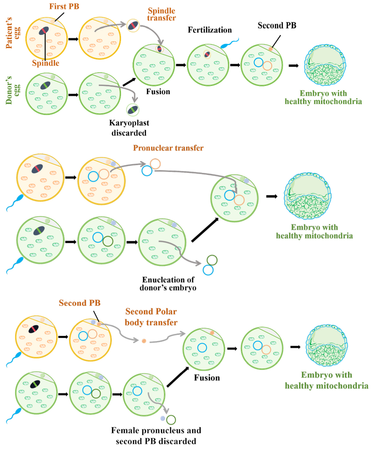
Figure 2. Mitochondrial replacement therapies or techniques (MRT). Created in BioRender.
Comparing the three methods, spindle transfer holds a clear advantage. PT starts with two fertilized eggs—meaning two embryos would be used for one baby—which raises ethical concerns. More importantly, the embryos are activated before nuclear transfer and time window for the transfer is very narrow, and the amount of carryover cytoplasm could be higher. In contrast, spindle transfer requires only one embryo per baby. As the technique advances, the carryover rate can be reduced to less than 1%[96]. Polar body transfer is also highly effective in minimizing carryover. However, since polar bodies are not typically involved in embryo development, their use remains uncertain. While animal studies show promise, other factors—such as DNA methylation and epigenetic modifications—should also be considered.
Although the successful application of MRT has identified one of the most effective means to block the inheritance of mtDNA mutations, despite therapeutic promise, almost all MRT approaches remain vulnerable to low-level mtDNA contamination and mutational reversion; consequently, their clinical use is outlawed in the majority of countries worldwide. Looking ahead, persistent clinical evaluation coupled with evolving ethical standards could position MRT as an essential and potentially irreplaceable remedy for such profoundly destructive conditions.
MST: The spindle-chromosome complex, which harbors the maternal nuclear genome, is extracted from the patient's oocyte and transferred into an enucleated donor egg. This reconstituted oocyte thereby retains the mother's nDNA while acquiring the donor's wild-type mitochondrial pool, and may subsequently be fertilized and implanted into the maternal uterus. PT: The maternal and donor oocytes are each fertilized with paternal sperm. Prior to the onset of cleavage, the pronuclei—containing the nuclear genome—are extracted from the maternal zygote and transferred into the enucleated donor zygote, from which the original nuclear material has previously been removed. PBT: A spindle-protrusion-retained PB2 separation technique was developed, which yielded a near-normal proportion of euploid blastocysts and further reduced mitochondrial carryover in both mice and humans.
CONCLUSION
Primary mitochondrial disorders exhibit profound, multi-system clinical heterogeneity alongside extraordinary genetic complexity in their underlying causative variants, and currently still lack specific treatments for most patients. Multidisciplinary symptomatic management is needed to optimize quality of life. While targeted therapies exist for select disorders, most patients receive empirical "cocktail" therapy, which aims at enhancing mitochondrial function and modulating dynamics.
DECLARATIONS
Acknowledgments
The Graphical Abstract was created with BioRender.
Authors’ contributions
Conceptualization, methodology, literature search, data extraction, writing - original draft: Hu C
Literature search, data extraction, visualization: Wang X
Conceptualization, review & editing: Fang F
Conceptualization, supervision, writing - review & editing, project administration: Huang T
All authors have read and agreed to the published version of the manuscript.
Availability of data and materials
Not Applicable.
AI and AI-assisted tools statement
Not applicable.
Financial support and sponsorship
None.
Conflicts of interest
Huang T is an Editorial Board Member of Journal of Translational Genetics and Genomics. Huang T was not involved in any steps of editorial processing, notably including reviewers' selection, manuscript handling, or decision-making, while the other authors have declared that they have no conflicts of interest.
Ethical approval and consent to participate
Not applicable.
Consent for publication
Not applicable.
Copyright
© The Author(s) 2026.
REFERENCES
1. Gorman GS, Schaefer AM, Ng Y, et al. Prevalence of nuclear and mitochondrial DNA mutations related to adult mitochondrial disease. Ann Neurol. 2015;77:753-9.
2. Mcfarland R, Taylor RW, Turnbull DM. A neurological perspective on mitochondrial disease. Lancet Neurol. 2010;9:829-40.
3. Heath O, Feichtinger RG, Achleitner MT, et al. Mitochondrial disorder diagnosis and management- what the pediatric neurologist wants to know. Eur J Paediatr Neurol. 2025;54:75-88.
4. Wen H, Deng H, Li B, et al. Mitochondrial diseases: from molecular mechanisms to therapeutic advances. Sig Transduct Target Ther. 2025;10:9.
5. Dimauro S, Schon EA, Carelli V, Hirano M. The clinical maze of mitochondrial neurology. Nat Rev Neurol. 2013;9:429-44.
6. Stewart JB, Chinnery PF. The dynamics of mitochondrial DNA heteroplasmy: implications for human health and disease. Nat Rev Genet. 2015;16:530-42.
7. Gorman GS, Chinnery PF, Dimauro S, et al. Mitochondrial diseases. Nat Rev Dis Primers. 2016;2:16080.
8. Smeitink J, Van Den Heuvel L, Dimauro S. The genetics and pathology of oxidative phosphorylation. Nat Rev Genet. 2001;2:342-52.
9. Calame DG, Emrick LT. Functional genomics and small molecules in mitochondrial neurodevelopmental disorders. Neurotherapeutics. 2024;21:e00316.
10. Klopstock T, Priglinger C, Yilmaz A, Kornblum C, Distelmaier F, Prokisch H. Mitochondrial disorders. Dtsch Arztebl Int. 2021;118:741-8.
11. Finsterer J. Genetic, pathogenetic, and phenotypic implications of the mitochondrial A3243G tRNALeu(UUR) mutation. Acta Neurol Scand. 2007;116:1-14.
12. Youle RJ, Van Der Bliek AM. Mitochondrial fission, fusion, and stress. Science. 2012;337:1062-5.
13. Zheng Y, Yang J, Li X, et al. Mitochondria at the crossroads: quality control mechanisms in neuronal senescence and neurodegeneration. Neurobiol Dis. 2025;208:106862.
14. Mclaughlin KL, Mcclung JM, Fisher-Wellman KH. Bioenergetic consequences of compromised mitochondrial DNA repair in the mouse heart. Biochem Biophys Res Commun. 2018;504:742-8.
15. Dautant A, Meier T, Hahn A, Tribouillard-Tanvier D, Di Rago J, Kucharczyk R. ATP synthase diseases of mitochondrial genetic origin. Front Physiol. 2018;9:329.
16. Bazil JN, Pannala VR, Dash RK, Beard DA. Determining the origins of superoxide and hydrogen peroxide in the mammalian NADH:ubiquinone oxidoreductase. Free Radic Biol Med. 2014;77:121-9.
17. Andreyev AY, Kushnareva YE, Murphy AN, Starkov AA. Mitochondrial ROS metabolism: 10 years later. Biochemistry. 2015;80:517-31.
19. Bhatti JS, Bhatti GK, Reddy PH. Mitochondrial dysfunction and oxidative stress in metabolic disorders — A step towards mitochondria based therapeutic strategies. Biochim Biophys Acta. 2017;1863:1066-77.
20. Panov AV, Dikalov SI. Cardiolipin, perhydroxyl radicals, and lipid peroxidation in mitochondrial dysfunctions and aging. Oxid Med Cell Longev. 2020;2020:1-14.
21. Zanfardino P, Amati A, Perrone M, Petruzzella V. The balance of MFN2 and OPA1 in mitochondrial dynamics, cellular homeostasis, and disease. Biomolecules. 2025;15:433.
22. Candé C, Cohen I, Daugas E, et al. Apoptosis-inducing factor (AIF): a novel caspase-independent death effector released from mitochondria. Biochimie. 2002;84:215-22.
23. Kim R, Emi M, Tanabe K. Role of mitochondria as the gardens of cell death. Cancer Chemother Pharmacol. 2005;57:545-53.
24. Ahola S. Mitochondria setting the stage for ferroptosis. Trends Endocrinol Metab. 2026:S1043276026001505.
25. Kolotova A, Shestopalov A, Kutsev S. The role of apoptosis and ferroptosis in primary mitochondrial diseases: mechanisms and pathogenesis. Int J Mol Sci. 2026;27:5931.
26. Kim J, Kim H, Chung JH. Molecular mechanisms of mitochondrial DNA release and activation of the cGAS-STING pathway. Exp Mol Med. 2023;55:510-9.
27. Horssen J, Van Schaik P, Witte M. Inflammation and mitochondrial dysfunction: a vicious circle in neurodegenerative disorders? Neurosci Lett. 2019;710:132931.
28. Aguilar K, Jakubek P, Zorzano A, Wieckowski MR. Primary mitochondrial diseases: the intertwined pathophysiology of bioenergetic dysregulation, oxidative stress and neuroinflammation. Eur J Clin Investig. 2024;54:e14217.
29. Barcelos I, Shadiack E, Ganetzky RD, Falk MJ. Mitochondrial medicine therapies: rationale, evidence, and dosing guidelines. Curr Opin Pediatr. 2020;32:707-18.
30. Parikh S, Goldstein A, Koenig MK, et al. Diagnosis and management of mitochondrial disease: a consensus statement from the Mitochondrial Medicine Society. Genet Med. 2015;17:689-701.
31. De Vries MC, Brown DA, Allen ME, et al. Safety of drug use in patients with a primary mitochondrial disease: an international Delphi‐based consensus. J Inherit Metab Dis. 2020;43:800-18.
32. Sue CM, Balasubramaniam S, Bratkovic D, et al. Patient care standards for primary mitochondrial disease in Australia: an Australian adaptation of the Mitochondrial Medicine Society recommendations. Intern Med J. 2021;52:110-20.
33. Pfeffer G, Majamaa K, Turnbull DM, Thorburn D, Chinnery PF; Cochrane Neuromuscular Group. Treatment for mitochondrial disorders. Cochrane Database Syst Rev. 2012:2012.
34. Reetz K, Lischewski SA, Dogan I, et al. Friedreich's ataxia—a rare multisystem disease. Lancet Neurol. 2025;24:614-24.
36. Lynch DR, Chin MP, Delatycki MB, et al. Safety and efficacy of omaveloxolone in friedreich ataxia (MOXIeStudy). Ann Neurol. 2020;89:212-25.
37. Reid Thompson W, Hornby B, Manuel R, et al. A phase 2/3 randomized clinical trial followed by an open-label extension to evaluate the effectiveness of elamipretide in Barth syndrome, a genetic disorder of mitochondrial cardiolipin metabolism. Genet Med. 2021;23:471-8.
38. Thompson WR, Manuel R, Abbruscato A, et al. Long-term efficacy and safety of elamipretide in patients with Barth syndrome: 168-week open-label extension results of TAZPOWER. Genet Med. 2024;26:101138.
39. Hornby B, Thompson WR, Almuqbil M, et al. Natural history comparison study to assess the efficacy of elamipretide in patients with Barth syndrome. Orphanet J Rare Dis. 2022;17:336.
41. Karaa A, Haas R, Goldstein A, Vockley J, Weaver WD, Cohen BH. Randomized dose-escalation trial of elamipretide in adults with primary mitochondrial myopathy. Neurology. 2018;90:e1212-21.
42. Karaa A, Haas R, Goldstein A, Vockley J, Cohen BH. A randomized crossover trial of elamipretide in adults with primary mitochondrial myopathy. J Cachexia Sarcopenia Muscle. 2020;11:909-18.
43. Karaa A, Bertini E, Carelli V, et al. Efficacy and safety of elamipretide in individuals with primary mitochondrial myopathy: the MMPOWER-3 randomized clinical trial. Neurology. 2023;101:e238-52.
44. Karaa A, Bertini E, Carelli V, et al. Genotype-specific effects of elamipretide in patients with primary mitochondrial myopathy: a post hoc analysis of the MMPOWER-3 trial. Orphanet J Rare Dis. 2024;19:431.
46. Domínguez-González C, Chiang C, Colson A, et al. Pyrimidine Nucleos(t)ide therapy in patients with thymidine kinase 2 deficiency: a multicenter retrospective chart review study. Neurology. 2025;105:e213908.
47. Bermejo-Guerrero L, Hernández-Voth A, Serrano-Lorenzo P, et al. Remarkable clinical improvement with oral nucleoside treatment in a patient with adult-onset TK2 deficiency: a case report. Mitochondrion. 2024;76:101879.
48. Berrahmoune S, Dassi C, Pekeles H, et al. Investigating the safety and efficacy of deoxycytidine/deoxythymidine in mitochondrial DNA depletion disorders: phase 2 open-label trial. J Neurol. 2025;272:307.
49. Johnson SC, Yanos ME, Kayser E, et al. mTOR inhibition alleviates mitochondrial disease in a mouse model of leigh syndrome. Science. 2013;342:1524-8.
50. Sage‐schwaede A, Engelstad K, Salazar R, et al. Exploring mTOR inhibition as treatment for mitochondrial disease. Ann Clin Transl Neurol. 2019;6:1877-81.
51. Repp BM, Mastantuono E, Alston CL, et al. Clinical, biochemical and genetic spectrum of 70 patients with ACAD9 deficiency: is riboflavin supplementation effective? Orphanet J Rare Dis. 2018;13:120.
52. Plantone D, Pardini M, Rinaldi G. Riboflavin in neurological diseases: a narrative review. Clin Drug Investig. 2021;41:513-27.
53. Xu C, Jin H, Li J, et al. Deficiency of the NAD(P)HX metabolic repair system: a treatable mitochondrial disease. Orphanet J Rare Dis. 2026;21:63.
54. Invernizzi F, Panteghini C, Chiapparini L, et al. Thiamine-responsive disease due to mutation of tpk1: importance of avoiding misdiagnosis. Neurology. 2017;89:870-1.
55. Alcázar-Fabra M, Trevisson E, Brea-Calvo G. Clinical syndromes associated with Coenzyme Q10 deficiency. Essays Biochem. 2018;62:377-98.
56. Montini G, Malaventura C, Salviati L. Early coenzyme Q10 supplementation in primary coenzyme Q10 deficiency. N Engl J Med. 2008;358:2849-50.
57. Distelmaier F, Corral-Sarasa J, Jiménez-Sánchez L, et al. Preclinical and first-in-human evidence of 4-hydroxybenzoic acid for mitochondrial COQ2 deficiency. Brain. 2026;149:1784-97.
58. Canda E, Kalkan Ucar S, Coker M. Biotinidase deficiency: prevalence, impact and management strategies. Pediatric Health Med Ther. 2020;11:127-33.
59. Jiang H, Alahmad A, Fu S, et al. Identification and characterization of novel MPC1 gene variants causing mitochondrial pyruvate carrier deficiency. J Inherit Metab Dis. 2022;45:264-77.
60. Pata S, Flores-Rojas K, Gil A, et al. Clinical improvements after treatment with a low-valine and low-fat diet in a pediatric patient with enoyl-CoA hydratase, short chain 1 (ECHS1) deficiency. Orphanet J Rare Dis. 2022;17:340.
61. Sato-Shirai I, Ogawa E, Arisaka A, et al. Valine-restricted diet for patients with ECHS1 deficiency: divergent clinical outcomes in two Japanese siblings. Brain Dev. 2021;43:308-13.
62. Yang H, Yu D. Clinical, biochemical and metabolic characterization of patients with short-chain enoyl-CoA hydratase(ECHS1) deficiency: two case reports and the review of the literature. BMC Pediatr. 2020;20:50.
63. Xu Y, Zhang J, Yu K, et al. A therapeutic regimen for 3-hydroxyisobutyryl-CoA hydrolase deficiency with exercise-induced dystonia. Eur J Paediatr Neurol. 2019;23:755-9.
64. Parikh S, Goldstein A, Karaa A, et al. Patient care standards for primary mitochondrial disease: a consensus statement from the Mitochondrial Medicine Society. Genet Med. 2017;19:1380-97.
65. Orsucci D, Caldarazzo Ienco E, Montano V, Siciliano G, Mancuso M. Mitochondrial stroke-like episodes: the search for new therapies. Pharmacol Res. 2022;180:106228.
66. Mancuso M, Papadopoulou MT, Ng YS, et al. Management of seizures in patients with primary mitochondrial diseases: consensus statement from the InterERNs Mitochondrial Working Group. Eur J Neurol. 2024;31:e16275.
67. Na J, Lee Y. Therapeutic approach to epilepsy in patients with mitochondrial diseases. Yonsei Med J. 2025;66:131.
68. Varughese R, Rahman S. Endocrine dysfunction in primary mitochondrial diseases. Endocr Rev. 2025;46:376-96.
69. Chaudhry A, Thompson DM, Chanoine JP. Diabetes management in maternally inherited diabetes and deafness (MIDD): a review and a proposed treatment algorithm. Diabetes Obes Metab. 2025;28:826-39.
70. Sue CM, Lipsett LJ, Crimmins DS, et al. Cochlear origin of hearing loss in MELAS syndrome. Ann Neurol. 2004;43:350-9.
71. O'toole J. Renal manifestations of genetic mitochondrial disease. Int J Nephrol Renovasc Dis. 2014;7:57-67.
72. Madsen S, Allender B, Blackford R, et al. Ketogenic diet therapy in pyruvate dehydrogenase deficiency: global clinical practice from literature and survey data. Mol Genet Metab. 2026;148:110190.
73. Huang L, Li H, Zhong J, et al. Efficacy and safety of the ketogenic diet for mitochondrial disease with epilepsy: a prospective, open-labeled, controlled study. Front Neurol. 2022;13:880944.
74. Keshavan N, Minczuk M, Viscomi C, Rahman S. Gene therapy for mitochondrial disorders. J Inherit Metab Dis. 2024;47:145-75.
75. Crystal RG, Weinsaft JW, Kaminsky SM, et al. AAVrh.10hFXN gene therapy for the cardiomyopathy of friedreich ataxia: a nonrandomized clinical trial. JAMA Cardiol. 2026;11:709.
76. Faria R, Boisguérin P, Sousa Â, Costa D. Delivery systems for mitochondrial gene therapy: a review. Pharmaceutics. 2023;15:572.
77. Chen BS, Yu-Wai-Man P. From bench to bedside—delivering gene therapy for leber hereditary optic neuropathy. Cold Spring Harb Perspect Med. 2022;12:a041282.
78. Sahel J, Vignal-Clermont C, Yu-Wai-Man P, Biousse V, Blouin L, Taiel M. Neuropathie optique héréditaire de Leber: amélioration bilatérale de la vision après thérapie génique par injection unilatérale. Med Sci. 2021;37:712-5.
79. Yu-Wai-Man P, Newman NJ, Biousse V, et al. Five-year outcomes of lenadogene nolparvovec gene therapy in leber hereditary optic neuropathy. JAMA Ophthalmol. 2025;143:99.
80. Newman NJ, Yu-Wai-Man P, Subramanian PS, et al. Randomized trial of bilateral gene therapy injection for m.11778G>A MT-ND4 Leber optic neuropathy. Brain. 2023;146:1328-41.
81. Newman NJ, Biousse V, Yu-Wai-Man P, et al. Meta-analysis of treatment outcomes for patients with m.11778G>A MT-ND4 Leber hereditary optic neuropathy. Surv Ophthalmol. 2025;70:283-95.
82. Mok BY, De Moraes MH, Zeng J, et al. A bacterial cytidine deaminase toxin enables CRISPR-free mitochondrial base editing. Nature. 2020;583:631-7.
83. Cho S, Lee S, Mok YG, et al. Targeted A-to-G base editing in human mitochondrial DNA with programmable deaminases. Cell. 2022;185:1764-76.e12.
84. Lei Z, Meng H, Liu L, et al. Mitochondrial base editor induces substantial nuclear off-target mutations. Nature. 2022;606:804-11.
85. Lee S, Lee H, Baek G, Kim J. Precision mitochondrial DNA editing with high-fidelity DddA-derived base editors. Nat Biotechnol. 2022;41:378-86.
86. Mok YG, Lee JM, Chung E, et al. Base editing in human cells with monomeric DddA-TALE fusion deaminases. Nat Commun. 2022;13:4038.
88. Zhang X, Zhang X, Ren J, et al. Precise modelling of mitochondrial diseases using optimized mitoBEs. Nature. 2025;639:735-45.
89. Chen L, Hong M, Luan C, et al. Efficient mitochondrial A-to-G base editors for the generation of mitochondrial disease models. Nat Biotechnol. 2025;44:418-29.
90. Chen L, Luan C, Hong M, et al. A mitochondrial disease model is generated and corrected using engineered base editors in rat zygotes. Nat Biotechnol. 2025;44:382-6.
91. Du S, Long Q, Zhou Y, et al. Transplantation of encapsulated mitochondria alleviates dysfunction in mitochondrial and Parkinson’s disease models. Cell. 2026;189:2821-33.e23.
92. Zhang J, Liu H, Luo S, et al. Live birth derived from oocyte spindle transfer to prevent mitochondrial disease. Reprod Biomed Online. 2017;34:361-8.
93. Hyslop LA, Blakely EL, Aushev M, et al. Mitochondrial donation and preimplantation genetic testing for mtDNA disease. N Engl J Med. 2025;393:438-49.
94. Mcfarland R, Hyslop LA, Feeney C, et al. Mitochondrial donation in a reproductive care pathway for mtDNA disease. N Engl J Med. 2025;393:461-8.
95. Li W, Liao X, Lin K, et al. Earlier second polar body transfer and further mitochondrial carryover removal for potential mitochondrial replacement therapy. MedComm. 2023;4:e217.
96. Liao X, Li W, Lin K, et al. Significant decrease of maternal mitochondria carryover using optimized spindle-chromosomal complex transfer. PLoS Biol. 2023;21:e3002313.
97. Sato Y, Nakagawa M, Higuchi I, Osame M, Naito E, Oizumi K. Mitochondrial myopathy and familial thiamine deficiency. Muscle Nerve. 2000;23:1069-75.
98. Bernsen P, Gabreëls F, Ruitenbeek W, Hamburger H. Treatment of complex I deficiency with riboflavin. J Neurol Sci. 1993;118:181-7.
99. Glover EI, Martin J, Maher A, Thornhill RE, Moran GR, Tarnopolsky MA. A randomized trial of coenzyme Q10 in mitochondrial disorders. Muscle Nerve. 2010;42:739-48.
100. Neugebauer J, Reinson K, Bellusci M, et al. Current global vitamin and cofactor prescribing practices for primary mitochondrial diseases: Results of a European reference network survey. J Inherit Metab Dis. 2024;48:e12805.
101. Panetta J, Smith LJ, Boneh A. Effect of high‐dose vitamins, coenzyme Q and high‐fat diet in paediatric patients with mitochondrial diseases. J Inherit Metab Dis. 2004;27:487-98.
102. Gimenes A, Bravo D, Nápolis L, et al. Effect of L-carnitine on exercise performance in patients with mitochondrial myopathy. Braz J Med Biol Res. 2015;48:354-62.
103. Ikawa M, Povalko N, Koga Y. Arginine therapy in mitochondrial myopathy, encephalopathy, lactic acidosis, and stroke-like episodes. Curr Opin Clin Nutr Metab Care. 2020;23:17-22.
104. Koga Y, Povalko N, Inoue E, et al. Therapeutic regimen of l-arginine for MELAS: 9-year, prospective, multicenter, clinical research. J Neurol. 2018;265:2861-74.
105. Ortigoza-escobar JD, Molero-luis M, Arias A, et al. Treatment of genetic defects of thiamine transport and metabolism. Expert Rev Neurother. 2016;16:755-63.
Cite This Article
 Open Access
How to Cite
Hu C, Wang X, Fang F, Huang T. Current and emerging therapeutic strategies for mitochondrial disorders. J Transl Genet Genom. 2026;10:524-44. https://dx.doi.org/10.20517/jtgg.2026.44
Download Citation
If you have the appropriate software installed, you can download article citation data to the citation manager of your choice. Simply select your manager software from the list below and click on download.
Export Citation File
Type of Import
Tips on Downloading Citation
Citation Manager File Format
Type of Import
Direct Import: When the Direct Import option is selected (the default state), a dialogue box will give you the option to Save or Open the downloaded citation data. Choosing Open will either launch your citation manager or give you a choice of applications with which to use the metadata. The Save option saves the file locally for later use.
Indirect Import: When the Indirect Import option is selected, the metadata is displayed and may be copied and pasted as needed.
About This Article
Copyright

Data & Comments
Data
0









Comments
Comments must be written in English. Spam, offensive content, impersonation, and private information will not be permitted. If any comment is reported and identified as inappropriate content by OAE staff, the comment will be removed without notice. If you have any queries or need any help, please contact us at [email protected].