

fig2
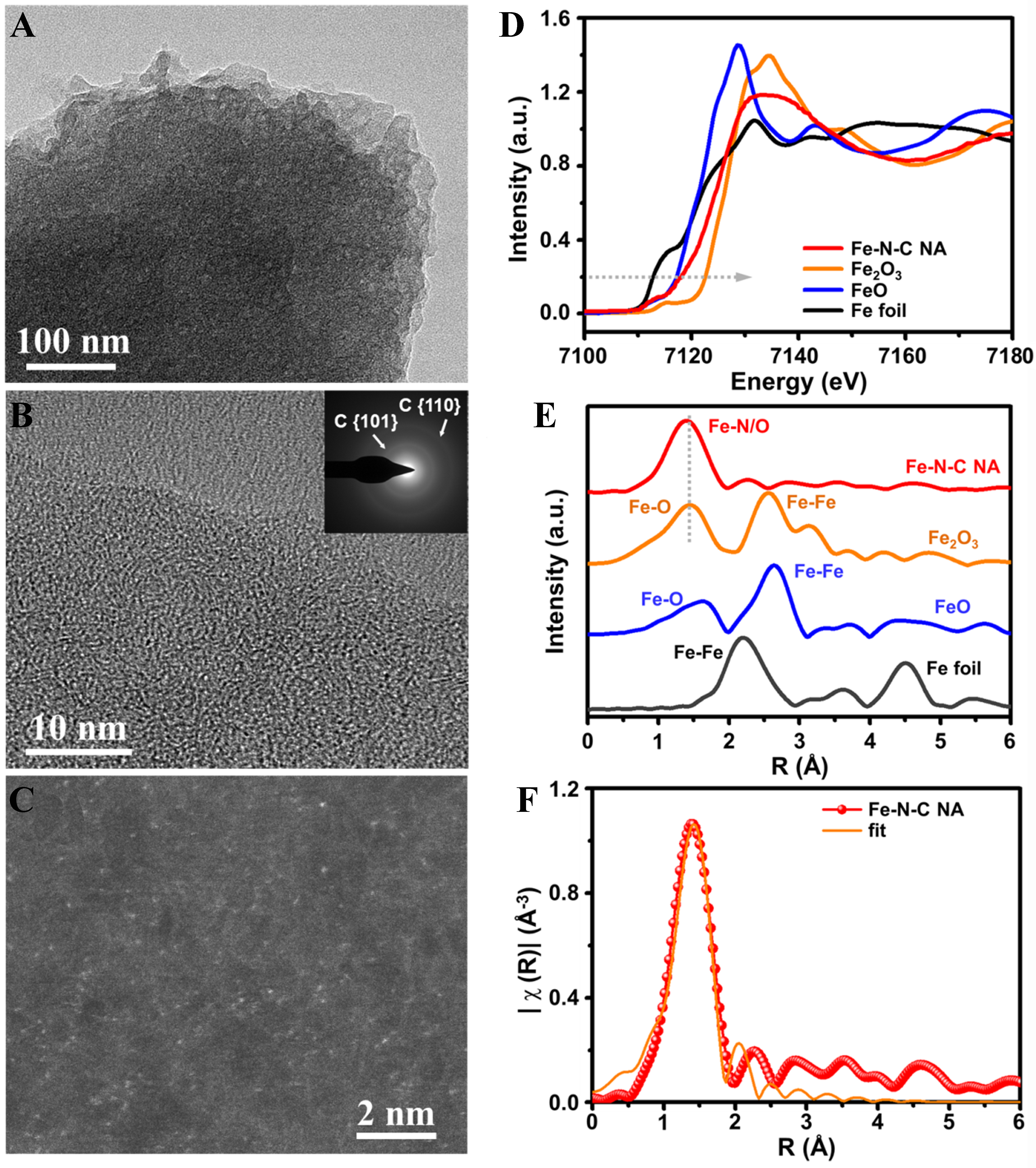
Figure 2. Analysis for the active sites in Fe-N-C NA. (A and B) HR-TEM images in low and high magnification, respectively. (B) the selected area electron diffraction. The d-spacings of the diffraction rings are 0.208 and 0.128 nm corresponding to graphite {101} and {110}, respectively; (C) AC-HR-HAADF-STEM image of Fe SA sites; (D) XANES of E space plots and (E) EXAFS of R space plots for Fe-K edge of Fe-N-C NA and Fe foil, FeO and Fe2O3 references; (F) R space fitting for Fe-K edge of Fe-N-C NA. NA: Nano-assembly; HR-TEM: high-resolution transmission electron microscopy; AC-HR-HAADF-STEM: aberration-corrected high-resolution high-angle annular dark-field scanning transmission electron microscopy; SA: single atom; XANES: X-ray absorption near edge structure; EXAFS: extended X-ray absorption fine structure.


