


From fibrosis to newly formed vessels: a new paradigm for heart failure recovery
 0
0 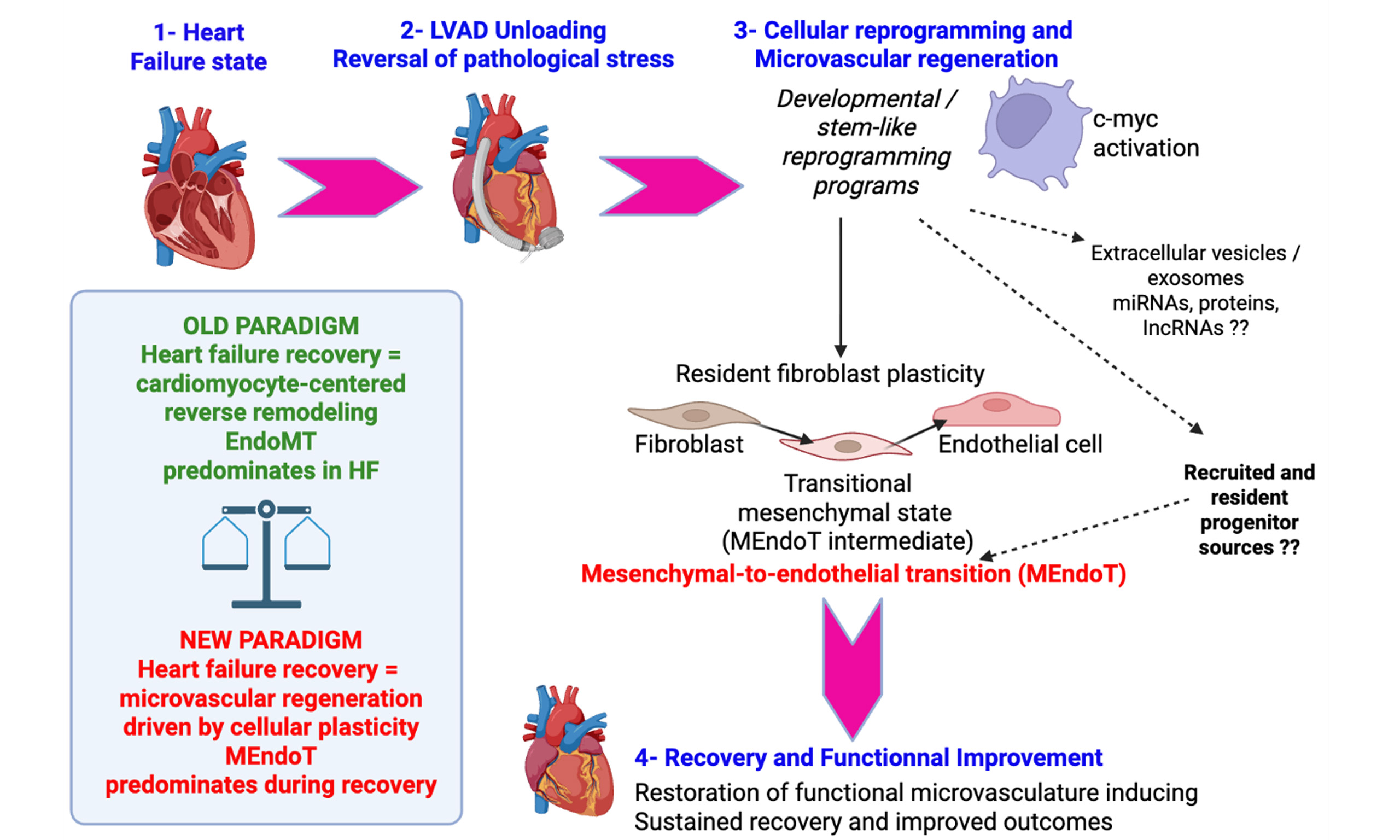
Despite major therapeutic advances, the biological mechanisms driving recovery from heart failure (HF) remain incompletely understood, particularly at the level of the cardiac microvasculature. The study by Li et al.[1] introduces a significant conceptual shift in our understanding of HF recovery, positioning microvascular restoration - not merely cardiomyocyte improvement - as a central determinant of functional reversal[1]. Traditionally, HF therapies have focused on modulating hemodynamics, neurohormonal signaling, or cardiomyocyte survival. However, the demonstration that left ventricular assist device (LVAD) support induces a coordinated reduction in fibrosis alongside increased capillary density highlights the microvasculature as an active and dynamic driver of myocardial recovery[2]. A key contribution of this work is the identification of mesenchymal-to-endothelial transition (MEndoT) as a mechanistic basis for vascular regeneration. Through single-nucleus RNA sequencing and lineage tracing, the authors show that fibroblast populations, classically viewed as terminal effectors of fibrosis, can acquire endothelial identity during recovery. This finding is particularly compelling given the well-established role of fibroblasts in HF pathophysiology, where they contribute to extracellular matrix deposition, increased stiffness, and impaired myocardial compliance. The inverse correlation between fibroblast and endothelial cell abundance observed in post-LVAD tissues underscores a dynamic balance between fibrotic and angiogenic programs. Importantly, this transition is not merely phenotypic but functionally relevant. Post-LVAD myocardial samples exhibit enhanced angiogenic capacity, increased proliferation of non-myocyte populations, and the formation of vascular-like structures in vitro. These observations suggest that the recovering heart reactivates latent plasticity programs in resident cells, enabling structural remodeling that restores perfusion. In this context, MEndoT may represent a reversal of the pathological endothelial-to-mesenchymal transition (EndoMT) known to contribute to fibrosis in HF, effectively rebalancing cellular states toward a regenerative phenotype. In addition to direct cellular plasticity, extracellular vesicles and exosome-mediated transfer of proteins, microRNAs, and other noncoding RNAs may contribute to endothelial-fibroblast communication and facilitate reparative reprogramming during heart failure recovery. The identification of the MYC proto-oncogene (c-Myc) as a regulator adds key mechanistic insight. c-Myc is one of the canonical Yamanaka reprogramming factors alongside OCT4, SOX2, and KLF4, originally identified as sufficient to induce the conversion of somatic cells into induced pluripotent stem cells (iPSCs)[3]. Beyond its role in enhancing reprogramming efficiency, c-Myc functions as a global regulator of transcription, metabolism, and chromatin accessibility, thereby facilitating the rapid proliferation and epigenetic remodeling required for the acquisition of pluripotency[4]. Although subsequent studies have demonstrated that iPSC generation can occur in the absence of exogenous c-Myc, its inclusion markedly accelerates reprogramming kinetics and improves overall efficiency, albeit with increased tumorigenic risk due to its oncogenic properties[3]. In addition to its role in the induction of pluripotency, Myc activity is critical for maintaining the biosynthetic and metabolic state of pluripotent stem cells[4]. Notably, depletion of Myc family proteins induces a reversible, diapause-like dormant state in pluripotent stem cells, characterized by reduced transcriptional output, suppressed anabolic metabolism, and global downregulation of biosynthetic processes[5]. This Myc-dependent regulation highlights a fundamental link among cellular metabolism, proliferation, and pluripotent identity, suggesting that Myc acts not only as a driver of reprogramming but also as a key modulator of stem cell state transitions between active proliferation and dormancy[5]. As a driver of proliferation, metabolic reprogramming, and cell fate plasticity, Myc’s upregulation in post-LVAD tissues links microvascular recovery to activation of developmental and regenerative gene programs, suggesting that HF recovery extends beyond structural remodeling toward cellular reprogramming. These findings also position the microvasculature as a limiting factor in recovery: capillary rarefaction and impaired perfusion sustain hypoxia and fibrosis, whereas restoration of vascular density correlates with improved function. Although increased capillary density is unlikely to be sufficient alone to restore cardiac function, improved perfusion may create a permissive microenvironment supporting cardiomyocyte survival, metabolic recovery, and reverse remodeling. Thus, perfusion appears not merely a consequence but a prerequisite of recovery, supporting therapeutic strategies targeting angiogenesis and vascular remodeling. These observations raise the possibility that microvascular recovery may extend beyond intrinsic fibroblast plasticity and involve the activation of broader regenerative cell programs. Importantly, the precise cellular origin of the transitional populations identified during recovery remains incompletely resolved and may involve multiple complementary sources. While Li et al. provide evidence supporting fibroblast-to-endothelial transition[1], at least a subset of these fibroblast-like intermediary cells may derive from recruited bone marrow-derived progenitors or resident primitive stem/progenitor populations activated under conditions of mechanical unloading and reversal of pathological stress. In this framework, both resident cardiac cellular plasticity and circulating progenitor recruitment could contribute to MEndoT-associated vascular regeneration, consistent with previous observations describing mobilization of stem-like populations during tissue repair and ischemic recovery. The capacity of human fibroblast-like cells or mesenchymal stromal/stem cells (MSCs) to differentiate into endothelial cells (ECs) remains a subject of ongoing debate. While early studies suggested a degree of endothelial plasticity, subsequent investigations have yielded inconsistent and sometimes contradictory findings, underscoring the heterogeneity of MSC populations and the context-dependent nature of their differentiation potential. Notably, immature or developmentally primitive mesenchymal populations, such as hemangioma-derived stem cells (Hem-SCs)[6] and Wharton’s jelly-derived MSCs (WJ-MSCs)[7], have been reported to exhibit endothelial differentiation capabilities under specific in vitro and in vivo conditions. In contrast, other mesenchymal-like populations, particularly those derived from cardiovascular tissues, appear to exhibit more restricted lineage potential. Valvular interstitial cells (VICs), especially those isolated from calcified aortic valve disease, have been characterized as phenotypically plastic and capable of osteogenic and myofibroblastic differentiation. However, despite this apparent plasticity, current evidence indicates that VICs lack the capacity to transdifferentiate into functional endothelial cells under standard in vitro conditions, highlighting lineage constraints linked to their tissue-specific origin[8]. Furthermore, the role of very small embryonic-like stem cells (VSELs) in endothelial differentiation has attracted attention. VSELs, often isolated from CD133+ cell fractions, have been proposed as a primitive pluripotent population capable of giving rise to mesenchymal progenitors. These MSC-like derivatives have, in turn, demonstrated the ability to differentiate into endothelial lineages, suggesting a hierarchical model in which early stem cell populations contribute indirectly to endothelial regeneration[9]. Collectively, these findings highlight that endothelial differentiation capacity is not a universal property of all mesenchymal populations but is instead restricted to specific subsets characterized by developmental immaturity or distinct molecular signatures. This underscores the importance of rigorous phenotypic characterization and standardized differentiation protocols when evaluating MSCs plasticity for vascular regenerative applications. VSELs are more primitive stem cell populations and have been described in adult tissues and potentially within the heart itself[10], and could participate in this regenerative process described by Li et al.[1]. This intermediate mesenchymal state is strikingly reminiscent of the fibroblast-like populations identified in the Li et al. dataset, suggesting that at least a subset of “fibroblasts” undergoing MEndoT may represent progenitor-derived mesenchymal intermediates[1]. This interpretation aligns with the concept that tissue injury mobilizes multipotent stem cells which can home to damaged myocardium and contribute to repair. Such behavior parallels the increase in capillary density and functional vascular networks observed in post-LVAD myocardium. The observed downregulation of TWIST1/2 during recovery is particularly intriguing given the established role of TWIST proteins in mesenchymal transition and fibrosis. Such modulation may favor endothelial differentiation and may parallel developmental regenerative programs observed in neonatal mammalian hearts, where angiogenesis and cardiomyocyte regeneration are closely coupled. Integrating these findings, one can envision a model in which LVAD-induced hemodynamic unloading and reversal of pathological stress create a permissive microenvironment that reactivates developmental or stem-like programs. Although LVAD support provides a robust model of mechanical unloading and recovery, it remains possible that similar microvascular regenerative programs may occur, at least partially, during pharmacological reverse remodeling induced by guideline-directed medical therapies. In addition, the nature of mechanical unloading itself may influence vascular remodeling responses, as continuous-flow and pulsatile-flow LVADs generate distinct hemodynamic and endothelial shear stress environments. Such differences could potentially modulate endothelial activation, angiogenesis, mechanotransduction pathways, and regenerative signaling programs, although these aspects were not specifically addressed by Li et al. and warrant further investigation[1]. In this setting, c-Myc activation identified by Li et al. as a key regulator of MEndoT may not only drive direct fibroblast reprogramming but also facilitate expansion or differentiation of resident or recruited progenitor cells[1]. This is consistent with the known role of c-Myc as a Yamanaka factor capable of inducing pluripotency and cellular plasticity, suggesting that the observed endothelial regeneration may be rooted in a broader reactivation of stemness pathways rather than a purely lineage-restricted transition[3,4]. Such a framework also offers a plausible explanation for the efficacy of expanded CD34+ cell therapy in ischemic heart disease[11]. Clinical and translational studies, including the EXCELLENT trial (Expanded Cell Endocardiac Transplantation; NCT02669810), demonstrate that expanded CD34+ cells improve vascularization, reduce fibrosis, and enhance functional recovery after myocardial infarction[11]. While traditionally attributed to endothelial progenitor activity and paracrine signaling, these effects may also reflect the presence of primitive progenitor subsets within the CD34+ compartment, potentially including VSEL-like cells. Indeed, CD34+ cells have been shown to promote angiogenesis, limit scar formation, and support myocardial repair through both direct differentiation and microenvironmental modulation. The observed increase in capillary density likely reflects a combination of angiogenesis from pre-existing vessels and vasculogenic mechanisms involving progenitor-like intermediates capable of integrating into the recovering microvascular network. This raises the intriguing possibility that the beneficial effects of CD34+ cell therapies may derive not solely from Endothelial Progenitor Cells (EPCs) but from a broader hierarchy of progenitors, including VSELs capable of transitioning through mesenchymal states before endothelial differentiation. Such a mechanism would mirror the fibroblast-to-endothelial trajectories described by Li et al., further supporting a unified model of vascular regeneration[1].
Overall, these findings support a paradigm in which heart failure recovery is fundamentally a process of microvascular regeneration driven by cellular plasticity and reactivation of developmental programs. Beyond fibroblast reprogramming alone, the potential contribution of progenitor-derived intermediates suggests that endothelial renewal may arise from a broader regenerative hierarchy. This integrated view not only refines our understanding of MEndoT but also provides a unifying framework linking mechanical unloading, transcriptional reprogramming, and cell-based therapies. Targeting these interconnected pathways may open new avenues to enhance durable vascular and functional recovery in heart failure.
DECLARATIONS
Acknowledgments
The Graphical Abstract was created with BioRender.com (Created in Smadja DM (2026) https://BioRender.com/bpyi2bf).
Authors’ contribution
The author contributed solely to the article.
Availability of data and materials
Not applicable.
AI and AI-assisted tools statement
Not applicable.
Financial support and sponsorship
None.
Conflicts of interest
The author declared no conflicts of interest.
Ethical approval and consent to participate
Not applicable.
Consent for publication
Not applicable.
Copyright
© The Author(s) 2026.
REFERENCES
1. Li S, Gupta K, Ranka RK, et al. Recovery from heart failure: microvascular mechanisms. Circulation. 2026;153:1661-78.
2. Drakos SG, Kfoury AG, Selzman CH, et al. Left ventricular assist device unloading effects on myocardial structure and function: current status of the field and call for action. Curr Opin Cardiol. 2011;26:245-55.
3. Yamanaka S. Two decades of induced pluripotent stem cell research: from discovery to diverse applications. Cell Stem Cell. 2026;33:372-81.
4. Nakagawa M, Karagiannis P, Yamanaka S. When Myc’s asleep, embryonic stem cells are dormant. EMBO J. 2016;35:801-2.
5. Scognamiglio R, Cabezas-Wallscheid N, Thier MC, et al. Myc depletion induces a pluripotent dormant state mimicking diapause. Cell. 2016;164:668-80.
6. Khan ZA, Boscolo E, Picard A, et al. Multipotential stem cells recapitulate human infantile hemangioma in immunodeficient mice. J Clin Invest. 2008;118:2592-9.
7. Wang SH, Lin SJ, Chen YH, et al. Late outgrowth endothelial cells derived from Wharton jelly in human umbilical cord reduce neointimal formation after vascular injury: involvement of pigment epithelium-derived factor. Arterioscler Thromb Vasc Biol. 2009;29:816-22.
8. Gendron N, Rosa M, Blandinieres A, et al. Human aortic valve interstitial cells display proangiogenic properties during calcific aortic valve disease. Arterioscler Thromb Vasc Biol. 2021;41:415-29.
9. Guerin CL, Loyer X, Vilar J, et al. Bone-marrow-derived very small embryonic-like stem cells in patients with critical leg ischaemia: evidence of vasculogenic potential. Thromb Haemost. 2015;113:1084-94.
10. El-Helw M, Chelvarajan L, Abo-Aly M, et al. Identification of human very small embryonic like stem cells (VSELS) in human heart tissue among young and old individuals. Stem Cell Rev Rep. 2020;16:181-5.
Cite This Article
 Open Access
How to Cite
Download Citation
Export Citation File:
Type of Import
Tips on Downloading Citation
Citation Manager File Format
Type of Import
Direct Import: When the Direct Import option is selected (the default state), a dialogue box will give you the option to Save or Open the downloaded citation data. Choosing Open will either launch your citation manager or give you a choice of applications with which to use the metadata. The Save option saves the file locally for later use.
Indirect Import: When the Indirect Import option is selected, the metadata is displayed and may be copied and pasted as needed.
About This Article
Copyright

Data & Comments
Data
0









Comments
Comments must be written in English. Spam, offensive content, impersonation, and private information will not be permitted. If any comment is reported and identified as inappropriate content by OAE staff, the comment will be removed without notice. If you have any queries or need any help, please contact us at [email protected].