


Intracardiac echocardiography in the diagnosis and treatment of atrial fibrillation
 0
0 Abstract
Intracardiac echocardiography (ICE) has become an indispensable real-time imaging modality in atrial fibrillation (AF) ablation procedures, offering distinct advantages across multiple domains, including pre-procedural exclusion of left atrial appendage (LAA) thrombus, real-time guidance for transseptal puncture, catheter localization during ablation, anatomical assessment for LAA closure, and monitoring of complications such as pericardial effusion. Recent investigations have further expanded its utility, particularly in pre-ablation LAA thrombus screening and in guiding emerging pulsed-field ablation techniques. Beyond procedural navigation, ICE enables early detection and timely management of potentially serious complications, while also substantially reducing radiation exposure during transseptal puncture. This review provides a comprehensive overview of the broadening applications of ICE in AF catheter ablation and related interventions, with a specific focus on its current roles in AF ablation and LAA occlusion. The synthesized evidence offers a valuable framework for optimizing future clinical strategies that integrate ICE into AF diagnosis and treatment. Given its considerable benefits in enhancing both safety and efficacy, the broader adoption of ICE in these procedures is strongly warranted.
Keywords
INTRODUCTION
Currently, catheter ablation and left atrial appendage (LAA) closure represent the two primary interventional strategies for atrial fibrillation (AF). Recent evidence suggests that catheter ablation offers superior clinical outcomes compared with pharmacological therapy in patients with AF[1-3], and current guidelines endorse it as a first-line treatment for those who are drug-refractory[2,3]. For patients at high bleeding risk who are unsuitable for anticoagulation, or those anticipated to respond poorly to ablation, LAA closure provides a safer alternative to long-term anticoagulation[4-7]. As the adoption of these two interventional approaches continues to expand, there is an increasing demand for enhanced procedural safety and efficacy.
Imaging plays an essential role throughout the entire workflow of AF interventional therapy[8,9]. Preoperative transesophageal echocardiography (TEE) remains the standard modality for excluding LAA thrombus prior to ablation; however, it frequently causes patient discomfort, including nausea and vomiting. Conventional fluoroscopy-guided transseptal puncture inevitably exposes both operators and patients to radiation[10]. Preoperative LAA computed tomography angiography (CTA) allows detailed assessment of LAA morphology, facilitating accurate occluder sizing before LAA occlusion and enabling postoperative evaluation of device positioning and endothelialization. Furthermore, integration of CTA data with electroanatomic mapping and fluoroscopic angiography can improve intraoperative ablation targeting. Nevertheless, these modalities primarily provide static anatomical images, which limits their real-time positional accuracy during procedures.
The rapid advancement of intracardiac echocardiography (ICE) has effectively addressed these limitations. ICE offers prominent clinical benefits across multiple key steps, including preoperative exclusion of LAA thrombus, real-time guidance for transseptal puncture, intraoperative positioning of the ablation catheter, anatomical evaluation during LAA closure, and early detection of procedural complications such as pericardial effusion. It enables high-resolution visualization of the left atrium (LA) and LAA fine structures, while also significantly reducing intraoperative fluoroscopy exposure[11,12] [Table 1].
Key parameters comparison of ICE, TEE, CTA and fluoroscopy
| Key parameters | ICE | TEE | CTA | Fluoroscopy |
| Sensitivity/specificity | Sensitivity 92%-96%, specificity 90%-95%[17,24]; enables real-time detection of thrombus, pericardial effusion, and catheter malposition | Sensitivity 95%-98%, specificity 93%-96%[19,21]; gold standard for LAA thrombus screening, only for preoperative use | Anatomical evaluation sensitivity > 98%[8]; provides static anatomical images and is poor for dynamic lesion diagnosis | Cannot identify soft tissue lesions or microthrombi; has low diagnostic sensitivity and specificity for indicators related to AF interventions |
| Patient comfort | High. Vascular access only; no nausea or pharyngeal discomfort | Poor. Esophageal probe placement often causes nausea, vomiting, and throat injury | Moderate. Non-invasive, but requires breath-holding during scanning | High. Fully non-invasive with no physical discomfort |
| Requirement for general anesthesia | Not required; performed under local anesthesia | Usually required or combined with deep sedation | Not required; mild sedation for agitated patients only | Not required; local anesthesia for routine use |
| Radiation exposure | No ionizing radiation | No ionizing radiation | Moderate to high ionizing radiation | Continuous low-dose ionizing radiation |
| Procedural workflow efficiency | High. Integrated with intraoperative procedures, no extra preoperative preparation | Low. Independent preoperative examination, unable to guide real-time operation | Low. Complicated preoperative preparation, only for anatomical reference | Moderate. Easy to operate, single-function, needs combination with other imaging modalities |
| Core application in AF interventions | Real-time intraoperative guidance for ablation and LAA closure | Preoperative LAA thrombus exclusion | Preoperative anatomical assessment and surgical planning | Basic intraoperative catheter positioning |
This review aims to summarize the latest clinical applications of ICE in AF interventional therapy, with particular emphasis on its value in improving overall procedural safety and therapeutic efficacy.
ICE probe types
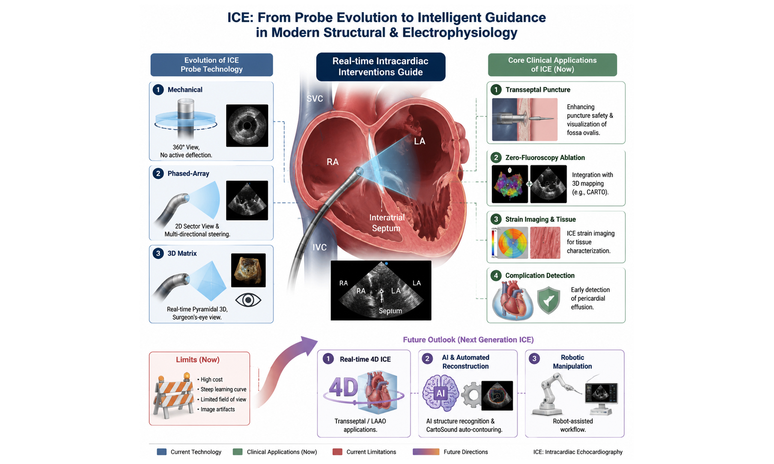
ICE probes can be classified into two main categories. Mechanical probes (e.g., Boston Scientific Ultra ICE) utilize a rotating crystal to generate a 360° radial view but lack active tip deflection. In contrast, electronic phased-array probes (e.g., CartoSound, AcuNav) employ beam-steering technology to produce fan-shaped images and offer multi-directional deflection as well as Doppler functionality[13]. However, 2D imaging still requires mental spatial reconstruction by the operator. 3D matrix probes address this limitation by using a matrix array of crystals to acquire pyramidal volume data in real time, thereby providing intuitive, surgeon’s-eye views. Their primary advantage lies in seamless integration with electroanatomical mapping systems (e.g., CARTO), which enhances navigational accuracy[13]. Additionally, ICE enables non-contact 3D reconstruction through catheter rotation. Some ICE catheters can be directly visualized on mapping systems, further clarifying their spatial position. Key anatomical structures can be identified from the right atrium (RA), right ventricle (RV), or LA[14].
CLINICAL INDICATIONS AND SPECIFIC APPLICATIONS
Pre-procedural LAA thrombus evaluation
The presence of LAA thrombus remains an absolute contraindication to AF ablation. TEE has traditionally been the gold standard for thrombus exclusion. Early studies from 2002 demonstrated that ICE could provide image quality comparable to that of TEE[15]. However, initial ICE catheter placement in the RA often failed to fully visualize the distal LAA[16]. Subsequent optimization of probe positioning-specifically within the right ventricular outflow tract or the supra-pulmonary valve region-enabled clear, multi-planar LAA views, with thrombus detection rates equivalent to those of TEE[17-20]. In selected patients and experienced centers, ICE may serve as an alternative or complementary imaging modality[21]. Nonetheless, operators using this type of ICE probe typically require additional training to master catheter-handling techniques. Some studies have even suggested that ICE may offer higher sensitivity in patients with negative or equivocal TEE findings[22,23]. A large retrospective study reported that ICE shortened hospital stays and reduced the incidence of post-procedural fever[24]. Most recently, a randomized controlled trial involving 1,810 patients demonstrated that ICE was noninferior to TEE in preventing peri-procedural thromboembolic events, while offering additional advantages in safety, efficiency, and patient comfort[25]. It should be noted that preoperative LAA thrombus assessment via ICE is generally performed during the procedure itself. Therefore, this approach may be more suitable for emergency surgeries or for patients who are unable to undergo CTA or TEE scanning [Table 2].
Summary of key representative clinical studies on intracardiac echocardiography-guided atrial fibrillation interventions
| References | Study type | Sample size | Main findings | Clinical implications |
| Hu et al.[25] | Prospective randomized controlled trial | 1,810 AF ablation patients | ICE was non-inferior to TEE in preventing peri-procedural thromboembolic events. ICE-guided procedures eliminated the need for general anesthesia, significantly improved patient comfort, and shortened total procedural duration without increasing adverse events | ICE can replace routine peri-procedural TEE screening for AF ablation, simplifying the preoperative workflow and reducing anesthesia-related risks |
| Wang et al.[24] | Prospective observational study (ICE-TEE trial) | 526 AF ablation patients | Intraoperative ICE exhibited consistent accuracy with TEE in LAA thrombus detection. ICE guidance reduced postoperative fever incidence and shortened hospital stay, with comparable procedural safety profiles | Intraoperative ICE-based LAA thrombus assessment is a reliable alternative to preoperative TEE, optimizing clinical workflow and postoperative recovery |
| Wang et al.[37] | Multicenter prospective trial (PAF-ICE trial) | 1,208 paroxysmal AF patients | Systematic ICE guidance enabled standardized minimal/zero-fluoroscopy AF ablation in Chinese multicenter practice. It drastically reduced fluoroscopy time and radiation exposure while maintaining identical acute procedural success and mid-term recurrence rates | ICE is feasible for widespread adoption of zero-fluoroscopy AF ablation in large-volume clinical centers, realizing radiation-free and high-precision intervention |
| Ahn et al.[50] | Prospective randomized controlled trial | 328 patients undergoing cryoballoon AF ablation | ICE-guided zero-fluoroscopy cryoablation achieved equivalent acute pulmonary vein isolation (PVI) success and 12-month AF recurrence rates compared with fluoroscopy-guided procedures. It significantly reduced radiation exposure and contrast agent consumption | ICE enables safe and efficient zero-fluoroscopy cryoballoon ablation, which is optimal for radiation-sensitive patients and contrast-intolerant populations |
| Dello Russo et al.[56] | Multicenter propensity-matched analysis | 896 AF patients undergoing pulsed field ablation (PFA) | Real-time ICE visualization ensured stable catheter-tissue contact and accurate lesion deployment during PFA. ICE-guided zero-fluoroscopy PFA achieved comparable acute success and short-term safety to conventional imaging-guided procedures | ICE addresses the navigation dilemma of incompletely integrated PFA catheters, promoting the clinical application of zero-fluoroscopy pulsed field ablation technology |
| Serpa et al.[64] | Updated systematic review and meta-analysis | 18 eligible studies (n = 2,468) for LAAO | ICE-guided LAAO demonstrated equivalent procedural success rate and peri-device leak rate versus TEE-guided approaches. ICE significantly shortened procedural time and reduced radiation and contrast usage without increasing major complications | ICE is a safe, efficient, and cost-effective alternative to TEE for routine LAAO guidance, optimizing procedural efficiency and reducing procedural trauma |
| Bottoni et al.[31] | Retrospective multicenter analysis | 1245 AF patients receiving transseptal puncture | Systematic ICE guidance reduced septum-related complications and accidental pericardial puncture risk during transseptal access. It showed unique advantages in complex anatomical conditions, including thickened interatrial septum and atrial septal malformation | ICE markedly improves the safety of transseptal puncture, especially for high-risk and complex anatomical AF interventional cases |
| Motoike et al.[42] | Prospective single-center study | 218 patients undergoing radiofrequency AF ablation | ICE-based real-time atrial wall thickness measurement enabled individualized energy titration. This personalized strategy improved acute PVI success rate and reduced 12-month AF recurrence compared with fixed energy protocols | ICE-mediated tissue characterization facilitates personalized ablation parameter adjustment, improving long-term efficacy of radiofrequency AF ablation |
| Xu et al.[43] | Meta-analysis of RCTs and observational studies | Included 22 studies (n = 3,892) | Compared with non-ICE guidance, ICE-guided AF ablation significantly reduced procedural time, fluoroscopy dosage, and major complication rates, with comparable acute success and long-term sinus rhythm maintenance rates | ICE guidance comprehensively improves the overall safety and efficiency of AF catheter ablation without compromising procedural efficacy |
| Pongratz et al.[49] | Prospective cohort subanalysis (FREEZE trial) | 412 cryoballoon ablation patients | Intraoperative ICE monitoring effectively detected early phrenic nerve traction and pericardial effusion during cryoablation, reducing irreversible phrenic nerve injury and severe complication rates | ICE provides real-time functional monitoring during cryoballoon ablation, enabling early intervention and improving procedural safety |
| Sularz et al.[65] | Prospective observational study | 196 patients undergoing Watchman FLX LAAO | 3D ICE-guided LAAO achieved high consistency with TEE in LAA sizing and occluder positioning. It reduced intraoperative contrast usage and avoided general anesthesia in most patients | Novel 3D ICE is a reliable imaging tool for precise deployment of LAAO devices, further optimizing minimally invasive procedural strategies |
| Baran et al.[19] | Prospective single-center study (Action-Ice I Study) | 208 AF ablation patients | ICE exhibited high sensitivity and specificity for LAA thrombus detection, with consistent diagnostic performance compared with TEE. ICE could identify marginal thrombus missed by conventional TEE in partial cases | ICE serves as a valid intraoperative alternative and complementary tool for LAA thrombus screening during AF ablation procedures |
Transseptal puncture guidance
ICE provides real-time, high-definition visualization of the interatrial septum and fossa ovalis, thereby enhancing puncture safety and substantially reducing radiation exposure compared with conventional fluoroscopy[26-28]. A 15-year multicenter retrospective analysis confirmed that ICE significantly reduced septal-related complications during left atrial procedures[29,30]. ICE is particularly valuable in complex anatomical scenarios, including thickened septum, atrial septal aneurysm, post-closure of atrial septal defect/patent foramen ovale (ASD/PFO), and congenital variants[13,26,27,29-34]. More recently, the integration of ICE with 3D electroanatomical mapping has enabled zero- or near-zero-fluoroscopy ablation, as confirmed by a randomized trial in which over half of 448 patients completely avoided X-ray exposure[35-37] [Table 2, Figure 1].
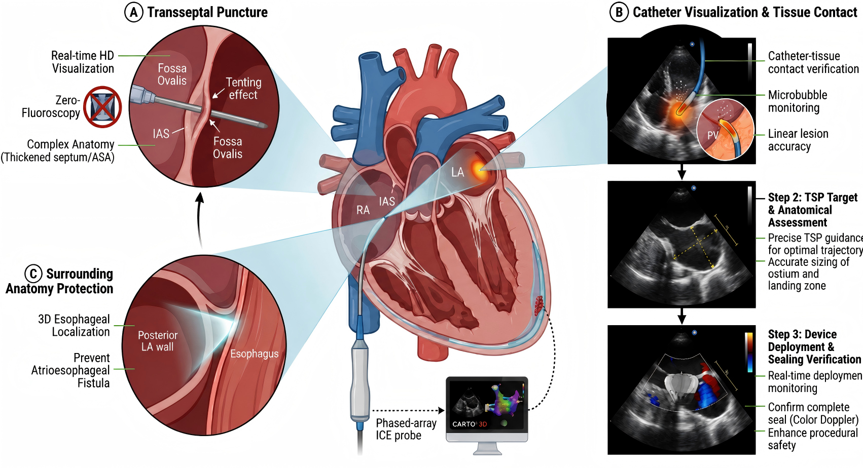
Figure 1. Schematic overview of the core clinical applications of intracardiac echocardiography (ICE) in atrial fibrillation ablation and left atrial appendage closure procedures. (A) Transseptal puncture (TSP) guidance. ICE provides real-time high-definition visualization of the interatrial septum (IAS) and fossa ovalis, enabling safe puncture even in complex anatomies (e.g., thickened septum or atrial septal aneurysm) by clearly visualizing the tenting effect, with the potential to reduce or eliminate fluoroscopy exposure. (B) Stepwise procedural guidance. Top panel: Direct visualization of catheter-tissue contact, microbubble formation, and linear lesion accuracy during pulmonary vein (PV) isolation. Middle panel: Target and anatomical assessment for TSP, including precise trajectory planning and accurate sizing of the left atrial ostium and landing zone. Bottom panel: Real-time monitoring of device deployment and immediate sealing verification using color Doppler, confirming complete occlusion and enhancing procedural safety. (C) Surrounding anatomy protection. ICE enables 3D localization of the esophagus relative to the posterior left atrial (LA) wall, facilitating risk mitigation strategies to prevent atrioesophageal fistula formation. RA: right atrium; LA: left atrium.
Catheter visualization and lesion assessment
During AF catheter ablation, ICE enables real-time visualization of catheters and target areas, as well as monitoring of ablation lesions, thereby enhancing both procedural safety and efficacy [Table 2, Figure 1].
Radiofrequency ablation
ICE clearly visualizes the pulmonary vein antrum and ostia to help prevent pulmonary vein stenosis[38], and provides real-time 3D localization of the esophagus to reduce the risk of atrioesophageal fistula[39]. It also assesses catheter-tissue contact to ensure effective lesion formation. Studies have shown that ICE guidance improves energy delivery and linear lesion accuracy, shortening procedure time and increasing acute success rates[40,41]. Additionally, ICE allows energy titration based on atrial wall thickness. An individualized ICE-guided energy strategy has been shown to improve acute success and reduce one-year AF recurrence compared with a fixed protocol[42]. Although long-term success rates may be comparable[43] , multiple analyses confirm that ICE significantly reduces fluoroscopy time, radiation exposure, and procedure duration without increasing complications[44].
Cryoballoon ablation
ICE also plays a significant role in cryoablation. It assists in selecting the appropriate balloon size, facilitates precise positioning at the pulmonary vein antrum, and employs color Doppler to assess complete pulmonary vein occlusion, thereby predicting acute ablation success and ruling out acute venous narrowing[45,46]. Two small randomized studies showed that ICE guidance shortened procedure time and reduced radiation and contrast agent use; although it did not significantly affect long-term recurrence rates, it improved procedural efficiency[47,48]. A large observational study further indicated that ICE-guided cryoablation achieved higher rates of acute pulmonary vein isolation, fewer complications, and lower radiation exposure[49,50]. Additionally, ICE can monitor diaphragmatic movement in real time during right-sided pulmonary vein ablation, providing early warning of phrenic nerve injury[51].
Pulsed field ablation (PFA)
As a novel non-thermal ablation technology, PFA relies heavily on adequate electrode-tissue contact for lesion effectiveness. Preclinical studies have shown that good contact enhances electric field penetration and creates deeper transmural lesions[52-54]. Therefore, for PFA catheters that are not yet fully integrated with 3D electroanatomical systems (e.g., the FARAPULSE™ system), ICE holds significant value for catheter navigation, positioning, and contact assessment. Preliminary clinical experience demonstrates that ICE enables zero-fluoroscopy guidance of PFA procedures, with acute success, safety, and short-term efficacy comparable to those of fluoroscopic guidance[55,56]. Research in this area is still accumulating, and standardized application pathways for ICE in PFA require further establishment.
Left atrial appendage occlusion (LAAO)
LAAO is indicated for AF patients at moderate-to-high stroke risk who are unsuitable for long-term oral anticoagulation[4-6,57]. TEE has been the traditional imaging guide, but ICE may serve as an alternative or complementary modality in selected patients and experienced centers. Studies have shown that ICE-guided LAAO is safe and effective, with comparable peri-device leak rates, shorter procedure times, and reduced radiation and contrast use[58,59]. Although 2D imaging is limited by LAA anatomical variability[60-62], ICE accurately measures LAA dimensions for device sizing[63,64]. Recent studies indicate that 3D ICE outperforms 2D ICE in device sizing and implantation guidance, showing high agreement with pre-procedural TEE[63]. Novel 3D ICE probes (e.g., VeriSight Pro) have been successfully used to guide Watchman FLX implantation[64,65] [Table 2, Figure 1].
Real-time complication detection and immediate management
ICE provides critical support for real-time complication monitoring and immediate management during AF ablation procedures, significantly enhancing overall procedural safety. Data show that the use of ICE can reduce complication rates and the need for repeat ablation by approximately 36% compared with conventional ablation[66]. Its core advantages lie in the early detection of various complications. For example, when the ICE probe is placed in the RV to image the left ventricular (LV) posterior wall and adjacent pericardium, it can detect even a trace amount of pericardial effusion around the LV at an early stage[67]. Simultaneously, ICE can sensitively detect thrombus formation on catheters or sheaths, guiding prompt adjustment of anticoagulation or thrombus removal to reduce embolic risk[68,69]. During right-sided ablation, it can also monitor phrenic nerve function by observing diaphragmatic movement, thereby preventing permanent injury[70]. Furthermore, ICE can visualize tissue microbubbles and spontaneous echo contrast generated during ablation in real time, aiding in the assessment of ablation response and hypercoagulable states to optimize procedural decisions. Thus, ICE facilitates a shift from reactive management to proactive prevention, establishing itself as a crucial imaging tool for ensuring the safety of AF interventions [Table 2, Figure 1].
Myocardial tissue characterization by ICE strain imaging
Beyond anatomical guidance, ICE enables functional assessment of the atrial myocardium. A study using ICE-derived endocardial and myocardial speckle-tracking strain in patients undergoing AF ablation revealed that left atrial tissue characteristics and function were significantly impaired, particularly in those with persistent AF. Although inter-observer variation in strain measurements was minimal, further software refinement is needed to enhance clinical applicability[71] [Table 2].
Limitations and future developments
Despite its clinical advantages, ICE still faces several practical and technical barriers that limit its widespread adoption. First, the technology is associated with a steep learning curve and substantial operator dependence. Operators require systematic training to master probe positioning, image interpretation, and intraoperative manipulation. Moreover, the assessment of ablation lesions lacks standardized quantitative criteria, remaining largely subjective and variable among different operators. In addition, both 2D and 3D ICE involve inherent trade-offs between imaging field of view and resolution, which further increase the complexity of operation. Second, evidence supporting novel applications remains incomplete. For emerging techniques such as PFA, as well as 3D and 4D ICE, relevant clinical data are still accumulating[72,73]. Standardized procedural protocols, along with robust long-term safety and efficacy data, have yet to be fully established, thereby limiting their routine use on a large scale[74]. Third, cost remains a prominent concern: disposable ICE catheters entail high procurement expenses, which raise overall procedural costs for both hospitals and patients[75,76]. Although catheter reprocessing may partially alleviate the economic burden, such practices have not been universally promoted[77-79] [Table 3].
Comparison of ICE and TEE in atrial fibrillation interventions
| Parameter | ICE | TEE |
| Invasiveness | Minimally invasive (intravascular catheter) | Invasive (transesophageal insertion) |
| Anesthesia requirement | Local anesthesia only; no general anesthesia | Usually requires general anesthesia or deep sedation |
| Patient comfort | Excellent; no nausea, gagging, or throat injury | Poor; high discomfort, nausea, sore throat, or mucosal injury |
| Real-time procedural guidance | Excellent; continuous real-time imaging during ablation/LAAO | Limited; mainly for preoperative screening |
| Transseptal puncture guidance | Superior; direct visualization of fossa ovalis | Limited; cannot provide real-time intraprocedural guidance |
| Left atrial appendage thrombus detection | High sensitivity/specificity; non-inferior to TEE | Gold standard for LAA thrombus diagnosis |
| Radiation exposure | No radiation | No radiation |
| Learning curve | A moderate learning curve for manipulation | Relatively operator-friendly |
| Cost | Relatively higher (disposable catheter) | Lower |
| Contraindications | Very few contraindications | Contraindicated in esophageal diseases, strictures, tumors, etc. |
| Main clinical role | Intraoperative imaging guidance for AF ablation and LAA closure | Preoperative thrombus screening; limited intraoperative use |
CONCLUSION
ICE enables real-time, direct intracardiac visualization, shifting practice from indirect imaging to direct guidance. It facilitates transseptal puncture, catheter navigation, tissue contact assessment, and early detection of complications like pericardial effusion. It also supports low-fluoroscopy or zero-fluoroscopy ablation.
However, widespread use is constrained by cost, learning curve, and operator-dependent interpretation. Future advancements toward AI integration, automated reconstruction, and robotic assistance may reduce complexity. In summary, ICE is a critical tool in current AF interventions and is evolving toward greater precision and safety.
DECLARATIONS
Authors’ contributions
Conceptualization, literature collation and synthesis, writing-original draft, visualization: Guo J
Writing-review and editing: Liu X
Conceptualization, supervision, project administration: Chen M
Literature retrieval, resource collection, writing-review and editing: Sun Y
Literature analysis and evaluation, writing-review and editing, supervision: Hu X
All authors have read and approved the final manuscript.
Availability of data and materials
Not applicable.
AI and AI-assisted tools statement
During the preparation of this manuscript, the AI tool DeepSeek (version V3, released 2024-12-26) and Nano Banana (version 2, released 2026-2-1) were used solely for language and figure editing. The tool did not influence the study design, data collection, analysis, interpretation, or the scientific content of the work. All authors take full responsibility for the accuracy, integrity, and final content of the manuscript.
Financial support and sponsorship
None.
Conflicts of interest
All authors declared that there are no conflicts of interest.
Ethical approval and consent to participate
Not applicable.
Consent for publication
Not applicable.
Copyright
© The Author(s) 2026.
REFERENCES
1. Marrouche NF, Brachmann J, Andresen D, et al. Catheter ablation for atrial fibrillation with heart failure. N Engl J Med. 2018;378:417-27.
2. Van Gelder IC, Rienstra M, Bunting KV, et al. 2024 ESC Guidelines for the management of atrial fibrillation developed in collaboration with the European Association for Cardio-Thoracic Surgery (EACTS). Eur Heart J. 2024;45:3314-414.
3. Joglar JA, Chung MK, Armbruster AL, et al. 2023 ACC/AHA/ACCP/HRS guideline for the diagnosis and management of atrial fibrillation: a report of the American College of Cardiology/American Heart Association Joint Committee on clinical practice guidelines. Circulation. 2024;149:e1-e156.
4. Reddy VY, Sievert H, Halperin J, et al. Percutaneous left atrial appendage closure vs warfarin for atrial fibrillation: a randomized clinical trial. JAMA. 2014;312:1988.
5. Wazni OM, Saliba WI, Nair DG, et al. Left atrial appendage closure after ablation for atrial fibrillation. N Engl J Med. 2025;392:1277-87.
6. Holmes DR, Kar S, Price MJ, et al. Prospective randomized evaluation of the watchman left atrial appendage closure device in patients with atrial fibrillation versus long-term warfarin therapy. J Am Coll Cardiol. 2014;64:1-12.
7. Tzikas A, Shakir S, Gafoor S, et al. Left atrial appendage occlusion for stroke prevention in atrial fibrillation: multicentre experience with the AMPLATZER cardiac plug. EuroIntervention. 2016;11:1170-9.
8. Sousonis V, Asvestas D, Vavouris E, Karanikas S, Ypsilanti E, Tzeis S. The use of intracardiac echocardiography in catheter ablation of atrial fibrillation. Curr Cardiol Rep. 2024;26:893-901.
9. Lin D, Callans DJ. Use of intracardiac echocardiography during atrial fibrillation ablation to avoid complications. Future Cardiol. 2015;11:683-7.
10. Luani B, Braun-Dullaeus RC. A practical guide and review of the literature on zero-fluoroscopy electrophysiology catheter navigation by intracardiac echocardiography. Int J Cardiovasc Imaging. 2024;41:397-407.
11. Alyesh D, Choe W, Demo H, Razminia M, Sundaram S. The advanced application of intracardiac echocardiography for cardiac electrophysiology ablation procedures. Curr Cardiol Rep. 2022;24:505-11.
12. Kautzner J, Haskova J, Lehar F. Intracardiac echocardiography to guide non-fluoroscopic electrophysiology procedures. Cardiac Electrophysiol Clin. 2021;13:399-408.
13. Enriquez A, Saenz LC, Rosso R, et al. Use of intracardiac echocardiography in interventional cardiology: working with the anatomy rather than fighting it. Circulation. 2018;137:2278-94.
14. Garg J, Kewcharoen J, Bhardwaj R, Contractor T, Jain S, Mandapati R. Intracardiac echocardiography from coronary sinus. J Cardiovasc Electrophysiol. 2022;33:2382-8.
15. Morton JB, Sanders P, Sparks PB, Morgan J, Kalman JM. Usefulness of phased-array intracardiac echocardiography for the assessment of left atrial mechanical “stunning” in atrial flutter and comparison with multiplane transesophageal echocardiography. Am J Cardiol. 2002;90:741-6.
16. Blendea D, Heist EK, Danik SB, Barrett C, Ruskin JN, Mansour M. Analysis of the left atrial appendage morphology by intracardiac echocardiography in patients with atrial fibrillation. J Interv Card Electrophysiol. 2011;31:191-6.
17. Sriram CS, Banchs JE, Moukabary T, Moradkhan R, Gonzalez MD. Detection of left atrial thrombus by intracardiac echocardiography in patients undergoing ablation of atrial fibrillation. J Interv Card Electrophysiol. 2015;43:227-36.
18. Tsyganov A, Shapieva A, Sandrikov V, et al. Transesophageal vs. intracardiac echocardiographic screening in patients undergoing atrial fibrillation ablation with uninterrupted rivaroxaban. BMC Cardiovasc Disord. 2017;17:171.
19. Baran J, Stec S, Pilichowska-Paszkiet E, et al. Intracardiac echocardiography for detection of thrombus in the left atrial appendage: comparison with transesophageal echocardiography in patients undergoing ablation for atrial fibrillation: the action-ice I study. Circ Arrhythm Electrophysiol. 2013;6:1074-81.
20. Nishiyama T, Katsumata Y, Inagawa K, et al. Visualization of the left atrial appendage by phased-array intracardiac echocardiography from the pulmonary artery in patients with atrial fibrillation. Europace. 2015;17:546-51.
21. Saksena S, Sra J, Jordaens L, et al. A prospective comparison of cardiac imaging using intracardiac echocardiography with transesophageal echocardiography in patients with atrial fibrillation: the intracardiac echocardiography guided cardioversion helps interventional procedures study. Circ Arrhythm Electrophysiol. 2010;3:571-7.
22. Ren JF, Marchlinski FE, Supple GE, et al. Intracardiac echocardiographic diagnosis of thrombus formation in the left atrial appendage: a complementary role to transesophageal echocardiography. Echocardiography. 2012;30:72-80.
23. Anter E, Silverstein J, Tschabrunn CM, et al. Comparison of intracardiac echocardiography and transesophageal echocardiography for imaging of the right and left atrial appendages. Heart Rhythm. 2014;11:1890-7.
24. Wang Y, Zhao Y, Zhou K, et al. Intracardiac echocardiography is a safe and effective alternative to transesophageal echocardiography for left atrial appendage thrombus evaluation at the time of atrial fibrillation ablation: the ICE-TEE study. Pacing Clin Electrophysiol. 2022;46:3-10.
25. Hu X, Jiang W, Wang X, et al. Intracardiac vs transesophageal echocardiography in atrial fibrillation ablation: a randomized clinical trial. JAMA Cardiol. 2025;10:1249.
26. Tzeis S, Andrikopoulos G, Deisenhofer I, Ho SY, Theodorakis G. Transseptal catheterization: considerations and caveats. Pacing Clin Electrophysiol. 2010;33:231-42.
27. Rosu R, Cismaru G, Muresan L, et al. Intracardiac echocardiography for transseptal puncture. A guide for cardiac electrophysiologists. Med Ultrason. 2019;21:183.
28. Razminia M, Manankil MF, Eryazici PL, et al. Nonfluoroscopic catheter ablation of cardiac arrhythmias in adults: feasibility, safety, and efficacy. J Cardiovasc Electrophysiol. 2012;23:1078-86.
29. Santangeli P, Di Biase L, Burkhardt JD, et al. Transseptal access and atrial fibrillation ablation guided by intracardiac echocardiography in patients with atrial septal closure devices. Heart Rhythm. 2011;8:1669-75.
30. Lakkireddy D, Rangisetty U, Prasad S, et al. Intracardiac echo-guided radiofrequency catheter ablation of atrial fibrillation in patients with atrial septal defect or patent foramen ovale repair: a feasibility, safety, and efficacy study. J Cardiovasc Electrophysiol. 2008;19:1137-42.
31. Bottoni N, Donateo P, Rossi L, et al. Impact of systematic use of intracardiac ultrasound during transseptal catheterization in the electrophysiology laboratory. J Cardiovasc Dev Dis. 2023;10:62.
32. Aldhoon B, Wichterle D, Peichl P, Čihák R, Kautzner J. Complications of catheter ablation for atrial fibrillation in a high-volume centre with the use of intracardiac echocardiography. EP Eur. 2013;15:24-32.
33. Bhatia NL, Humphries J, Chandrasekaran K, Srivathsan K. Atrial fibrillation ablation in cor triatriatum: value of intracardiac echocardiography. J Interv Card Electrophysiol. 2009;28:153-5.
34. Karimianpour A, Cai AW, Cuoco FA, Sturdivant JL, Litwin SE, Wharton JM. Catheter ablation of atrial fibrillation in patients with cor triatriatum sinister; case series and review of literature. Pacing Clin Electrophysiol. 2021;44:2084-91.
35. Pantano A. Fluoroless catheter ablation of atrial fibrillation: integration of intracardiac echocardiography and cartosound module. J Atr Fibrillation. 2021;14:20200477.
36. Jan M, Žižek D, Kuhelj D, et al. Combined use of electro-anatomic mapping system and intracardiac echocardiography to achieve zero-fluoroscopy catheter ablation for treatment of paroxysmal atrial fibrillation: a single centre experience. Int J Cardiovasc Imaging. 2019;36:415-22.
37. Wang Y, Long D, Xiao F, et al. Multicenter practice of non/minimized fluoroscopy ablation for paroxysmal AF in China. JACC Asia. 2025;5:1540-8.
38. Verma A, Marrouche NF, Natale A. Pulmonary vein antrum isolation: intracardiac echocardiography-guided technique. J Cardiovasc Electrophysiol. 2004;15:1335-40.
39. Bunch TJ, May HT, Crandall BG, et al. Intracardiac ultrasound for esophageal anatomic assessment and localization during left atrial ablation for atrial fibrillation. J Cardiovasc Electrophysiol. 2012;24:33-9.
40. Olgin JE, Kalman JM, Chin M, et al. Electrophysiological effects of long, linear atrial lesions placed under intracardiac ultrasound guidance. Circulation. 1997;96:2715-21.
41. Jackson LR, Holmqvist F, Parish A, Green CL, Piccini JP, Bahnson TD. Safety of continuous left atrial phased-array intracardiac echocardiography during left atrial ablation for atrial fibrillation. Heart Rhythm O2. 2022;3:673-80.
42. Motoike Y, Harada M, Ito T, et al. Wall thickness-based adjustment of ablation index improves efficacy of pulmonary vein isolation in atrial fibrillation: Real-time assessment by intracardiac echocardiography. J Cardiovasc Electrophysiol. 2021;32:1620-30.
43. Xu J, Gao Y, Liu C, Wang Y. Radiofrequency ablation for treatment of atrial fibrillation with the use of intracardiac echocardiography versus without intracardiac echocardiography: a meta-analysis of observational and randomized studies. J Cardiovasc Electrophysiol. 2022;33:897-907.
44. Goya M, Frame D, Gache L, et al. The use of intracardiac echocardiography catheters in endocardial ablation of cardiac arrhythmia: meta-analysis of efficiency, effectiveness, and safety outcomes. J Cardiovasc Electrophysiol. 2020;31:664-73.
45. NÖlker G, Heintze J, Gutleben KJ, et al. Cryoballoon pulmonary vein isolation supported by intracardiac echocardiography: integration of a nonfluoroscopic imaging technique in atrial fibrillation ablation. J Cardiovasc Electrophysiol. 2010;21:1325-30.
46. Makino Y, Mizutani Y, Yamashita D, et al. Cryoballoon ablation for atrial fibrillation without the use of a contrast medium: a combination of the intracardiac echocardiography and pressure wave monitoring guided approach. Heart Vessels. 2021;37:765-74.
47. Catanzariti D, Maines M, Angheben C, Centonze M, Cemin C, Vergara G. Usefulness of contrast intracardiac echocardiography in performing pulmonary vein balloon occlusion during cryo-ablation for atrial fibrillation. Ind Pacing Electrophysiol J. 2012;12:237-49.
48. Schmidt M, Daccarett M, Marschang H, et al. Intracardiac echocardiography improves procedural efficiency during cryoballoon ablation for atrial fibrillation: a pilot study. J Cardiovasc Electrophysiol. 2010;21:1202-7.
49. Pongratz J, Kuniss M, Wu L, et al. Impact of intracardiac echocardiography usage on the safety of cryoballoon atrial fibrillation ablation: suban alysis of the prospective FREEZE cluster cohort study. J Cardiovasc Electrophysiol. 2023;34:2029-39.
50. Ahn J, Shin DG, Han S, Lim HE. Safety and efficacy of intracardiac echocardiography-guided zero-fluoroscopic cryoballoon ablation for atrial fibrillation: a prospective randomized controlled trial. Europace. 2023;25:euad086.
51. Lakhani M, Saiful F, Bekheit S, Kowalski M. Use of intracardiac echocardiography for early detection of phrenic nerve injury during cryoballoon pulmonary vein isolation. J Cardiovasc Electrophysiol. 2012;23:874-6.
52. Howard B, Verma A, Tzou WS, et al. Effects of electrode-tissue proximity on cardiac lesion formation using pulsed field ablation. Circ Arrhythm Electrophysiol. 2022;15:e011110.
53. Mattison L, Verma A, Tarakji KG, et al. Effect of contact force on pulsed field ablation lesions in porcine cardiac tissue. J Cardiovasc Electrophysiol. 2023;34:693-9.
54. Nakagawa H, Castellvi Q, Neal R, et al. Effects of contact force on lesion size during pulsed field catheter ablation: histochemical characterization of ventricular lesion boundaries. Circ Arrhythm Electrophysiol. 2024;17:e012026.
55. Rauber M, Manninger M, Eberl AS, Scherr D. Zero-fluoroscopy ablation with multielectrode pulse field ablation system: case series. Pacing Clin Electrophysiol. 2023;47:117-20.
56. Dello Russo A, Tondo C, Schillaci V, et al. Intracardiac echocardiography-guided pulsed-field ablation for successful ablation of atrial fibrillation: a propensity-matched analysis from a large nationwide multicenter experience. J Interv Card Electrophysiol. 2023;67:1257-66.
57. Daimee UA, Wang Y, Masoudi FA, et al. Indications for left atrial appendage occlusion in the united states and associated in-hospital outcomes: results from the NCDR LAAO registry. Circ Cardiovasc Qual Outcomes. 2022;15:e008418.
58. Korsholm K, Jensen JM, Nielsen-Kudsk JE. Intracardiac echocardiography from the left atrium for procedural guidance of transcatheter left atrial appendage occlusion. JACC Cardiovasc Interv. 2017;10:2198-206.
59. Gianni C, Horton RP, Della Rocca DG, et al. Intracardiac echocardiography- versus transesophageal echocardiography-guided left atrial appendage occlusion with Watchman FLX. J Cardiovasc Electrophysiol. 2021;32:2781-4.
60. Berti S, Paradossi U, Meucci F, et al. Periprocedural intracardiac echocardiography for left atrial appendage closure. JACC Cardiovasc Interv. 2014;7:1036-44.
61. Matsuo Y, Neuzil P, Petru J, et al. Left atrial appendage closure under intracardiac echocardiographic guidance: feasibility and comparison with transesophageal echocardiography. J Am Heart Assoc. 2016;5:e003695.
62. Ayhan H, Mohanty S, Gedikli Ö, et al. A simple method to detect leaks after left atrial appendage occlusion with Watchman. J Cardiovasc Electrophysiol. 2020;31:2338-43.
63. Della Rocca DG, Magnocavallo M, Gianni C, et al. Three-dimensional intracardiac echocardiography for left atrial appendage sizing and percutaneous occlusion guidance. Europace. 2024;26:euae010.
64. Serpa F, Rivera A, Fernandes JM, et al. Intracardiac vs transesophageal echocardiography for left atrial appendage occlusion: an updated systematic review and meta-analysis. Heart Rhythm. 2025;22:786-95.
65. Sularz A, Chavez Ponce A, Al-Abcha A, et al. Safety and feasibility of 3D intracardiac echocardiography in guiding left atrial appendage occlusion with WATCHMAN FLX. JACC Adv. 2025;4:101570.
66. Pimentel RC, Rahai N, Maccioni S, Khanna R. Differences in outcomes among patients with atrial fibrillation undergoing catheter ablation with versus without intracardiac echocardiography. J Cardiovasc Electrophysiol. 2022;33:2015-47.
67. Miller JD, Brinker JA, Spragg DD. First report of dabigatran reversal in iatrogenic pericardial tamponade during catheter ablation of atrial fibrillation. HeartRhythm Case Rep. 2017;3:566-7.
68. Ren J, Marchlinski FE, Callans DJ. Left atrial thrombus associated with ablation for atrial fibrillation: identification with intracardiac echocardiography. J Am Coll Cardiol. 2004;43:1861-7.
69. Maleki K, Mohammadi R, Hart D, Cotiga D, Farhat N, Steinberg JS. Intracardiac ultrasound detection of thrombus on transseptal sheath: incidence, treatment, and prevention. J Cardiovasc Electrophysiol. 2005;16:561-5.
70. Liu X, Lin R, Peng X, et al. Visualization and mapping of the right phrenic nerve by intracardiac echocardiography during atrial fibrillation ablation. EP Europace. 2023;25:1352-60.
71. Lakkireddy D. ICE-derived left atrial and left ventricular endocardial and myocardial speckle tracking strain patterns in atrial fibrillation at the time of radiofrequency ablation. J Atr Fibrillation. 2021;13:2509.
72. Sharma A, Bertog S, Tholakanahalli V, Mbai M, Chandrashekhar Y. 4D intracardiac echocardiography-guided LA appendage closure under conscious sedation. JACC Cardiovasc Imaging. 2021;14:2254-9.
73. Ranard LS, Khalique OK, Donald E, et al. Transcatheter left atrial appendage closure using preprocedural computed tomography and intraprocedural 4-dimensional intracardiac echocardiography. Circ Cardiovasc Interv. 2021;14:e010686.
74. Akerström F, Drca N, Jensen-Urstad M, Braunschweig F. Feasibility of a novel algorithm for automated reconstruction of the left atrial anatomy based on intracardiac echocardiography. Pacing Clin Electrophysiol. 2022;45:1288-94.
75. Hemam ME, Kuroki K, Schurmann PA, et al. Left atrial appendage closure with the Watchman device using intracardiac vs transesophageal echocardiography: procedural and cost considerations. Heart Rhythm. 2019;16:334-42.
76. Alkhouli M, Chaker Z, Alqahtani F, Raslan S, Raybuck B. Outcomes of routine intracardiac echocardiography to guide left atrial appendage occlusion. JACC Clin Electrophysiol. 2020;6:393-400.
77. Winkle RA, Mead RH, Engel G, Kong MH, Patrawala RA. Physician-controlled costs: the choice of equipment used for atrial fibrillation ablation. J Interv Card Electrophysiol. 2013;36:157-65.
78. Duncker D, Svetlosak M, Guerra F, et al. Reprocessing of electrophysiology material in EHRA countries: an EHRA Young EP survey. EP Eur. 2021;23:479-85.
Cite This Article
 Open Access
How to Cite
Download Citation
Export Citation File:
Type of Import
Tips on Downloading Citation
Citation Manager File Format
Type of Import
Direct Import: When the Direct Import option is selected (the default state), a dialogue box will give you the option to Save or Open the downloaded citation data. Choosing Open will either launch your citation manager or give you a choice of applications with which to use the metadata. The Save option saves the file locally for later use.
Indirect Import: When the Indirect Import option is selected, the metadata is displayed and may be copied and pasted as needed.
About This Article
Special Topic
Copyright

Data & Comments
Data
0









Comments
Comments must be written in English. Spam, offensive content, impersonation, and private information will not be permitted. If any comment is reported and identified as inappropriate content by OAE staff, the comment will be removed without notice. If you have any queries or need any help, please contact us at [email protected].