


fig5
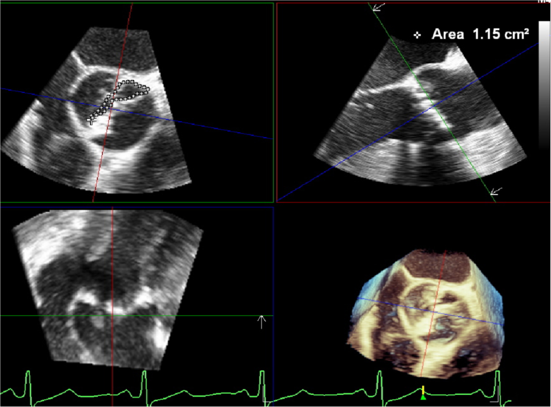
Figure 5. 3D image planes are carefully repositioned towards AV leaflet tips during the mid-systolic phase of the cardiac cycle. This repositioning allows for generating a short axis MPR, where planimetry can be precisely traced to determine the AV area. 3D: Three-dimensional; AV: aortic valve; MPR: multiplanar reconstruction.


