fig2

From single debulking techniques to combined approaches: a review of bailout strategies in heavily calcified coronary lesions

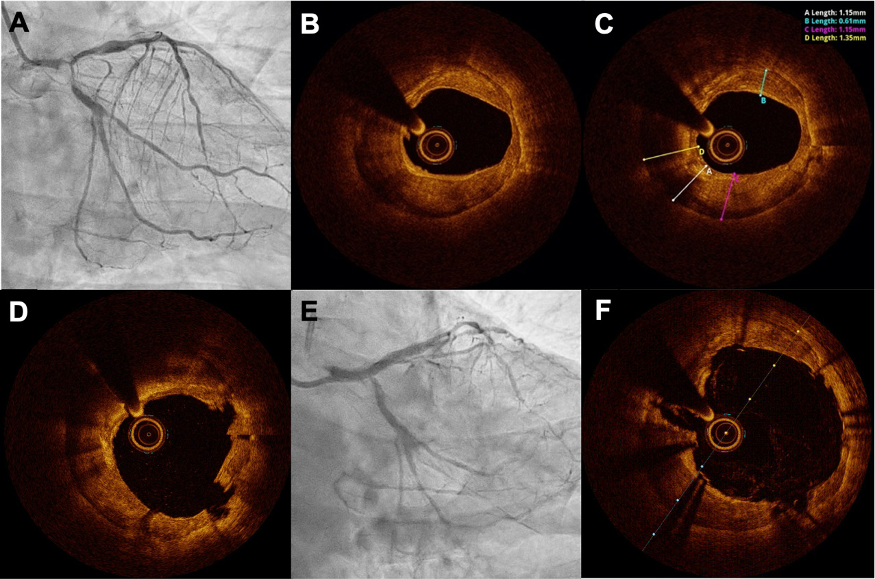
Figure 2. A 74-year-old male patient with severe calcific three-vessel disease presented with critical stenosis at the left main bifurcation (Medina 1,1,1), proximal LAD, OM2, and CTO of the mid RCA (A), who refused cardiac surgery. As the first step, OCT-guided LM PCI was performed; OCT showed severe circumferential calcification (B) with a maximum thickness of 1.35 mm (C). Due to the incomplete expansion of NC balloons (up to 3.0 mm), accompanied by a dog-bone effect, effective plaque debulking was achieved using a 1.5 mm RotaPro burr, followed by coronary lithotripsy (Shockwave 3.5 mm balloon); Subsequent OCT imaging demonstrated both circumferential and longitudinal calcium fractures (D); after stent implantation (E), OCT confirmed an optimal increase in lumen area, with excellent stent expansion and strut opposition (F). RCA: Right coronary artery; LAD: left anterior descending artery, LM: left main; PCI: percutaneous coronary intervention; OM: obtuse marginal; OCT: optical coherence tomography; NC: non-compliant balloon.

Vessel Plus
ISSN 2574-1209 (Online)
Follow Us

Portico

All published articles are preserved here permanently:

https://www.portico.org/publishers/oae/

Portico

All published articles are preserved here permanently:

https://www.portico.org/publishers/oae/