


fig1
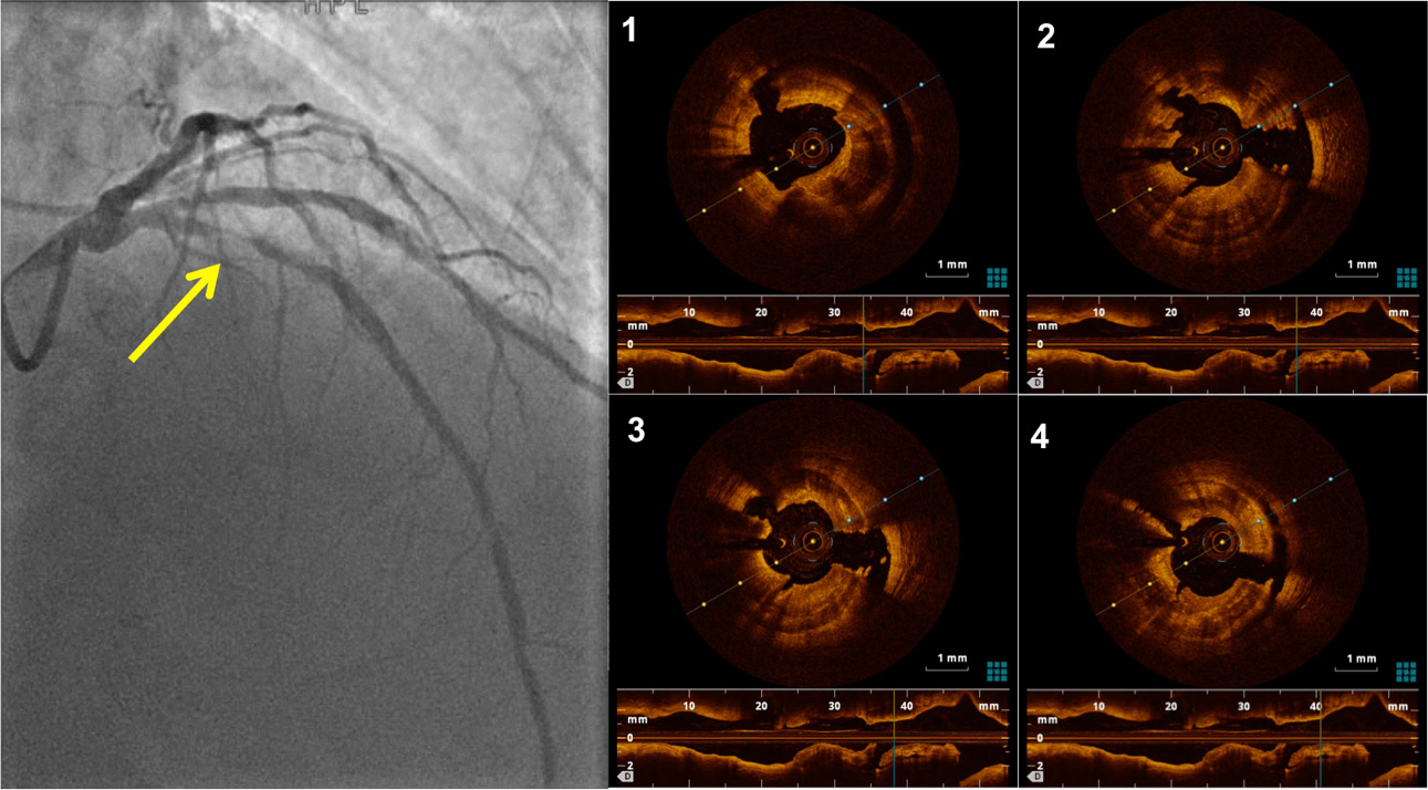
Figure 1. Left panel: Left coronary angiogram showing severe concentric stenosis in the proximal LAD (yellow arrow). Right panel: OCT images of a severely calcific concentric plaque, presenting deep radial and longitudinal (long-axis view shown below) fractures after combined treatment with Rotational Atherectomy (Rotablator) and IntraVascular Lithotripsy (Shockwave). LAD: Left anterior descending artery, OCT: optical coherence tomography.


