


Autologous fat transplantation technology: traditional therapies and emerging applications
 0
0 Abstract
In recent years, autologous fat transplantation technology has been widely applied in tissue repair and regenerative medicine. Autologous fat grafts offer several advantages: they are easy to harvest, possess favorable biocompatibility and excellent tissue integration capacity, and serve as an abundant source of stem cells. Refined techniques for fat extraction, processing, and transplantation, not only can morphological improvements be achieved, but the regeneration and functional recovery of damaged tissues can also be promoted. This review summarizes the wide-ranging clinical applications of fat grafting, covering tissue reconstruction, alopecia management, local drug delivery, and other novel indications. Combined with multidisciplinary strategies and advanced technologies, this technique can better satisfy the structural and functional requirements of recipient sites. Autologous fat grafting is evolving toward personalized and precise therapeutic regimens, providing patients with more diverse and effective treatment options. In the future, therapies based on adipose tissue are expected to become more tissue-specific and emerge as a new platform for regenerative medicine.
Keywords
INTRODUCTION
In recent decades, the acquisition and transplantation of autologous fat grafts have become increasingly important in medical practice. Adipose tissue is a complex connective tissue, primarily composed of mature adipocytes, extracellular matrix components, and the stromal vascular fraction (SVF)[1]. Mature adipocytes and rich extracellular matrix can provide sufficient tissue volume to support soft tissue augmentation. Initially, autologous fat grafts were frequently utilized as a soft tissue filler in conventional reconstructive surgeries, including cosmetic surgery, breast reconstruction, and traumatic defect repair[2].
With in-depth research on the biological properties and functional mechanisms of adipose-derived cells, fat grafting has achieved continuous technological innovation. Multiple strategies for adipose-derived stem cells (ADSCs) enrichment have been proposed one after another[3]. These new technologies not only broaden the application scope of autologous adipose tissue transplantation techniques but also highlight their significance in regenerative medicine and tissue engineering reconstruction. Consequently, the application scenarios of autologous fat transplantation technology are no longer confined to traditional soft tissue filling. Researchers are now exploring its potential in emerging fields, including cutaneous fibrosis, local drug delivery, and pain management[4].
This review mainly summarizes the conventional clinical applications and novel therapeutic potentials of autologous fat grafting, and discusses its future development trends and prospects.
COLLECTION, PROCESSING, AND PURIFICATION OF ADIPOSE TISSUE
The first report of fat tissue surgery dates back to 1893 when German surgeon Gustav Neuber performed direct excision to harvest fat tissue from the upper arm and implanted the graft beneath the lower eyelid to revise sunken deformity secondary to osteomyelitis. This approach leads to severe donor-site trauma, prolonged postoperative recovery, and unstable graft survival, which restricts its broad clinical use. Thus, the advancements in nearly all autologous fat transplantation techniques during the early stages concentrated on purifying the fat tissue before transplantation to minimize adipocyte damage and improve overall fat graft survival[5].
Unlike the traditional method of direct excision and transfer, in 1910, Hollander first adopted injection techniques to transplant fat into the face, marking a radical change in the practice of fat grafting[6]. Starting in the 1970s, with the advancement of anesthesia and surgical techniques, pump-assisted liposuction was proposed, and modern liposuction techniques gradually matured. Subsequently, a series of suction-assisted liposuction techniques, such as ultrasound-assisted liposuction, tissue liquefaction liposuction, and laser-assisted liposuction, were developed, further improving the efficiency of fat extraction and enhancing the outcomes for grafts and donor areas[7].
In 1997, Coleman et al. established a systematic fat grafting and purification process and first introduced the term “structural fat grafting.” This method is widely recognized as the gold standard for autologous fat grafting, marking an important milestone in the field of modern fat grafting techniques[8].
In 2001, Zuk et al. discovered that adipose tissue is a rich source of mesenchymal stem cells (MSCs). ADSCs have the capacity for self-renewal and multi-directional differentiation, which can facilitate tissue remodeling through proliferation and directed differentiation. Additionally, they exert strong paracrine effects and modulate the local microenvironment by secreting multiple bioactive factors[9]. This discovery heralded a new era for fat grafting research in the field of regenerative medicine. Subsequently, the application of stem cell-assisted transplantation strategies based on ADSCs has reduced the inflammatory response of the grafts, promoted vascular reconstruction, and thereby effectively improved the survival rate of the grafts[10].
In 2013, the introduction of the zonal survival theory for fat grafting overturned traditional fat graft survival theories[11]. Researchers gradually shifted their focus from minimizing damage to fat cells to actively destroying mature fat cells, while preserving ADSCs with regenerative potential. Following this principle, the field of fat grafting proposed innovative product concepts such as nanofat and adipose matrix-derived stromal vascular fraction gel (SVF-gel)[12].
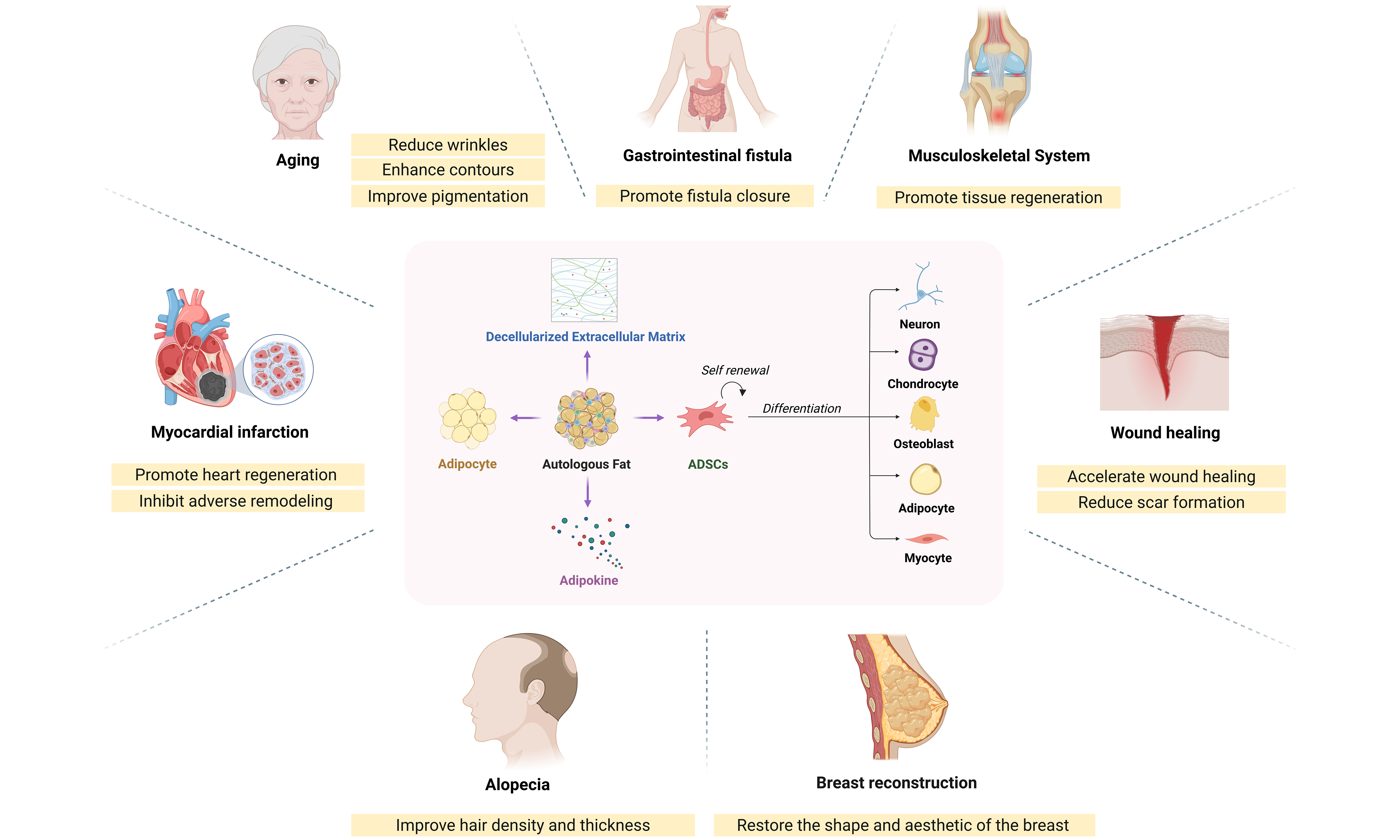
Currently, the rapid advancement of tissue engineering technology has led to strategies that combine various growth factors to enhance regenerative performance and cross-link polymer scaffolds to improve physical or mechanical properties, further optimizing transplantation procedures. The injectable vascularized preadipose tissue (iPAT), created by combining mature adipocytes, ADSCs, and human umbilical vein endothelial cells (HUVECs) within a fibrin gel matrix, not only largely maintains the activity of mature adipocytes but also significantly improves the retention rate of the grafts[13]. By cross-linking adipose tissue active microfragments with fibrin to create bio-ink, the stiffness of the adipose tissue is increased, and it can be shaped to meet the personalized soft tissue augmentation needs of individual patients[14]. Additionally, in-depth research into the adipose tissue extracellular matrix has broadened the scope of fat grafting applications, offering new insights and strategies for treating a range of diseases [Figure 1][15,16].
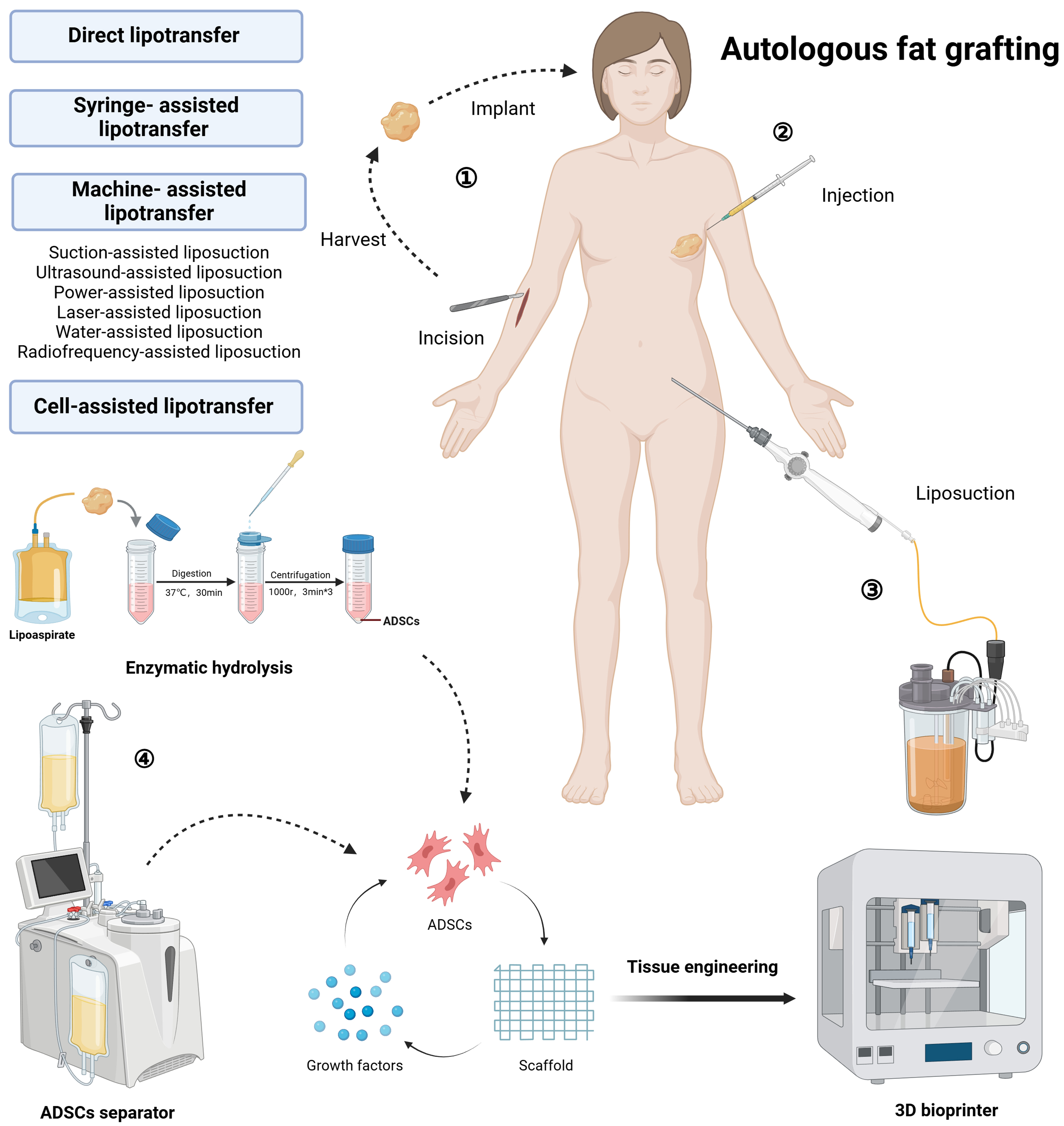
Figure 1. Schematic diagram of fat tissue acquisition and application. (1) Direct lipotransfer after excision; (2) Syringe-assisted lipotransfer; (3) Machine-assisted lipotransfer; (4) Cell-assisted lipotransfer. Created in BioRender. https://BioRender.com/76g1ew8. ADSCs: Adipose-derived stem cells.
APPLICATIONS OF FAT GRAFTING
Traditional therapy
Repair and reconstruction surgery
Regenerative medicine focuses on repairing or replacing tissues damaged by genetic, traumatic, or degenerative injuries. The introduction of grafts into the damaged area provides not only an alternative tissue structure for volume augmentation but also active seed cells and growth factors that facilitate tissue repair[4]. As a result, autologous fat transplantation has shown superior therapeutic effects in many reconstructive surgeries, including soft tissue augmentation, breast reconstruction, and wound healing.
Surface soft tissue depression deformity
Multiple congenital disorders, including spina bifida, clubfoot, and progressive hemifacial atrophy (PHA), manifest as progressive atrophy of the skin, soft tissue, muscle, and underlying bone, which commonly leads to facial or limb asymmetry. Through precise fat injection techniques, doctors can repair and reconstruct defective areas, restoring symmetry and filling in indentations caused by developmental abnormalities, thereby improving the patient’s mental health[17]. The application of muscle fascial composite flaps or cartilage grafts can lead to better long-term results and functional recovery[18].
Breast reconstruction
Breast cancer is the most prevalent form of cancer among women globally, affecting one in eight. With ongoing advances in early diagnosis and treatment technologies for breast cancer, the survival rate of patients is steadily improving[19]. Consequently, there is a pressing clinical need to address the physical deformities resulting from the disease and its treatment in breast cancer survivors, to further enhance their quality of life and prognosis. In recent years, autologous fat transplantation has emerged as the primary method for local breast reconstruction post-cancer surgery. Numerous cases have demonstrated that autologous fat transplantation effectively improves breast satisfaction, social mental health, and sexual health in patients undergoing breast reconstruction[20]. Although its oncological safety has aroused concerns, to date, no clinical evidence has confirmed that autologous fat transplantation increases the risk of tumor recurrence[21].
As this procedure is widely carried out in clinical settings, technological innovations are also taking place. Currently, methods such as enriching ADSCs in the grafts[22] or using vacuum-based external tissue expanders (i.e., the Brava device) for pre-expansion[23] can effectively enhance the graft’s retention rate, reduce the need for secondary surgeries, and demonstrate significantly better enlargement and aesthetic results.
Wound healing
The skin serves as a natural protective barrier against the external environment. Refractory wounds, including late-stage wounds from extensive burns, diabetic foot ulcers, and skin defects caused by pressure sores, have long posed a challenging issue in the medical field. ADSCs have demonstrated unique therapeutic effects. Studies have shown that they can differentiate into specific cell types, such as fibroblasts, within particular microenvironments, directly participating in the tissue repair process. Additionally, exosomes derived from ADSCs promote local angiogenesis, enhance the local microenvironment, decrease cell apoptosis, and thereby accelerate wound healing[24].
Furthermore, studies have shown that autologous fat transplantation can effectively prevent and inhibit the development of pathological scars[25]. After chylous fat injection, the density and number of fibroblasts in scar tissue decline, while the distribution and morphology of type III collagen fibers return to normal. In vitro and animal experiments have demonstrated that ADSCs can inhibit the proliferation of fibroblasts and the secretion of collagen by secreting abundant antifibrotic factors[26]. When used in conjunction with other scar prevention and treatment methods, such as scar tissue release surgery, laser therapy, and platelet-rich plasma, it has shown superior improvement in scars, including elasticity, color, and softness[27].
Cosmetic surgery
Adipose tissue, with its loose connective tissue structure, offers soft mechanical properties that enable it to mimic the deformation contours of native tissue. Consequently, fat grafting has been regarded as an ideal filling material for soft tissue repair and contour remodeling, exhibiting significant potential for applications in facial rejuvenation and body contour reshaping.
Facial rejuvenation
As we age, the volume of fat compartments decreases, soft tissues descend, and the skin becomes thinner, leading to the typical signs of aging. Although lifting surgeries can provide a more youthful appearance by removing excess skin, they cannot address the loss of facial volume. Autologous fat transplantation, with its significant effects and relatively safe treatment method, has become increasingly favored by those seeking beauty. Facial rejuvenation procedures commonly include filling hollowed areas of the temples and cheeks, restoring diminished apple cheeks, and addressing pigmentation spots, among other concerns. Autologous fat transplantation technology precisely augments the depleted fat volume in the face, not only effectively enhancing the three-dimensionality of the facial contours but also significantly improving skin laxity, wrinkles, and pigmentation issues associated with aging, resulting in a high level of clinical satisfaction[28]. Histological evidence shows that after transplantation, skin quality is also improved, specifically manifesting as skin thickening, increased type I collagen content, and increased vascular density[29]. An interesting observation is that the therapeutic effect of using ADSCs in conjunction with fat tissue is more meaningful than the arithmetic sum of the effects of each treatment alone. This indicates that there is an interaction between ADSCs and fat grafts, which can synergistically affect collagen synthesis and the formation of new blood vessels.
Contour reshaping
The human body curve is an important aspect of female attractiveness. To achieve an ideal and harmonious body contour, fat transplantation has become the choice of many. As early as the 20th century, fat transplantation for cosmetic purposes. Beyond its established role in breast reconstruction, this technique extends to the volumetric enhancement and refined sculpting of the legs, buttocks, and hips[30-32]. Through refined surgical techniques and advanced fat processing technologies, such as stem cell enrichment, doctors can perform contour reshaping surgeries more effectively[33].
Emerging applications
Drug delivery
Drug molecules currently available on the market face issues such as short half-lives, poor absorption, low specificity, rapid degradation, and the development of resistance. In contrast to systemic administration, local drug delivery minimizes systemic exposure and mitigates the loss of drug activity due to the “first-pass effect”. However, employing foreign materials like microbeads and hydrogels as drug carriers introduces a significant risk of adverse immune reactions. Autologous adipose tissue, as an ideal characteristic of a drug delivery system, offers low cost, biocompatibility, and the ability to effectively remain at the application site. Studies have shown that adipose tissue can serve as a natural carrier for lipophilic drugs, such as insulin-sensitizing thiazolidinediones, including pioglitazone and rosiglitazone[34].
Most anticancer compounds exert potent cytotoxic effects on patients because they target not only cancer cells but also normal, healthy tissues and organs. As nutrients, tumor tissues actively uptake fatty acids (FA) due to their increased energy demands[35]. Hence, fat grafting has a unique advantage in the specific delivery of therapeutic drugs to cancer cells, as drugs can be naturally released locally through lipolysis induced by cancer cells, effectively reducing the systemic side effects of anticancer drugs. Numerous studies have confirmed that using adipose tissue to carry anticancer drugs helps reduce the growth rate and recurrence risk of tumors [Table 1].
Adipose tissue-based local drug delivery strategies for tumor therapy
| Source of adipose tissue/adipocytes | Drug | In vitro | In vivo | Ref. |
| Human subcutaneous fat aspirate | Paclitaxel | Inhibits the proliferation of human pancreatic cancer cells (CFPAC-1); Promotes apoptosis and necrosis in human glioblastoma cell lines (IMR-32 cells and U87MG cells); Anti-angiogenesis | Inhibit or delay the recurrence of neuroblastoma | [36] |
| Human subcutaneous fat aspirate | Paclitaxel | Inhibit the proliferation of liver cancer cells (Hep-3B); Promote apoptosis and necrosis of cancer cells | Inhibit liver tumor growth | [37] |
| Autologous lumbar lateral wing liposuction material | Paclitaxel | - | Control the progression of mesothelioma; Reduce the formation of effusions | [38] |
| Human thigh or abdominal fat aspirate | Fulvestrant | Inhibit the proliferation of breast cancer cells (MCF7 cells); Downregulate the expression and activity of estrogen receptors | - | [39] |
| Primary human omental mature adipocytes | Paclitaxel | Inhibit the proliferation of ovarian cancer cells (SK-OV-3 cells and OVCAR-5 cells); Induce cell death within tumor organoids | Inhibit the growth of ovarian tumors | [40] |
| 3T3-L1 cell line | Rumenic acid and a doxorubicin prodrug | Inhibits the growth of melanoma cells (B16F10 cells) and breast cancer cells (E0771 cells); Downregulates the expression of PD-L1 within tumor cells; Promotes the effector function of infiltrating T cells | Reduce the growth rate of B16F10 melanoma in mice; Promote tumor cell apoptosis and T cell infiltration; Delay post-surgical tumor recurrence | [41] |
| hADSCs | BMP4 | Suppress the tumorigenic potential of BTICs | Extend the survival period of intracranial brain tumor models | [42] |
| mADSCs | Cisplatin | Inhibit the activity of breast cancer cells (4T1 cancer cells) | Reduce the risk of metastasis and recurrence after breast cancer surgery; Increase the survival time of mice. Reduce the rate of tumor growth | [43] |
Unlike premixing, which relies on the principle of physical adsorption, coupling drug molecules through chemical means or genetic tools aids in further enhancing the pharmacodynamics/pharmacokinetics, thereby improving therapeutic efficacy[44]. Bo et al. innovatively applied azide groups for the metabolic labeling of adipocytes. By transplanting fat grafts to the surgical site to capture circulating DBCO drugs and achieve sustained release, they effectively prevented tumor recurrence and metastasis in a triple-negative breast cancer model[43]. Thanks to the unique tumor-homing properties of ADSCs, Mangraviti et al. genetically engineered them with a bone morphogenetic protein-4 (BMP-4) plasmid. Following simple intranasal or systemic intravenous administration, ADSCs can penetrate the blood-brain barrier and secrete engineered BMP-4, which effectively improves the survival rate of rats with brain tumor models[42]. Thus, this adipocyte-based, targetable, and refillable drug reservoir presents a novel and unique opportunity for drug delivery, with a broad prospect.
Skin fibrosis
Subcutaneous fat plays a crucial regulatory role in skin tissue, participating in both the physiological regulation of skin homeostasis and the pathological regulation of diseased skin[45]. Numerous reports have confirmed the effective role of fat transplantation in fibrosis associated with pathological skin conditions such as systemic sclerosis (SSc), radiation dermatitis, and vulvar lichen sclerosus (VLS).
SSc
SSc is a rare connective tissue disease that affects multiple systems, characterized by significant fibrosis and vasculopathy of the skin and internal organs. To date, there is no recognized gold standard treatment[46].In recent years, autologous fat transplantation has been experimentally applied to SSc. Preliminary clinical observations suggest that various adipose tissue derivatives, including fresh adipose tissue[47], ADSCs[48], and SVF-gel[49], contribute to both immediate and lasting benefits in aesthetic and functional improvements for the skin sclerosis of SSc patients. Pathological analysis has confirmed collagen reconstruction in damaged skin areas, improved capillary density, and regeneration of skin appendages such as subcutaneous fat and hair follicles post-treatment. A comparative study indicates that autologous fat transplantation even exhibits a therapeutic effect superior to that of hyaluronic acid filling[50]. Studies have shown that autologous fat transplantation not only utilizes the mechanical filling properties of adipose tissue to improve skin elasticity in patients with scleroderma but may also improve skin texture and function by releasing bioactive factors with regenerative and anti-fibrotic properties from ADSCs[51,52]. Moreover, autologous fat transplantation has demonstrated favorable therapeutic outcomes for complications frequently neglected in clinical practice, including perioral sclerosis, finger ulcers, and hand dysfunction[53,54].
However, in clinical practice, the fat retention rate of patients with scleroderma is often lower than that of normal individuals. Further research indicates that there are inherent abnormalities in the patients’ ADSC population, with a relative reduction in CD55high ADSCs, thereby exhibiting enhanced fibrogenesis, reduced anti-inflammatory properties, and increased oxidative stress[55]. At the same time, given that SSc patients are typically thin and have a higher sensitivity to skin ulcers, factors such as insufficient volume or exacerbated pain response must be considered when performing fat tissue aspiration.
Radiation dermatitis
Radiation dermatitis is an inflammatory condition affecting the skin and mucous membranes due to radiation exposure, which can be categorized into acute and chronic phases. In the acute phase, symptoms such as erythema and ulceration manifest, whereas in the chronic phase, the skin becomes hardened, scars develop, and there is contraction, along with permanent hair loss or loss of pigmentation. In 2007,
VLS
VLS is a chronic inflammatory skin disease that primarily affects the skin of the genital and anal areas. It is characterized by pain, itching, loss of libido, vulvar fibrosis, and scarring, significantly impacting the quality of life of patients[60]. In cases of severe complications, a vulvectomy may be performed. Fat grafting has been proposed as a novel treatment for patients with VLS. In a cohort of VLS patients undergoing autologous fat grafting, 88.7% reported satisfaction, with significant improvements in pruritus, burning, dyspareunia, psychosexual well-being, and histological inflammation lasting up to 11 years post-surgery[61]. Furthermore, the combination therapy of nanofat mixed with platelet-rich plasma has demonstrated positive outcomes in patients with lichen sclerosus[62]. ADSCs and growth factors abundant in platelet-rich plasma work synergistically to alleviate inflammatory responses and promote tissue repair[63].
Musculoskeletal system
The musculoskeletal system plays a crucial role in providing structural support, protecting vital organs, and facilitating movement. Trauma, congenital malformations, infections, tumors, and other factors can disrupt the integrity and overall physiological function of the musculoskeletal system[64]. A promising approach to repair is tissue engineering technology carrying ADSCs.
Bone defect
Currently, autologous free bone grafting and vascularized bone flap repair are the gold standards for restoring bone defects in clinical practice. However, new defects can form at the donor site, and the risks of rejection and infection associated with allogeneic bone tissue transplantation also limit its use. How to further optimize transplantation protocols to effectively restore the function of the skeletal system has always been a significant issue in bone regeneration research and discussion. The bio-material system constructed by combining ADSCs with instructive molecules has shown promising regenerative effects in repairing the skeletal system[65]. Joint cartilage is a crucial structural component of the skeletal system, providing a smooth and lubricated surface for joint movement and load distribution. However, due to the difficulty in spatially controlling the stratification or differentiation of stem cells between cartilage and subchondral bone, the co-regeneration of stratified osteochondral tissue remains a challenge. Lee et al. prepared two types of composite spheroids using ADSCs and nanofibers coated with transforming growth factor-β3 (TGF-β3) or bone morphogenetic protein-2 (BMP-2), respectively, for chondrogenesis or osteogenesis. Subsequently, each type of spheroid was layered and assembled in a spatial arrangement within 3D-printed microchambers, mimicking the hierarchical microstructure of biomimetics, thus achieving the directional regeneration of osteochondral tissue[66].
Muscle injury
Muscle injury is a prevalent musculoskeletal disorder characterized by the dysregulation of the collagen matrix, chronic low-grade inflammation, and tissue degeneration. In severe cases, limbs may become functionally impaired due to the formation of fibrous scars and inadequate muscle regeneration. Although current clinical treatment protocols can reduce the degree of tissue fibrosis, they cannot yet achieve complete restoration of muscles.
Sastourné-Arrey et al. discovered in a mouse model that when the muscle is damaged, ADSCs migrate from subcutaneous adipose tissue and actively enhance the repair of injured skeletal muscle[67]. ADSCs can promote post-injury muscle regeneration and restore limb function by spontaneous myotube formation, differentiating into muscle tissue, secreting angiogenic and anti-apoptotic cytokines, and reducing inflammatory responses. The combination of ADSCs and basic fibroblast growth factor (bFGF) hydrogel can achieve functional recovery, revascularization, and nerve innervation in lacerated muscles[68]. Furthermore, studies have indicated that exosomes secreted by skeletal muscle cells encompass growth factors associated with muscle formation, including insulin-like growth factor (IGF), hepatocyte growth factor (HGF), basic fibroblast growth factor 2 (FGF-2), and platelet-derived growth factor-AA (PDGF-AA). These factors can direct ADSCs to differentiate into skeletal muscle cells[69]. Consequently, the combined recellularization strategy, which involves co-perfusing ADSCs and myogenic cells through vascular pedicles into the adipose-derived decellularized extracellular matrix (adECM), demonstrates superior muscle regeneration and functional recovery compared to the strategy of recellularization with ADSCs alone[70].
Pain
Pain is not merely a physical torment; it can also significantly affect a patient’s mental health, leading to emotional issues, sleep disorders, and limitations in daily activities. In recent years, numerous case reports have documented the beneficial effects of autologous fat transplantation in treating pain syndromes, although the degree of pain relief varies among patients. For instance, post-mastectomy pain syndrome (PMPS)[71,72], post-traumatic pain[73], and post-amputation stump pain[74]. The mechanism of fat transplantation for pain management may involve multiple aspects. First, fat transplantation can increase the volume of subcutaneous tissue, providing mechanical protection and reducing nerve compression, thereby alleviating pain. Second, this effect may also be linked to the pain-relieving properties of adipokines secreted by ADSCs[75,76]. Further research indicates that the abundance of CD304+ CD10+ CD73+ ADSCs derived from adipose tissue is closely related to the improvement of pain. This subset of cells may alleviate local pain by degrading pain mediators such as adenosine triphosphate (ATP) and bradykinin or by secreting endogenous opioids[77]. Therefore, the isolation and purification of this specific subset of cells may contribute to providing specific therapeutic tools for pain relief.
Obesity
Obesity is a disease caused by an imbalance between energy intake and expenditure, and it is considered a key pathogenic factor leading to metabolic syndrome (including diabetes, insulin resistance, etc.). As the largest endocrine organ in the human body, adipose tissue plays a dominant role in regulating energy homeostasis. The pathological expansion of white adipose tissue (WAT) and the whitening of brown adipose tissue (BAT) have caused obesity-related metabolic dysfunction[78]. As early as 2013, Liu et al. reported the potential benefits of BAT transplantation in enhancing systemic energy metabolism, suggesting that it could prevent or reverse obesity induced by a high-fat diet (HFD)[79]. Further research indicates that the beneficial effects of exogenous BAT transplantation are not entirely dependent on the supplementation of protective brown adipokines, but are partly attributed to the activation of endogenous BAT function by the transplanted tissue[80]. To obtain a stable and substantial volume of BAT, the technology for producing BAT-like tissue based on biomaterial scaffolds and stem cell assistance is continuously evolving. Among these, hydrogels are considered one of the most promising materials for the development of BAT engineering[81]. Wang et al. developed a 3D brown adipose microtissue based on PEG hydrogel-supported suspension culture of brown adipose aggregates for alleviating HFD-induced obesity and metabolic syndrome[82]. This study is the first to report the use of human brown adipose progenitor cells to induce differentiation into BAT microtissues and validate their safety and efficacy in a type 2 diabetes mellitus (T2DM) mouse model. Compared to traditional tissue transplantation methods, engineered BAT shows significant advantages in terms of product scalability, storage stability, purity, safety, survival rate, and efficacy, indicating that it may become a new strategy for obese patients to control weight.
Gastrointestinal fistula
Crohn’s disease (CD) is a chronic, full-thickness inflammation of the gastrointestinal tract of unknown cause that can destroy the integrity of the intestinal mucosa from the mouth to the anus, thereby causing the formation of fistulas[83]. Although antibiotics, immunosuppressants, and biologics are effective options for treating perianal fistulas in CD, they primarily focus on controlling intestinal inflammation and cannot achieve complete closure of the fistulas or reduce recurrence. Surgical treatment remains the preferred therapy for gastrointestinal perforation, using adjacent omental adipose tissue as a patch to close the perforation site, which has shown promising therapeutic effects[84]. Inspired by this, people began to attempt to use freshly collected autologous adipose tissue for the repair of various fistulas. Research results indicate that autologous adipose tissue can serve as a durable graft, persisting and integrating with the host tissue, while secreting growth factors to promote the repair of the defect area[85,86]. In a study involving 21 patients with CD and perianal fistulas, approximately 57% of the patients achieved complete clinical healing of the fistulas[87].
To achieve a higher mitigation rate, new treatment strategies, including stem cell therapy, have been developed. By expanding ADSCs in vitro, long-term effective treatment has been provided for complex perianal fistulas that are difficult to cure with conventional or biological therapies[88]. Park et al. demonstrated that the closure rate of patients receiving autologous ADSC transplantation was significantly faster, and the closure time was significantly shorter compared to those treated with anti-tumor necrosis factor (TNF) drugs[89].
Hair loss
Hair loss is a skin condition that affects patients’ appearance, self-esteem, and quality of life. It can be classified into reversible non-scarring alopecia and irreversible scarring alopecia. Follicular unit extraction (FUE) hair transplant surgery is the preferred treatment for hair loss. Autologous fat injection is a potential technique that can be used to promote hair growth[90]. Some surgeons have also reported positive changes in hair regeneration after transplantation. Autologous fat tissue transplantation not only provides the necessary nutritional support for hair follicles, promoting hair growth, but also improves the appearance of the scalp through its filling effect, making the hair appear denser. In a case series study, 9 patients with alopecia caused by lichen planopilaris underwent fat grafting treatment. The results indicated significant improvements in hair density and diameter, a reduction in scalp inflammation and erythema, and enhancements in the hair pull test LPPAI score[91]. Additionally, fat grafting has demonstrated considerable potential in treating recurrent folliculitis, post-traumatic alopecia, and scar alopecia by promoting hair regeneration and modulating inflammation[92,93]. Pre-injected adipose tissue can effectively enhance the mechanical and vascular characteristics of the recipient area, providing an optimal environment for hair follicles to produce new, healthy hair.
Lymphedema
Lymphedema, triggered by lymphatic dysfunction, manifests as interstitial fluid retention, swelling, chronic inflammation, lipid deposition, and fibrosis. This condition is categorized as either congenital primary lymphedema or secondary lymphedema resulting from trauma or postoperative obstruction; currently, no definitive cure exists[94]. Extracellular vesicles derived from ADSCs markedly ameliorate lymphedema in mice, concurrently boosting the density of both capillaries and lymphatic vessels[95]. Adipose-derived microvascular fragment transplantation markedly promotes lymphangiogenesis and angiogenesis in mouse lymphedema models[96]. Furthermore, multiple clinical trials have demonstrated that the combined application of ADSCs and autologous fat grafting is safe and feasible for alleviating breast cancer‑related lymphedema, showing promising therapeutic potential[97].
Cutting-edge research into the sorting and genetic modification of functional ADSC subpopulations has unveiled novel strategies to enhance lymphatic vascular regeneration. Notably, the podoplanin-positive ADSC subset exhibits high expression of the lymphangiogenic factor vascular endothelial growth factor C (VEGF-C), demonstrating significantly superior efficacy in promoting lymphatic regeneration compared to unselected, mixed ADSCs[98]. Genetic modification of key pathway genes further amplifies these cellular therapeutic effects. Crucially, regulation of the Hippo/YAP signaling pathway markedly boosts the differentiation potential of ADSCs into lymphatic endothelial cells while concurrently inhibiting subcutaneous fibrosis, with edema resolution efficiency nearly doubling in animal models[99].
Other diseases
Fat transplantation technology is increasingly widely applied in the medical field, particularly in the treatment of vocal cord paralysis, dural repair, tympanic membrane repair, and temporomandibular joint reconstruction [Table 2], demonstrating its unique therapeutic effects and potential.
Application scenario of autologous fat transplantation in other types of diseases
| Type of disease | Treatment effect | Ref. |
| Dural repair | Improve the quality of scar tissue; Promote angiogenesis; Prevent fibrosis and adhesion | [100,101] |
| Tympanic membrane perforation | Promote neovascularization and tissue repair | [102] |
| Vocal cord paralysis | Increase the volume of the vocal cords; Help restore normal vocal cord closure; Improve voice quality; Enhance vocal efficiency; Alleviate difficulty in breathing | [103-105] |
| Temporomandibular Joint reconstruction | Achieve a greater postoperative mouth opening; Reduce the occurrence of heterotopic ossification | [106,107] |
| Chronic myocardial infarction | Promote cardiac regeneration; Inhibit adverse remodeling | [108] |
| Polycystic ovary syndrome | Enhanced fertility in rats; Improved and stabilized menstrual irregularities; Increased insulin sensitivity | [109-111] |
| Neural injury | Enhance the regeneration of axons and myelin sheaths; Promote functional recovery | [16,112-115] |
CONCLUSIONS
As the unique biological properties of adipose tissue are gradually elucidated, autologous fat transplantation technology has shown tremendous potential in the field of regenerative medicine. Autologous fat tissue transplantation can be obtained in large quantities through minimally invasive procedures and allows for repeated harvesting, effectively overcoming the source limitations of autologous tissue transplantation. At the same time, autologous fat transplantation has unparalleled biocompatibility and lower immune rejection compared to allogeneic tissue transplantation.
In addition to serving as a filler, ADSCs and their rich adipokines play a crucial role in promoting tissue repair and regeneration. As adult MSCs, ADSCs hold distinct clinical translational advantages over pluripotent ESCs and iPSCs: accessible harvest free of ethical issues, standardized culture techniques, low immunogenicity, and negligible teratoma formation risk. With the rapid advancement of tissue engineering, biomaterial scaffolds provide ADSCs with the necessary physical and mechanical support to facilitate their adhesion and proliferation. Furthermore, by incorporating specific cytokines, they offer critical chemical signals that promote the proliferation and directed differentiation of ADSCs. Currently, researchers have developed various engineered tissue grafts that combine ADSCs with biomaterials, achieving significant success in enhancing stem cell function and improving the local microenvironment. Here, we present the diverse applications of current fat transplantation techniques. In addition to traditional areas such as cosmetic plastic surgery, breast reconstruction, and wound repair, we also highlight emerging uses such as drug delivery, obesity treatment, and pain management, offering new insights into the therapeutic potential of autologous fat transplantation.
While autologous fat transplantation has demonstrated positive therapeutic effects on various complex tissue injuries, some studies have only been preliminarily confirmed in animal models. For ADSC-based therapies, standardized protocols and automated workflows for cell collection, isolation, and large-scale in vitro expansion remain lacking. Furthermore, the molecular and cellular mechanisms governing ADSC-mediated tissue regeneration remain poorly characterized, substantially confounding the predictability of clinical efficacy. In light of this, rigorous clinical trials and in-depth mechanistic studies are indispensable to drive future translational progress. With continuous technological innovation and more in-depth research, autologous fat transplantation technology is expected to bring hope to a broader patient population and become a bright star in the field of regenerative medicine in the future.
DECLARATIONS
Acknowledgments
The Graphical Abstract was created using BioRender (https://BioRender.com/mw39hm1). We sincerely acknowledge Ascsa Dsbsdgv for professional assistance in drawing the Graphical Abstract and Figure 1 using BioRender.
Authors’ contributions
Made substantial contributions to conception and design of the study and performed data analysis and interpretation: Liao Y
Drafted the manuscript or revised it critically for important intellectual content: Zhang Q
Provided funding acquisition: Lu F, Cai J
Availability of data and materials
Not applicable.
AI and AI-assisted tools statement
Not applicable.
Financial support and sponsorship
This work was supported by the National Nature Science Foundation of China (81901976, 82472585, 82372544), Guangdong Provincial Natural Science Foundation (2023A1515011741), Guangzhou Science and Technology Program (2023A04J2348), Nanfang Hospital’s Outstanding Young Talent Cultivation Program (K50906009, K50304020), Nanfang Hospital’s Scientific Research Development Fund (K51701101).
Conflicts of interest
All authors declared that there are no conflicts of interest.
Ethical approval and consent to participate
Not applicable.
Consent for publication
Not applicable.
Copyright
© The Author(s) 2026.
REFERENCES
1. Gandolfi S, Lupon E, Varin A, et al. Evolution of cell therapies derived from adipose tissue: historical perspectives, current development trends and future directions. Biol Direct. 2025;20:95.
2. Hasiba-Pappas S, Opriessnig E, Nischwitz SP, et al. Optimizing autologous fat grafting: a systematic review of enhancement strategies and graft survival. Aesthet Surg J. 2025;Epub ahead of print.
3. Choudhery MS, Mahmood R, Harris DT. Pre-enrichment of adipose tissue grafts: potential and limitations. In: Haider KH, Editor. Handbook of stem cell applications. Singapore: Springer Nature Singapore; 2023. pp. 1-43.
4. Firriolo JM, Condé-Green A, Pu LLQ. Fat grafting as regenerative surgery: a current review. Plast Reconstr Surg. 2022;150:1340e-7.
5. Varghese J, Mosahebi A. Historical overview of stem cell biology and fat grafting. Aesthet Surg J. 2017;37:S1-3.
6. Mazzola RF, Mazzola IC. The fascinating history of fat grafting. J Craniofac Surg. 2013;24:1069-71.
7. Urbonas T, Kievišas M, Petrikaitė V, et al. Assessing adipocyte viability and surgeons’ work efficiency by comparing different liposuction methods. Plast Reconstr Surg Glob Open. 2023;11:e5190.
9. Zuk PA, Zhu M, Mizuno H, et al. Multilineage cells from human adipose tissue: implications for cell-based therapies. Tissue Eng. 2001;7:211-28.
10. Shen S, Huo H, Ren H, Shao Y. Comparative efficacy and safety of cell-assisted and conventional lipotransfer in facial filling: a systematic review and meta-analysis. Aesthetic Plast Surg. 2024;48:1444-56.
11. Kato H, Mineda K, Eto H, et al. Degeneration, regeneration, and cicatrization after fat grafting: dynamic total tissue remodeling during the first 3 months. Plast Reconstr Surg. 2014;133:303e-13.
12. Yang Z, Dong L, Jin S, Han X, Li F. Comparison of microfat, nanofat, and extracellular matrix/stromal vascular fraction gel for skin rejuvenation: basic animal research. Aesthet Surg J. 2023;43:NP573-86.
13. Louis F, Sowa Y, Irie S, et al. Injectable prevascularized mature adipose tissues (iPAT) to achieve long-term survival in soft tissue regeneration. Adv Healthc Mater. 2022;11:e2201440.
14. Jeong W, Son J, Choi J, et al. Clinically relevant and precisely printable live adipose tissue-based bio-ink for volumetric soft tissue reconstruction. Adv Healthc Mater. 2025;14:e2402680.
15. Kang Y, Yeo M, Derman ID, et al. Intraoperative bioprinting of human adipose-derived stem cells and extra-cellular matrix induces hair follicle-like downgrowths and adipose tissue formation during full-thickness craniomaxillofacial skin reconstruction. Bioact Mater. 2024;33:114-28.
16. Feng J, Fu S, Luan J. Regenerated fat induced by a decellularized adipose matrix can survive long-term in vivo. Acta Biomater. 2025;191:233-43.
17. Xiang X, Jiang Z, Che D, et al. Application of free serratus anterior muscle-fascial composite tissue flap and facial lipofilling in repairing progressive hemifacial atrophy. Asian J Surg. 2024;47:973-81.
18. He L, Liu X, Khatter NJ, Yu X, Washington KM, Shu M. Treatment of progressive hemifacial atrophy by cartilage graft and free adipofascial flap combined with three-dimensional planning. Plast Reconstr Surg. 2024;153:679-88.
19. Miller KD, Nogueira L, Devasia T, et al. Cancer treatment and survivorship statistics, 2022. CA Cancer J Clin. 2022;72:409-36.
20. Piatkowski AA, Wederfoort JLM, Hommes JE, et al. ; Breast Reconstruction With External Preexpansion & Autologous Fat Transfer vs Standard Therapy (BREAST) Trial Investigators. Effect of total breast reconstruction with autologous fat transfer using an expansion device vs implants on quality of life among patients with breast cancer: a randomized clinical trial. JAMA Surg. 2023;158:456-64.
21. Kronowitz SJ, Mandujano CC, Liu J, et al. Lipofilling of the breast does not increase the risk of recurrence of breast cancer: a matched controlled study. Plast Reconstr Surg. 2016;137:385-93.
22. Giannakaki AG, Stylianaki A, Giannakaki MN, et al. Stem-cell-assisted lipotransfer and platelet-rich plasma in breast reconstruction: a literature review. Aesthetic Plast Surg. 2025;49:5764-73.
23. Moltaji S, Hoffbauer S, Brown MH. Large-volume fat grafting to the breast with external expansion assist. Aesthet Surg J. 2023;43:NP401-12.
24. Uhrbom M, Muhl L, Genové G, et al. Adipose stem cells are sexually dimorphic cells with dual roles as preadipocytes and resident fibroblasts. Nat Commun. 2024;15:7643.
25. Dai Q, Yu Q, Xiao R, Cai L. Early intervention with autologous fat grafting to reduce hypertrophic scar formation: a prelimilary study. Aesthetic Plast Surg. 2021;45:2409-16.
26. Yu Q, Dai Q, Huang Z, et al. Microfat exerts an anti-fibrotic effect on human hypertrophic scar via fetuin-A/ETV4 axis. J Transl Med. 2023;21:231.
27. Alfarafisa NM, Chou Y, Santika R, Riestiano BE, Soedjana H, Syamsunarno MRAA. Adipose-derived stem cell products and combination therapies for the treatment of pathological scars: a review of current preclinical and clinical studies. Clin Cosmet Investig Dermatol. 2025;18:1309-37.
28. Strong AL, Rohrich RJ, Tonnard PL, Vargo JD, Cederna PS. Technical precision with autologous fat grafting for facial rejuvenation: a review of the evolving science. Plast Reconstr Surg. 2024;153:360-77.
29. Tan PC, Wang YW, Xie Y, et al. Enhancing skin regeneration during expansion: a multicenter randomized controlled trial of stromal vascular fraction and fat grafting. Plast Reconstr Surg. 2026;157:360-71.
30. Añorve Borquez IR. A new technique for remodelling and increasing the diameter of the legs through lipotransfer and vertical subcutaneous fasciotomy of the posterior tibial aponeurosis. Aesthetic Plast Surg. 2021;45:1642-50.
31. Mendes FH, Viterbo F. Fragmented fat transfer for massive weight loss gluteal contouring. Plast Reconstr Surg. 2022;149:624-7.
32. Cortes W, Mather T, Logiudice J. Hip expansion with fat grafting combined with abdominoplasty: an evolution of body contouring technique. Plast Reconstr Surg Glob Open. 2023;11:11.
33. Troell RJ. Breast augmentation in body contouring using autologous stem cell-enriched fat grafting: fifteen-year clinical experience. Aesthet Surg J Open Forum. 2025;14:5607.
34. Ostberg JE, Ahmed Z, Saatchi K, et al. Adipose tissue-targeted delivery of rosiglitazone with iron oxide nanoparticles ameliorates insulin resistance in male mice. Obesity. 2026;34:1068-80.
35. Ibanez S, Giolito MV, Al-Siyabi S, et al. Acidosis forces fatty acid uptake and metabolism in cancer cells regardless of genotype. Adv Sci. 2025;12:e05436.
36. Alessandri G, Coccè V, Pastorino F, et al. Microfragmented human fat tissue is a natural scaffold for drug delivery: potential application in cancer chemotherapy. J Control Release. 2019;302:2-18.
37. Alessandri G, Pessina A, Paroni R, et al. Single-shot local injection of microfragmented fat tissue loaded with paclitaxel induces potent growth inhibition of hepatocellular carcinoma in nude mice. Cancers. 2021;13:5505.
38. Zeira O, Ghezzi E, Pettinari L, et al. Case report: microfragmented adipose tissue drug delivery in canine mesothelioma: a case report on safety, feasibility, and clinical findings. Front Vet Sci. 2020;7:585427.
39. Thomas S, Chen S, Sbitany H, et al. Autologous fat grafting as a novel antiestrogen vehicle for the treatment of breast cancer. Plast Reconstr Surg. 2017;140:537-44.
40. Andele PK, Palazzolo S, Corona G, et al. Human omental mature adipocytes used as paclitaxel reservoir for cell-based therapy in ovarian cancer. Adv Healthc Mater. 2024;13:e2304206.
41. Wen D, Wang J, Van Den Driessche G, et al. Adipocytes as anticancer drug delivery depot. Matter. 2019;1:1203-14.
42. Mangraviti A, Tzeng SY, Gullotti D, et al. Non-virally engineered human adipose mesenchymal stem cells produce BMP4, target brain tumors, and extend survival. Biomaterials. 2016;100:53-66.
43. Bo Y, Wang Y, Han J, et al. Primary adipocytes as targetable drug depot to prevent post-surgical cancer recurrence. Mater Today Bio. 2024;25:101020.
44. Brysgel TV, Hood ED, Milosavljevic A, et al. Decoupling physisorption from chemisorption in clickable lipid nanoparticles. ACS Nanosci Au. 2026;6:348-56.
45. Ziadlou R, Pandian GN, Hafner J, et al. Subcutaneous adipose tissue: implications in dermatological diseases and beyond. Allergy. 2024;79:3310-25.
46. Lescoat A, Huang S, Carreira PE, et al. ; EUSTAR collaborators. Cutaneous manifestations, clinical characteristics, and prognosis of patients with systemic sclerosis sine scleroderma: data from the international EUSTAR database. JAMA Dermatol. 2023;159:837-47.
47. Wang J, Cai J, Zhang Q, Wen J, Liao Y, Lu F. Fat transplantation induces dermal adipose regeneration and reverses skin fibrosis through dedifferentiation and redifferentiation of adipocytes. Stem Cell Res Ther. 2022;13:499.
48. Khanna D, Caldron P, Martin RW, et al. Adipose-derived regenerative cell transplantation for the treatment of hand dysfunction in systemic sclerosis: a randomized clinical trial. Arthritis Rheumatol. 2022;74:1399-408.
49. Liu J, Wang J, Zhang Q, Lu F, Cai J. Clinical, histologic, and transcriptomic evaluation of sequential fat grafting for morphea: a nonrandomized controlled trial. JAMA Dermatol. 2024;160:425-33.
50. Del Papa N, Sambataro D, Zaccara E, et al. SAT0446 autologous fat transplantation has a long term efficacy on scleroderma skin fibrosis: results from a controlled study versus hyaluronic acid filler. Ann Rheum Dis. 2015;74:822.
51. Wang L, Li T, Ma X, et al. Exosomes from human adipose-derived mesenchymal stem cells attenuate localized scleroderma fibrosis by the let-7a-5p/TGF-βR1/Smad axis. J Dermatol Sci. 2023;112:31-8.
52. Wang HC, Sun ET, Zhao RC, et al. Adipose-derived stem cells attenuate skin fibrosis and improve fat retention of a localized scleroderma mouse model. Plast Reconstr Surg. 2023;151:97-107.
53. Perrier A, Pugnet G, Chaput B, Gandolfi S. Hand surgical and injectable treatments in systemic sclerosis: a systematic review of published cases. J Scleroderma Relat Disord. 2025;10:212-23.
54. Almadori A, Griffin M, Hyun J, Hansen E, Denton CP, Butler PEM. Long-term outcome of fat grafting to treat facial systemic sclerosis: a prospective cohort study. Plast Reconstr Surg. 2025;156:611-21.
55. Liu X, Li Z, Wang HC, et al. Single-cell RNA sequencing identifies inherent abnormalities of adipose-derived stem cells from nonlesional sites of patients with localized scleroderma. Cell Mol Biol Lett. 2024;29:115.
56. Rigotti G, Marchi A, Galiè M, et al. Clinical treatment of radiotherapy tissue damage by lipoaspirate transplant: a healing process mediated by adipose-derived adult stem cells. Plast Reconstr Surg. 2007;119:1409-22.
57. Kenny EM, Egro FM, Ejaz A, Coleman SR, Greenberger JS, Rubin JP. Fat grafting in radiation-induced soft-tissue injury: a narrative review of the clinical evidence and implications for future studies. Plast Reconstr Surg. 2021;147:819-38.
58. Luan A, Duscher D, Whittam AJ, et al. Cell-assisted lipotransfer improves volume retention in irradiated recipient sites and rescues radiation-induced skin changes. Stem Cells. 2016;34:668-73.
59. Yu P, Yang X, Qi Z. Deferoxamine preconditioning of irradiated tissue improves perfusion and fat graft retention. Plast Reconstr Surg. 2018;142:977e.
60. Sivalingam V, Tamber K. Understanding the impact of life with vulval lichen sclerosus. Br J Dermatol. 2022;187:840.
61. Boero V, Brambilla M, Di Loreto E, et al. Fat grafting in vulvar lichen sclerosus: long term follow-up. J Low Genit Tract Dis. 2023;27:365-72.
62. Gutierrez-Ontalvilla P, Giner F, Vidal L, Iborra M. The effect of lipofilling and platelet-rich plasma on patients with moderate-severe vulvar lichen sclerosus who were non-responders to topical clobetasol propionate: a randomized pilot study. Aesthetic Plast Surg. 2022;46:2469-79.
63. Paganelli A, Contu L, Condorelli A, et al. Platelet-rich plasma (PRP) and adipose-derived stem cell (ADSC) therapy in the treatment of genital lichen sclerosus: a comprehensive review. Int J Mol Sci. 2023;24:16107.
64. Qi L, Zhang F, Wang K, et al. Advancements in skeletal muscle tissue engineering: strategies for repair and regeneration of skeletal muscle beyond self-repair. Regen Biomater. 2025;12:rbaf050.
65. Song Y, Wang N, Shi H, et al. Biomaterials combined with ADSCs for bone tissue engineering: current advances and applications. Regen Biomater. 2023;10:rbad083.
66. Lee J, Lee S, Huh SJ, Kang BJ, Shin H. Directed regeneration of osteochondral tissue by hierarchical assembly of spatially organized composite spheroids. Adv Sci. 2022;9:e2103525.
67. Sastourné-Arrey Q, Mathieu M, Contreras X, et al. Adipose tissue is a source of regenerative cells that augment the repair of skeletal muscle after injury. Nat Commun. 2023;14:80.
68. Mitsui R, Matsukawa M, Nakagawa K, et al. Efficient cell transplantation combining injectable hydrogels with control release of growth factors. Regen Ther. 2021;18:372-83.
69. Choi JS, Yoon HI, Lee KS, et al. Exosomes from differentiating human skeletal muscle cells trigger myogenesis of stem cells and provide biochemical cues for skeletal muscle regeneration. J Control Release. 2016;222:107-15.
70. Liang W, Han M, Li G, et al. Perfusable adipose decellularized extracellular matrix biological scaffold co-recellularized with adipose-derived stem cells and L6 promotes functional skeletal muscle regeneration following volumetric muscle loss. Biomaterials. 2024;307:122529.
71. Sollie M, Toyserkani NM, Bille C, Thomsen JB, Sørensen JA. Autologous fat grafting as treatment of postmastectomy pain syndrome: a randomized controlled trial. Plast Reconstr Surg. 2022;149:295-305.
72. Sowa Y, Nakayama I, Toyohara Y, Higai S, Yoshimura K. Pain-relieving effects of autologous fat grafting in breast cancer surgery: a scoping review. Plast Reconstr Surg Glob Open. 2024;12:e5909.
73. Pietramaggiori G, Ricci F, L’Erario S, Bassetto F, Scherer S. Minimally invasive scar release by autologous adipose tissue transfer for post-traumatic neuropathic pain. Regen Ther. 2024;25:302-7.
74. Loder SJ, Kim JS, Moroni EA, Peter Rubin J, Spiess AM. QS4: fat grafting for the dysesthetic digit: a discussion of technique and pilot data. Plast Reconstr Surg Glob Open. 2021;9:18.
75. Amodeo G, Niada S, Moschetti G, et al. Secretome of human adipose-derived mesenchymal stem cell relieves pain and neuroinflammation independently of the route of administration in experimental osteoarthritis. Brain Behav Immun. 2021;94:29-40.
76. Gao SJ, Liu DQ, Li DY, et al. Adipocytokines: emerging therapeutic targets for pain management. Biomed Pharmacother. 2022;149:112813.
77. Rezzonico Jost T, Lozito A, Mangani D, et al. CD304 + adipose tissue-derived mesenchymal stem cell abundance in autologous fat grafts highly correlates with improvement of localized pain syndromes. Pain. 2024;165:811-9.
78. Rangel-Azevedo C, Santana-Oliveira DA, Miranda CS, Martins FF, Mandarim-de-Lacerda CA, Souza-Mello V. Progressive brown adipocyte dysfunction: Whitening and impaired nonshivering thermogenesis as long-term obesity complications. J Nutr Biochem. 2022;105:109002.
79. Liu X, Zheng Z, Zhu X, et al. Brown adipose tissue transplantation improves whole-body energy metabolism. Cell Res. 2013;23:851-4.
80. Zhang Y, Cai Y, Zhang H, et al. Brown adipose tissue transplantation ameliorates diabetic nephropathy through the miR-30b pathway by targeting Runx1. Metabolism. 2021;125:154916.
81. Karanfil AS, Louis F, Matsusaki M. Brown adipose tissue engineering: advances, challenges, and future directions. Trends Biotechnol. 2026;44:957-74.
82. Wang O, Han L, Lin H, et al. Fabricating 3-dimensional human brown adipose microtissues for transplantation studies. Bioact Mater. 2023;22:518-34.
84. Pang YF, Shu L, Xia CW. Retrospective comparative study of different surgical methods for gastric ulcer perforation: Efficacy and postoperative complications. World J Gastrointest Surg. 2025;17:101896.
85. Hourneaux de Moura DT, Bestetti AM, Fernandes TR, Hojaij FC, Hourneaux de Moura EG. Autologous abdominal fat transplantation for treating complex GI fistulas: a promising new approach. Gastrointest Endosc. 2025;101:672-4.
86. Sadiq KO, Dobke MK, Boland BS, Lopez N, Eisenstein S. Autologous fat grafting for perianal fistula in Behçet’s disease: a case report. Inflamm Bowel Dis. 2025;31:601-2.
87. Dige A, Hougaard HT, Agnholt J, et al. Efficacy of injection of freshly collected autologous adipose tissue into perianal fistulas in patients with Crohn’s disease. Gastroenterology. 2019;156:2208-16.e1.
88. Aldakhil M, Albarrak RI, Alomar JA, Alnasr LI, Alassaf RM, Alhumaid NK. The efficacy of adipose-derived stem cell therapy for complex perianal fistulas in Crohn’s disease patients: a systematic review. Surg Open Sci. 2026;29:22-8.
89. Park MY, Yoon YS, Lee JL, et al. Comparative perianal fistula closure rates following autologous adipose tissue-derived stem cell transplantation or treatment with anti-tumor necrosis factor agents after seton placement in patients with Crohn’s disease: a retrospective observational study. Stem Cell Res Ther. 2021;12:401.
90. Kim SJ, Kim MJ, Lee YJ, et al. Innovative method of alopecia treatment by autologous adipose-derived SVF. Stem Cell Res Ther. 2021;12:486.
91. Nilforoushzadeh MA, Heidari-Kharaji M, Baiat Tork B, et al. The first study on autologous adipose tissue transfer in the treatment of lichen planopilaris. J Cosmet Dermatol. 2022;21:5295-7.
92. Evin N, Guray Evin S. Camouflage of postburn scarring alopecia using nanofat grafting and follicular unit hair transplantation. Plast Reconstr Surg. 2024;154:207-15.
93. Sadeghi S, Ghane Y, Hajizadeh N, Goodarzi A. Autologous adipose tissue injection in the treatment of alopecia: a mini-review. J Cosmet Dermatol. 2024;23:758-65.
94. Brown S, Dayan JH, Kataru RP, Mehrara BJ. The vicious circle of stasis, inflammation, and fibrosis in lymphedema. Plast Reconstr Surg. 2023;151:330e-41.
95. Tashiro K, Yoshioka Y, Ochiya T. Extracellular vesicles from adipose-derived stem cells relieve extremity lymphedema in mouse models. Plast Reconstr Surg. 2023;152:1011-21.
96. Frueh FS, Gassert L, Scheuer C, et al. Adipose tissue-derived microvascular fragments promote lymphangiogenesis in a murine lymphedema model. J Tissue Eng. 2022;13:20417314221109957.
97. Jørgensen MG, Jensen CH, Hermann AP, et al. No clinical efficacy of adipose-derived regenerative cells and lipotransfer in breast cancer-related lymphedema: a double-blind placebo-controlled phase II trial. Plast Reconstr Surg. 2024;154:1172-82.
98. Dai T, Jiang Z, Cui C, et al. The roles of podoplanin-positive/podoplanin-negative cells from adipose-derived stem cells in lymphatic regeneration. Plast Reconstr Surg. 2020;145:420-31.
99. Hu L, Zhang N, Zhao C, Pan J. Engineering ADSCs by manipulating YAP for lymphedema treatment in a mouse tail model. Exp Biol Med. 2024;249:10295.
100. Pruttikul P, Sutthiwongkit T, Kunakornsawat S, Paiboonsirijit S, Pongpirul K. Enhanced technique of dural closure using autologous fat graft and Gelfoam for effective management of dural tear following interlaminar endoscopic lumbar spine surgery. Eur Spine J. 2024;33:2886-91.
101. Luzzi S, Doğruel Y, Güngör A, et al. Clival dural reconstruction via transnasal approaches: fat graft fixation technique. Neurosurg Focus. 2025;58:E4.
102. Diaz AR, Reina CO, Plaza G, Posadas ER, Arevalo FV, Iriarte MTG. Long-term follow-up after fat graft myringoplasty: do size and location matter? Ear Nose Throat J 2021;100:229S-34.
103. Chen AW, Chen CH, Lin TM, Chang AC, Tsai TP, Chang SY. Office-based structural autologous fat injection laryngoplasty for unilateral vocal fold paralysis. J Clin Med. 2022;11:4806.
104. Chang WD, Chen SH, Tsai MH, Tsou YA. Autologous fat injection laryngoplasty for unilateral vocal fold paralysis. J Clin Med. 2021;10:5034.
105. Haddad R, Ismail S, Khalaf MG, Matar N. Lipoinjection for unilateral vocal fold paralysis treatment: a systematic review and meta-analysis. Laryngoscope. 2022;132:1630-40.
106. Roychoudhury A, Yadav P, Alagarsamy R, Bhutia O, Goswami D. Outcome of stock total joint replacement with fat grafting in adult temporomandibular joint ankylosis patients. J Oral Maxillofac Surg. 2021;79:75-87.
107. Kilinskaite G, Kilinskaite N, Leketas M. The effectiveness of using autologous fat in temporomandibular joint ankylosis treatment with interposition arthroplasty method: a systematic literature review. Healthcare. 2025;13:2241.
108. Adelino Recasens R, Galvez-monton C, Martinez-falguera D, et al. Cardiac reparative therapy with adipose graft transposition procedure (AGTP) improves electrophysiological remodeling of chronic myocardial infarction. Eur Heart J. 2021;42:ehab724.3258.
109. Yao L, Wang Q, Zhang R, et al. Brown adipose transplantation improves polycystic ovary syndrome-involved metabolome remodeling. Front Endocrinol. 2021;12:747944.
110. Ye R, Yan C, Zhou H, et al. Brown adipose tissue activation by cold treatment ameliorates polycystic ovary syndrome in rat. Front Endocrinol. 2021;12:744628.
111. Yuan X, Hu T, Zhao H, et al. Brown adipose tissue transplantation ameliorates polycystic ovary syndrome. Proc Natl Acad Sci U S A. 2016;113:2708-13.
112. Sundaram VK, Schütza V, Schröter NH, et al. Adipo-glial signaling mediates metabolic adaptation in peripheral nerve regeneration. Cell Metab. 2023;35:2136-52.e9.
113. Saffari TM, Saffari S, Vyas KS, Mardini S, Shin AY. Role of adipose tissue grafting and adipose-derived stem cells in peripheral nerve surgery. Neural Regen Res. 2022;17:2179-84.
114. Sun Y, Chi X, Meng H, et al. Polylysine-decorated macroporous microcarriers laden with adipose-derived stem cells promote nerve regeneration in vivo. Bioact Mater. 2021;6:3987-98.
Cite This Article
 Open Access
How to Cite
Download Citation
Export Citation File:
Type of Import
Tips on Downloading Citation
Citation Manager File Format
Type of Import
Direct Import: When the Direct Import option is selected (the default state), a dialogue box will give you the option to Save or Open the downloaded citation data. Choosing Open will either launch your citation manager or give you a choice of applications with which to use the metadata. The Save option saves the file locally for later use.
Indirect Import: When the Indirect Import option is selected, the metadata is displayed and may be copied and pasted as needed.
About This Article
Copyright

Data & Comments
Data
0









Comments
Comments must be written in English. Spam, offensive content, impersonation, and private information will not be permitted. If any comment is reported and identified as inappropriate content by OAE staff, the comment will be removed without notice. If you have any queries or need any help, please contact us at [email protected].