
Vascular graft infections

 , ...
, ... Abstract
Vascular graft infections are rare complications after surgical and endovascular treatment of aortic diseases. This condition is characterized by complexity in diagnosis and medico-surgical management. Moreover, even if properly treated, morbidity and mortality rates are high. Although several advances have been made over the years and guidelines of treatment have been published, there is still debate on the optimal care for this disease. With local microbiological patterns and multiresistant strains conditioning antimicrobial treatment as well as several surgical debridement techniques in the armamentarium, it is difficult to offer recommendations that can be generalized for every single case. In this review, we aim at describing thoracic and abdominal vascular graft infections and providing current information on diagnosis, medical treatment, and surgical management.
Keywords
INTRODUCTION
In the late 1950s, synthetic material was introduced in aortic and limb surgery as an alternative to homografts[1]. Vascular graft infections (VGI) were observed and reported in the following years[2] as a result of broader use. Advances in surgical and medical management as well as in diagnostic tools have allowed for optimized treatment strategies and improved results. Nevertheless, aortic graft infections are still associated with high morbidity and mortality. Furthermore, the introduction of endovascular aneurysm repair (EVAR) and thoracic endovascular aortic repair (TEVAR) in the 1990s have contributed to higher incidental rates ranging from 0.5% to 4% depending on the anatomical localization. Since it is a rare complication, randomized controlled trials and studies with high patient numbers do not exist. Evidence on how to manage this condition is based on small case series with approaches varying from center to center.
VGI is a complex disease and clinicians might have difficulty diagnosing it timely and offering optimal treatment. Therefore, a multidisciplinary approach which is already contemplated in current clinical practice guidelines[3,4] is recommended to offer the best medical care.
The aim of this review of the literature is to provide an overview of abdominal and thoracic vascular graft infections, their management, and current challenges in diagnostics and therapeutical options.
EPIDEMIOLOGY
Classification
According to the American Clinical Practice Guidelines, VGI can be divided into extracavitary and intracavitary infections[5]. The focus of this document is on the latter, abdominal and thoracic aortic graft infections[5].
Incidence
VGIs have a varying incidence depending on the site of infection. Whereas thoracic VGIs appear in 1%-4% of cases after implantation[6,7], incidental rates are reported to be up to 4% following open abdominal aortic surgery and below 1% after endovascular graft implantation[4,8-11].
Pathophysiology and risk factors
VGI is the result of a complex interplay between multifactorial patient-specific environmental and surgical factors[4,12,13]. Risk factors can be divided into preoperative, intraoperative, and postoperative, but they can also be patient-specific. These are summarized in Table 1.
Risk factors for vascular graft infections: adapted from Chakfe et al. and Anagnostopoulos et al.[4,13]
| Preoperative |
| Prolonged preoperative hospitalization Remote or adjacent site infection Percutaneous arterial access Emergency/urgent procedure Re-operation/intervention Lower limb infection (ulcer, gangrene, cellulitis) Groin incision Inadequate perioperative antimicrobial prophylaxis |
| Intraoperative |
| Breach in sterility Prolonged operation/intervention time Concomitant gastrointestinal or genitourinary procedure |
| Postoperative |
| Postoperative wound complications Graft thrombosis |
| Patient-specific |
| Malnutrition Diabetes mellitus/perioperative hyperglycemia Chronic renal insufficiency/end-stage renal disease Malignancy Liver disease/cirrhosis Lymphoproliferative disorder Immune disorders Medication (corticosteroid, chemotherapy, immunosuppression) |
Route of infection
Dividing early and late graft infections is controversial from a solely time-to-event point of view. Current European Clinical Practice Guidelines of treatment define early infections as those occurring within the first four months after implantation, whereas late infections are those occurring after this period[4]. Conversely, American Clinical Practice Guidelines suggest setting the threshold at two months[5]. Different routes of infection are found depending on anatomical localization and timing of infection.
Breach of surgical sterility during intervention and bacterial wound colonization, contiguous spread from surrounding tissues, and bacterial colonization from atherosclerotic plaques or thrombus in the aneurysmal sack might be responsible for the majority of early infections[14]. Other infections might originate from a hematogenous focus during, e.g., catheter placement or dental, urological, or other invasive procedures[14].
Microbiology
Several factors such as advances in surgical and endovascular techniques and the implementation of antibiotic prophylaxis have changed the microbiological spectrum over the last years. Furthermore, the fact that more interventions are performed on an urgent basis on older and more comorbid patients might also explain why more drug-resistant strains can now be found. Overall, more than half of the infections are caused by Gram-positive cocci (mainly staphylococci), 34% by Gram-negative bacteria, and anaerobes account for almost 10%[4,5]. Moreover, methicillin-resistant Staphylococcus aureus (MRSA), other drug-resistant strains, polymicrobial infections, and less common microorganisms such as Mycobacterium chimaera and fungi are part of the thoracic graft infection flora[5,15,16]. Revest et al.[17] suggested that the biological spectrum can be described as follows: S. aureus in 20%-53%; coagulase-negative staphylococci in 15%; Pseuudomonas aeruginosa, streptococci spp, and enterococci spp in 10%-15%; polymicrobial infections in 20%; obligate anaerobia in 5%; and yeast in 1%-2%. Bacteria such as staphylococci can produce biofilms, which hinder antibiotic treatment penetration and delay and decrease host reaction. Microorganisms found within biofilm layers have different metabolic characteristics: whereas bacteria on the surface have an active metabolism and are sensitive to antibiotics, embedded bacteria have a slow metabolism with a decreased sensibility to antibiotic agents[17]. An overview of the most common microorganisms is provided in Figure 1.
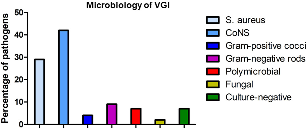
Figure 1. Microbiology of VGI.
Different microorganisms can be identified depending on the site of the infection. Thoracic VGIs are caused by similar microorganisms to those in left-sided infective endocarditis, predominantly Gram-positive bacteria (S. aureus, coagulase-negative staphylococci, enterococci, and streptococci). Gram-negative bacteria and polymicrobial infections are more likely found in abdominal VGIs.
Furthermore, local epidemiology and resistant patterns are important factors that cannot be ignored[17].
Mortality
Although several medical advances have been made over the last decades, morbidity and mortality rates remain high. Patients often have other adjacent diseases that hinder treatment and worsen prognosis[18-20]. Mortality depending on the clinical presentation can be as high as 88%-100%, emphasizing the severity of this disease[15,18,19,21-23]. Fortunately, overall mortality can be reduced with a proper approach to approximately 20%[4,5].
DIAGNOSIS
Clinical presentation
Timely diagnosis remains one of the main challenges. Clinical presentation varies from patient to patient with a broad range of symptoms depending on the acuity of infection: local manifestations such as discharge of pus or inflammation; nonspecific symptoms such as fever, fatigue, or bacteremia; or even severe findings including bleeding and septic shock. Clinicians often must rely on these non-pathognomonic signs to suspect infection. Furthermore, clinical practice varies from center to center due to the lack of standardization and therefore inconsistency in diagnosis and treatment strategies, as pointed out by some authors[24]. Although the vicinity of wounds makes superficial and deep surgical site infections common findings, these are not always present. Szilagyi et al.[25], Samson et al.[26], and Bunt et al.[27] suggested classifications for wound and peripheral VGIs, which help classify and assess the extent of the disease[28]. The non-specific Fitzgerald criteria[24] for abdominal or peripheral grafts or the modified Duke criteria[29] for composite grafts are also applied in this regard. To solve this problem, the Management of Aortic Graft Infection Collaboration (MAGIC)[30] suggested in 2016 a classification dividing clinical/surgical, radiological, and laboratory findings into major and minor criteria [Table 2], similar to the modified Duke criteria[29] in infective endocarditis. A suspected infection is defined as having one major criterion from any category or minor criteria from two of the three categories; confirmed infection requires one major criterion plus any other (major or minor) from another category. The importance of this classification, which is recommended in current European Clinical Practice Guidelines[4], is that of a formal case definition that can be applied with high sensitivity and specificity in cases of definite infections. However, reduced specificity for suspected thoracic VGIs, as confirmed by our group[31], leads to an overestimation of suspected infections, suggesting that additional assessment is required. Modification of the MAGIC criteria should be considered to improve diagnostic accuracy, mainly in the setting of suspected infections. The modified Duke criteria, which have shown high sensitivity and specificity, should be used in thoracic VGI to avoid overdiagnosis.
The MAGIC classification[30]
| Criterion | Clinical/surgical | Radiology | Laboratory |
| Major | Open wound with exposed graft or communicating sinus | Perigraft fluid on CT scan ≥ 3 months after insertion | Organisms recovered from an explanted graft |
| Open wound with exposed graft or communicating sinus | Perigraft gas on CT scan ≥ 7 weeks after insertion | Organisms recovered from an intraoperative specimen | |
| Fistula development, e.g., aorto-enteric or aorto-bronchial | Increase in perigraft gas volume demonstrated on serial imaging | Organisms recovered from a percutaneous, radiologically guided aspirate of perigraft fluid | |
| Graft insertion in an infected site, e.g., fistula, mycotic aneurysm, or infected pseudoaneurysm | |||
| Minor | Localized clinical features of graft infection, e.g., erythema, warmth, swelling, purulent discharge, pain | Other, e.g., suspicious perigraft gas/fluid soft tissue inflammation; aneurysm expansion; pseudoaneurysm formation; focal bowel wall thickening; discitis/ osteomyelitis; suspicious metabolic activity on FDG PET/CT; radiolabeled leukocyte uptake | Blood culture(s) positive and no apparent source except graft infection |
| Fever ≥ 38 °C with graft infection as the most likely cause | Abnormally elevated inflammatory markers with graft infection as the most likely cause, e.g., erythrocyte sedimentation rate, C reactive protein, white cell count |
Imaging modalities
Ultrasound
The use of ultrasound (US) in thoracic and abdominal VGIs is mainly limited by its high interrater variability and patient’s habitus leading to low sensitivity. However, it is easy to perform, and, for thoracic and especially composite grafts, it is used to identify valvular dysfunction, hematoma, abscess, or fistula formation.
Computed tomography angiography
Computed tomography angiography (CTA) remains the gold standard even though pooled sensitivity and specificity rates are 67% and 63%, respectively[32]. Moreover, in low-grade infections, sensitivity descends to 55%, resulting in high false negatives[33]. Nevertheless, CTA can show perigraft fluid, gas [Figure 2], or other findings such as pseudoaneurysm or anastomotic leaks, included in the MAGIC criteria[30]. Due to the aforementioned facts, a second imaging modality is often required to support diagnosis, especially in cases where the infection is suspected[3,4].
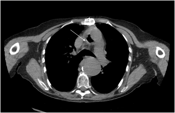
Figure 2. Gas collection (arrow) around aortic graft.
Magnetic resonance angiography
The use of magnetic resonance angiography (MRA) is limited to a small number of investigations[33]. Shahidi et al.[34] reported a sensitivity of 68% and a specificity of 98% in 40 patients with suspected abdominal aortic graft infections. Compared to CTA, MRA is less available, and it requires longer examination times, resulting in motion artifacts. It is, hence, not recommended as a first-choice imaging modality[4,5].
111In or 99m Tc- white-blood-cell-scintigraphy
The use of radiolabeled white-blood-cell (WBC)-scintigraphy is well established in diagnosing infection and inflammation. Reinders Folmer et al.[32] reported in a meta-analysis sensitivity and specificity rates of 90% and 88%, respectively, and even higher rates when combined with single-photon emission computed tomography (SPECT). Other authors have reported similar findings[35,36]. Unfortunately, similar to MRA, the procedure is time-consuming, requiring multiple timepoints for imaging acquisition[33]. WBC-scintigraphy is not recommended as a first-choice diagnostic tool for VGIs due to the elimination of the tracer via the intestine, its uptake in the bone marrow, and the aforementioned reasons[4].
18F-Fluorodeoxyglucose positron emission tomography
Fluorodeoxyglucose positron emission tomography (FDG-PET) alone or in combination with CTA plays an important role in diagnosing VGIs[4,5]. With an approximately 2 h scan time for a whole-body examination, FDG-PET/CT scans have been standardized by the European Association of Nuclear Medicine (EANM) and the Society of Nuclear Medicine and Molecular Imaging (SNMMI)[37] to avoid interhospital discrepancies. Based on the uptake of radiolabeled glucose in cells with enhanced metabolism [Figure 3], FDG-PET scans can be scored and interpreted using several systems[38]. Visual methods include the uptake intensity of FDG or the diffuse or focal uptake pattern; others include quantification of the maximum standardized uptake value (SUVmax) and calculation of the tissue-to-background ratio (TBR)[38]. Despite the high sensitivity of 96%, specificity remains relatively low at 74%[39], resulting in a high false positive rate especially within 6-8 weeks after surgery[40]. In a recent meta-analysis including 13 investigations, Reinders Folmer et al.[41] studied different scoring systems used for the interpretation of FDG-PET scans. They concluded that the uptake pattern was the most accurate assessment method for the diagnosis of graft infection and that the addition of two or more methods could enhance diagnostic accuracy. Nevertheless, it has been suggested that semiquantitative criteria should be used for diagnosis[39,42]. Tokuda et al.[40] suggested a SUVmax cut-off value higher than 8 for infected grafts, Berger et al.[43] set the threshold at 5.5, and Mitra et al.[44] used a cut-off at 6.3, comparing suspected infected with non-infected grafts. Unfortunately, there is no standardized SUVmax cut-off value that can be applied to diagnose VGIs. Nonetheless, FDG-PET scans can also be used to steer antibiotic treatment at follow-up, as suggested by Husmann et al.[45].
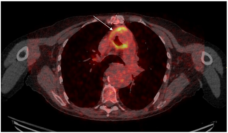
Figure 3. FDG-PET scan depicting (arrow) enhanced metabolic activity around an infected aortic graft.
Microbiological diagnosis
Identification of microorganisms is paramount to achieving optimal treatment. Through different available sampling techniques, microorganisms can be identified in up to 98% of the cases[4]. Sampling from surgical wounds or adjacent tissues is often insufficient since these usually represent colonizing flora. Thus, it is of utmost importance to obtain biopsies from the infected graft, surrounding tissues, perigraft fluid, or abscesses[24]. Specimens can be obtained directly during surgical intervention [Figures 4 and 5 ] or aspirated under US or CT guidance. Aside from direct sampling, blood and wound cultures cannot be forgotten since they can provide valuable information when biopsies are not available. However, microorganisms detected in blood cultures do not always correlate to those found in excised vascular grafts or perigraft tissue cultures in abdominal VGIs[46]. Regarding negative-pressure wound-therapy foams, Scherrer et al.[47] found no improvement in diagnostics from bacterial cultures.
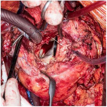
Figure 4. Intraoperative finding of an infected composite graft (arrow).
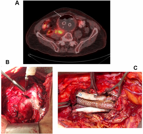
Figure 5. A 61-year-old patient with infected endograft after EVAR for abdominal aortic aneurysm after five years. (A) FDG-PET scan shows gas (arrow) within the aneurysm sac and retroperitoneal fluid collection. (B) Aspiration of pus from the aneurysm sac perioperatively to prevent massive dissemination into abdominal cavity. (C) After debridement and partial aneurysm sac resection, the use of white foam (star) over the endograft (arrow) followed by vacuum-assisted therapy aiming to preserve an endograft.
Broad-range polymerase chain reaction (PCR) and enhanced recovery techniques such as sonication of the graft or vortex mixing are used to identify microorganisms, especially in biofilm-producing bacteria[46,48].
MANAGEMENT
A multidisciplinary team approach including specialists in infectious diseases, cardiovascular surgery, radiology, nuclear medicine, and cardiac imaging is recommended in Clinical Practice Guidelines[4,5]. Additionally, other specialists should be involved in the care team in terms of reducing the risk of infection and enhancing wound healing or providing outpatient management. Due to its complexity, its high morbidity and mortality, and the involvement of such mentioned specialists, patients with VGIs should be referred to a tertiary center where optimal management can be offered[4,5].
Antimicrobial treatment
The antimicrobial treatment represents one of the cornerstones when dealing with VGIs, although there are no universal recommendations on what antimicrobial agent to use. Especially during the acute phase when an infection is suspected, broad-spectrum intravenous antimicrobial treatment is mandatory to control the infection and prevent or cure septicemia. Biofilm-producing bacteria as well as local epidemiology and resistance patterns must be taken into consideration when choosing treatment. Therefore, initial empirical treatment should cover staphylococci and Gram-negative species. This can be achieved by using a beta-lactam or a glycopeptide plus an aminoglycoside[14,17]. For thoracic VGIs, antimicrobial treatment resembles the approach in left-sided infective endocarditis. Abdominal VGIs, on the other hand, require a better coverage of Gram-negative species and anaerobes. After isolation of the causative microorganism, resistance and susceptibility must be tested to optimize treatment.
Duration of antimicrobial therapy is a matter of debate with different recommendations depending on the extent of surgical debridement[17]. Table 3 provides an overview of current recommendations[4,5,17], while Table 4 lists the empirical and targeted therapy in abdominal and thoracic VGI.
| European clinical practice guidelines[4] | Complete resection • 2 weeks i.v. + 2-4 weeks oral • If replaced by a new vascular graft: 4-6 weeks | Lifelong suppressive AB treatment for patients in poor conditions deemed unfit for surgery |
| American clinical practice guidelines[5] | Intraabdominal • 6 weeks i.v. + 3-6 months oral • If extensive perigraft infection or MRSA, Pseudomonas, multidrug-resistant strain: consider lifelong suppressive | Intrathoracic • 4-6 weeks i.v. • Oral AB or suppressive course in selected cases and MDT discussion |
| Revest et al.[17] | Complete resection • 6 weeks i.v. | Partial removal • MRSA, MSSA, Enterobacteriaceae: 6 weeks + suppressive • Streptococci: 6 weeks • Enterococci: 6 weeks + oral extended time • Pseudomonas: MDT decision • Obligate anaerobia: suppressive oral |
Antimicrobial treatment
| Empirical therapy VGI | Targeted therapy VGI* | |||
| No allergy | Allergy to penicillin | No allergy | Allergy to penicillin | |
| Abdominal VGI | ||||
| MSSA | Piperacillin/tazobactam + vancomycin | Cefepime + metronidazole + vancomycin | Parenteral treatment Flucloxacillin + gentamicin 3 days Followed by Flucloxacillin + Rifampicin1,2 Followed by Oral treatment Ciprofloxacin + rifampicin1,3 | Parenteral treatment Cefazolin, vancomycin, or daptomycin + gentamicin 3 days Followed by Cefazolin, vancomycin, or daptomycin + rifampicin1,2 Followed by Oral treatment Ciprofloxacin + rifampicin1,3 |
| MRSA | Parenteral treatment Vancomycin or daptomycin + gentamicin 14 days Followed by Vancomycin or daptomycin + gentamicin 14 days + rifampicin1,2 Followed by Oral treatment Ciprofloxacin + rifampicin1,3 | Parenteral treatment Daptomycin4 + gentamicin 14 days Followed by Daptomycin4 + gentamicin 14 days + rifampicin1,2 Followed by Oral treatment Ciprofloxacin + rifampicin1,3 | ||
| Coagulase-negative staphylococci (CONS) | Parenteral treatment Flucloxacillin, vancomycin, or daptomycin + gentamicin 14 days Followed by Flucloxacillin, vancomycin, or daptomycin + gentamicin 14 days + rifampicin1,2 Followed by Oral treatment Ciprofloxacin + rifampicin1,3 | Parenteral treatment Vancomycin or daptomycin + gentamicin 14 days Followed by Vancomycin or daptomycin + Gentamicin 14 days + rifampicin1,2 Followed by Oral treatment Ciprofloxacin + rifampicin1,3 | ||
| Enterobacteriaceae | Parenteral treatment Ceftriaxone or cefotaxime + gentamicin 14 days2 Followed by Oral treatment Ciprofloxacin3 | Parenteral treatment Cefepime + metronidazole + gentamicin 14 days2 Followed by Oral treatment Ciprofloxacin3 | ||
| Polymicrobial infection | Parenteral treatment Piperacillin/tazobactam + vancomycin Followed by Oral treatment According to pathogens | Parenteral treatment Cefepime + metronidazole + vancomycin Followed by Oral treatment According to pathogens | ||
| Thoracic VGI | ||||
| MSSA | Amoxicillin + flucloxacillin+ gentamicin5 or Vancomycin + gentamicin ± rifampicin1,6 | Vancomycin + gentamicin ± rifampicin1,6 | Parenteral treatment Flucloxacillin + gentamicin 3 days Followed by Flucloxacillin + rifampicin1,8 Followed by Oral treatment Ciprofloxacin + rifampicin1,3 | Parenteral treatment Cefazolin, vancomycin, or daptomycin + gentamicin 3 days Followed by Cefazolin, vancomycin, or daptomycin + rifampicin1,8 Followed by Oral treatment Ciprofloxacin + rifampicin1,3 |
| MRSA | Parenteral treatment Vancomycin or daptomycin + gentamicin 14 days Followed by Vancomycin or daptomycin + gentamicin 14 days + rifampicin1,8 Followed by Oral treatment Ciprofloxacin + rifampicin1,3 | Parenteral treatment Daptomycin 4 + gentamicin 14 days Followed by Daptomycin4 + Gentamicin 14 days + rifampicin1,8 Followed by Oral treatment Ciprofloxacin + rifampicin1,3 | ||
| Coagulase-negative staphylococci (CONS) | Parenteral treatment Flucloxacillin, vancomycin, or daptomycin + gentamicin 14 days Followed by Flucloxacillin, vancomycin, or daptomycin + gentamicin 14 days + rifampicin1,8 Followed by Oral treatment Ciprofloxacin + rifampicin p.o1,3 | Parenteral treatment Vancomycin or daptomycin + gentamicin 14 days Followed by Vancomycin or daptomycin + gentamicin 14 days + rifampicin1,8 Followed by Oral treatment Ciprofloxacin + rifampicin1,3 | ||
| Enterococcus SPP | Parenteral treatment Amoxicillin or ampicillin + gentamicin 14 days8 or Amoxicillin + ceftriaxone7,8 Followed by Oral treatment Amoxicillin p.o. | Parenteral treatment Vancomycin + gentamicin8 Followed by Oral treatment Doxycyclin or linezolid | ||
| Streptococcus SPP | Parenteral treatment Penicillin G + Gentamicin 14 days8 or Ceftriaxone + Gentamicin 14 days8 Followed by Oral treatment Amoxicillin or clindamycin | Parenteral treatment Vancomycin + gentamicin8 Followed by Oral treatment Clindamycin | ||
| HACEK | Parenteral treatment Ceftriaxone + or Amoxicillin + gentamicin 14 days8 Followed by Oral treatment Ciprofloxacin | Parenteral treatment Ceftriaxone or Ciprofloxacin orally Followed by Oral treatment Ciprofloxacin | ||
| Non-HACEK organisms | Parenteral treatment Ceftriaxone + Gentamicin 14 days Followed by Oral treatment Ciprofloxacin | Parenteral treatment Cefepime or ertapenem + gentamicin days Followed by Oral treatment Ciprofloxacin | ||
Surgery
Indication and timing for surgery
VGI is per definition an indication for surgery since the probability to cure a prosthetic graft infection only with antibiotics is highly uncertain[17]. However, although results compared to conventional excisional surgery are worse, patients who are unfit for surgery may be treated with a conservative or palliative approach that includes percutaneous drainage, endovascular techniques, and suppressive antibiotic treatment[4,49-51]. High-risk patients could also benefit from these alternatives where surgery foreshadows fatal outcomes[52]. More data in this regard are needed to properly evaluate these treatment options in this specific scenario. Patients with infected endografts could also benefit from a partial resection of the infected aneurysmal sac, wound irrigation, and regular changes of vacuum-assisted dressings. Finally, a conservative surgical approach including re-opening the chest/abdomen and treatment with vacuum-assisted drainage has to be discussed.
Timing for surgery is in close relationship to the patient’s clinical condition. In hemodynamically unstable patients with acute bleeding or septic shock, a “bridging to surgery” endovascular approach to treat aorto-bronchial or -esophageal fistula can be attempted. Timing for surgery is not provided in current clinical practice guidelines for vascular graft infections[4,5], but it could be possible to extrapolate, adapt, and use those for left-sided infective endocarditis[3].
Extra-anatomical bypass[53] and thorough debridement with complete removal of the infected prosthesis were historically the gold standard. However, due to the aggressiveness of the operation and subsequent high perioperative mortality[54], alternatives have emerged to achieve infection control. Techniques with selective debridement and irrigation are an example of such procedures. Considering that there is no “one-size-fits-all” solution, surgeons must master debridement techniques and select the most suitable approach to individualize treatment strategy. Figure 6 provides a suggested algorithm for patients deemed fit for surgery.
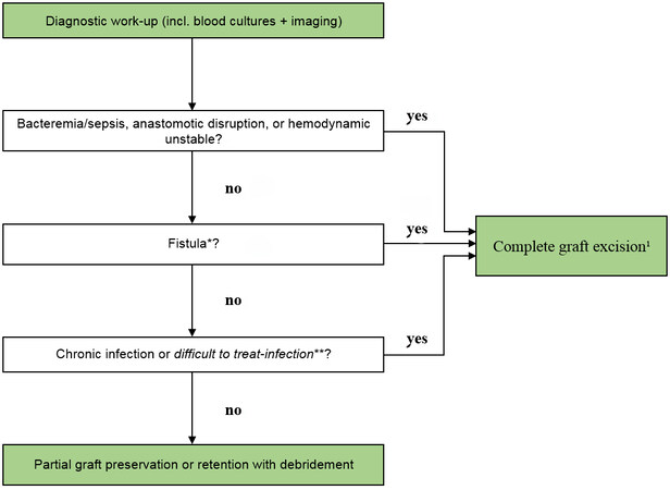
Figure 6. Suggested algorithm in patients deemed fit for surgery.
Abdominal VGI
If hemodynamically stable, percutaneous CT-guided drainage of periprosthetic abscess or fluid collection can be safely performed to isolate microorganisms and reduce infective load[55].
Two-stage axillo-femoral or axillo-popliteal EAB followed by infrarenal resection of the prosthetic graft and in-situ reconstruction[37] with different vascular conduits and partial graft removal are some of the surgical techniques used in this setting. A recent Swedish nationwide study with 126 abdominal graft infections[56] showed no significant differences between EAB and ISR in 30-day (81.7% vs. 76.4%), 1-year (66.2% vs. 72.7%), and 5-year survival (48.2% vs. 48.9%). Oderich et al. found no significant differences between the two strategies at five years[57]. Contrary to these findings, Batt et al.[58] showed that ISR performed better than EAB at one (20% vs. 63%) and five years (10% vs. 41%). Similarly, a recent meta-analysis[59] revealed better 30-day (12% vs. 26.7%), 1-year (54.3% vs. 78.7%), and 5-year (32.4% vs. 57.7%) survival rates for ISR over EAB. Thus, current clinical practice guidelines recommend ISR over EAB[4,5]. In patients with partial or complete removal of infected graft/endograft, it is recommended to cover the new graft with viable tissue[60]. Several biological alternatives exist such as pedicled omentum flap, muscle (intercostal or latissimus), fascia, or retroperitoneal tissue[4]. Reinforcing the newly implanted graft with viable tissue provides a biological barrier from other organs, protecting the prosthesis from re-infection. Furthermore, suture lines and anastomotic stumps (after EAB) are protected from leakage, rupture, or blowout[57,61,62].
Several conduits are available for revascularization with differing advantages one each other: cryopreserved allografts represent the most biological solution with a low risk for re-infection (0%-7%)[58,63-66]. Rifampicin-bonded and silver-coated grafts have the lowest limb amputation rate of around 3% and are an excellent solution for emergency procedures, mainly in the abdominal and pelvic location[67]. Major drawbacks are the limited availability and graft-related complications from degeneration of cryopreserved allografts[58,64] and the relatively high rate of re-infection (> 10%) in rifampicin-bonded and silver-coated grafts.
Self-made xenopericardial (bovine) tube-grafts are an outstanding off-the-shelf solution that can be tailored to form straight or bifurcated tubes[68,69]. Alonso et al.[70] reported in a recent analysis of the use of xenopericardial tubes in 21 patients (17 VGIs and 4 native aorta infections) with a 30-day mortality of 4.8 % and a mean follow-up of 14 months. Graft patency was 95%, and at follow-up, three patients presented with re-infections. Similar results were reported by Weiss et al. and Kreibich et al.[68,71]. Although the results are promising, longer follow-up data are needed to properly assess this type of reconstruction material.
Special situations: fistula and partial graft resection
Aortoenteric-fistula (AEF) is reported in up to 30% of VGIs[72] with poor outcomes and increased morbidity and mortality[73]. Gavali et al.[56] reported no significant difference between EAB and ISR in patients with or without fistulation. Similarly, in a recent multicenter contribution including 182 patients with AEF, Janko et al.[72] reported no superiority in mortality of ISR over EAB. Kakkos et al.[74] in a multicenter study compared endovascular and open repair techniques in AEF. Although perioperative outcomes were better, mortality was higher during follow-up. Other authors have also addressed this important topic[54,75]. In this specific scenario where patients often are hemodynamically unstable due to bleeding or sepsis, an endovascular treatment might be used as a stabilization strategy and “bridge to surgery”. It seems clear that an endovascular treatment without debridement portends a dismal prognosis with recurrent AEF, re-infection, and high mortality. Therefore, surgery should be performed in a second step.
Partial graft resection has been reported to provide better outcomes than complete graft removal when an infection is not located in the main body of bifurcated grafts and no fistula is observed[76].
Thoracic VGI
Surgical principles are the same as those applied to abdominal VGIs. Patients deemed unfit for surgery might be treated with endovascular techniques, irrigation, vacuum-assisted drainage on the prosthetic graft, and/or antibiotics. Due to its anatomical localization characteristic, intraoperative management might be required such as the use of cardiopulmonary bypass, left heart bypass, hypothermic circulatory arrest, or cerebrospinal fluid drainage.
Descending aorta grafts
EAB includes axillofemoral revascularization and bypass between the ascending and descending or supra-celiac aorta, and it can be used in cases where at the time of surgery ISR is not feasible[77]. For the so-called “ventral aorta”, McKellar et al.[78] reported an excellent graft patency rate at follow-up in the setting of aorta coarctation, while Hargrove et al.[77] in 1984 performed EAB in two patients with VGIs showing excellent results.
In general, the literature regarding the management of thoracic VGIs is scanty, and most of the available documents are surgical case series or retrospective studies with a limited number of patients[21,64,77,79]. Currently, and as a natural consequence of broader use of TEVAR compared to open repair, infected endografts are more commonly observed and reported[80]. Kahlberg et al.[60] studied outcomes for open surgical grafts and endograft infections. Mortality rates at 30 days, 1 year, and 5 years were 33%, 57%, and 82% after open repair, whereas after TEVAR they were 26%, 54%, and 81%, respectively. Rustum et al.[81] reported in 2021 outcomes for 20 patients with infected grafts of the descending aorta with mortality rates at 30 and 90 days of 35% and 45%, respectively. Conversely, Sandhu et al.[82] documented a 30-day mortality of 15%. Interestingly, they compared follow-up survival with a control group of reoperated patients without infection with similar cumulative survival rates. Although not statistically significant due to the small sample size, survival probabilities diverged from each other with a non-neglectable risk of 10%-15%. In a metanalysis, Moulakakis et al.[49] reported outcomes of patients with TEVAR infection with either endograft explantation or endograft preservation. Overall mortality rates were 46.3% (explantation) and 81.8% (preservation) with an odds ratio favoring graft explantation over preservation.
Special situations: airway and esophageal fistula
Aorto-esophageal (AEF) or aorto-bronchial fistula (ABF) are reported in up to 1.5% of treated patients[80,83]. Czerny et al.[84,85] from an international collaboration group found 1.5% AEF and 0.56% ABF. In these contributions, the highest survival was achieved through a radical approach including explantation of the infected graft and esophagectomy or pulmonary/bronchial parenchymal repair. In a metanalysis[49], the authors concluded that patients with fistula had worse outcomes than those without fistula. Differences in terms of management and outcomes exist between AEF and ABF, and they should be treated individually as different entities. In the emergency setting, endovascular treatment can be considered to overcome the initial unstable phase in AEF and ABF[83,86,87]. After this initial stabilizing step, excisional surgery with complete explantation of the infected prosthesis should be performed in AEF non-high-risk patients to properly address the disease. In patients with airway fistula, it is recommended to close the airway defect, preserving or not the infected prosthesis. In this high-risk setting, covering the newly implanted or preserved prosthesis with viable tissue is extremely important[4].
Aortic root, ascending aorta, and aortic arch
The paucity of this condition, the even lower limited number of surgical series, and the absence of endovascular treatment options in this region can explain the fact that current clinical practice guidelines do not provide specific information on VGIs in this localization[3-5]. One of the largest series reported to date comprising 68 patients with different treatment strategies from Japan[15] documented a 35.3% in-hospital mortality rate. Similarly, Shah et al.[19], Khaladj et al. [88], Takano et al.[23], Umminger et al.[89], Ramos et al.[18], and Coselli et al.[21] documented 13.3%, 24%, 25%, 25%, 28.6%, and 42% in-hospital mortality rates, respectively. Oda et al.[15] reported overall one- and three-year survival rates of 58.6% and 56.2%, which correlate to those of Shah et al.[19] at one year. At five years, Sandhu et al.[82] documented a 45.4% survival rate.
The use of cryopreserved allografts has been studied by several authors[64,88,90,91] and has been accepted as a viable solution in infected scenarios due to their resistance to infection. At a median of 2.4 years after implantation, Preventza et al.[90] achieved 100% freedom from reinfection, and Lytle et al.[92] documented only one patient with an infected homograft. Concordantly, freedom from reinfection after surgery for valve endocarditis with homograft implantation at 10 years was 92%[93].
Other authors have adopted the use of self-made xenopericardial conduits (with or without an integrated tissue valve) to compensate for the limited availability of allografts[68,71,94]. Kreibich et al.[68] reported a 2% re-infection rate after 11 months. Weiss et al.[71] observed no re-infections at four years. In addition to ISR and EAB, graft preservation strategies with irrigation, selective debridement, the use of negative pressure wound therapy, and omental or muscle wrapping have been described with acceptable survival outcomes[52,77,89,95,96]. Covering the dead space of an infected graft with vital tissue such as omentum or muscle flap provides an exceptional barrier against microbes[22,77].
LIMITATIONS
This document is a review of the literature, and a systematic protocolled search for documents describing this condition was not performed. Therefore, the information provided herein should be critically analyzed since it might be skewed.
CONCLUSION
Abdominal and thoracic graft infections are still associated with high mortality rates. The challenges are obvious not only from a diagnostic perspective but also from a therapeutical approach. The combination of antimicrobial treatment and surgery remains the gold standard. What appears to be clear is that survival does not only depend on the selected surgical strategy. More importantly, the patient’s clinical condition at the time of surgery, acuity of operation, fistula formation, and the virulence of responsible microorganisms are key factors that influence results. Moreover, there are patients who could benefit from a more conservative approach without radical excisional debridement. The available literature is limited by the lack of randomized trials and the restricted number of patients.
DECLARATIONS
Authors’ contributionsResearched data, discussed its content, wrote, reviewed, and edited the document: Van Hemelrijck M
Researched data, wrote, reviewed, and edited the document: Hasse B, Carrel TP
Discussed the content and reviewed and edited the manuscript before submission: Sromicki J, Husmann L, Rancic Z
Availability of data and materialsNot applicable.
Financial support and sponsorshipNone.
Conflicts of interestAll authors declared that there are no conflicts of interest.
Ethical approval and consent to participateNot applicable.
Consent for publicationNot applicable.
Copyright© The Author(s) 2022.
REFERENCES
1. Crawford ES, De Bakey ME, Colley DA. Clinical use of synthetic arterial substitutes in three hundred seventeen patients. AMA Arch Surg 1958;76:261-70.
2. Carter SC, Cohen A, Whelan TJ. Clinical experience with management of the infected dacron graft. Ann Surg 1963;158:249-55.
3. Habib G, Lancellotti P, Antunes MJ, et al. ESC Scientific Document Group. 2015 ESC Guidelines for the management of infective endocarditis: the Task force for the management of infective endocarditis of the European Society of Cardiology (ESC). Endorsed by: European Association for Cardio-Thoracic Surgery (EACTS), the European Association of Nuclear Medicine (EANM). Eur Heart J 2015;36:3075-128.
4. Chakfé N, Diener H, Lejay A, et al. Editor’s choice - European Society for Vascular Surgery (ESVS) 2020 clinical practice guidelines on the management of vascular graft and endograft infections. Eur J Vasc Endovasc Surg 2020;59:339-84.
5. Wilson WR, Bower TC, Creager MA, et al. American Heart Association Committee on Rheumatic Fever. Vascular graft infections, mycotic aneurysms, and endovascular infections: a scientific statement from the American Heart Association. Circulation 2016;134:e412-60.
6. Luehr M, Peterss S, Zierer A, et al. Aortic events and reoperations after elective arch surgery: incidence, surgical strategies and outcomes. Eur J Cardiothorac Surg 2018;53:519-24.
7. Vikholm P, Astudillo R, Thelin S. Long-term survival and frequency of reinterventions after proximal aortic surgery: a retrospective study†. Eur J Cardiothorac Surg 2019;56:722-30.
8. Vogel TR, Symons R, Flum DR. The incidence and factors associated with graft infection after aortic aneurysm repair. J Vasc Surg 2008;47:264-9.
9. Shiraev T, Barrett S, Heywood S, et al. Incidence, management, and outcomes of aortic graft infection. Ann Vasc Surg 2019;59:73-83.
10. Berger P, Vaartjes I, Moll FL, De Borst GJ, Blankensteijn JD, Bots ML. Cumulative incidence of graft infection after primary prosthetic aortic reconstruction in the endovascular era. Eur J Vasc Endovasc Surg 2015;49:581-5.
11. Langenberg JCM, Kluytmans JAJW, de Groot HGW, et al. Surgical site and graft infections in endovascular and open abdominal aortic aneurysm surgery. Surg Infect (Larchmt) 2018;19:424-9.
13. Anagnostopoulos A, Ledergerber B, Kuster SP, et al. VASGRA Cohort Study. Inadequate perioperative prophylaxis and postsurgical complications after graft implantation are important risk factors for subsequent vascular graft infections: prospective results from the vascular graft infection cohort study. Clin Infect Dis 2019;69:621-30.
14. Hasse B, Husmann L, Zinkernagel A, Weber R, Lachat M, Mayer D. Vascular graft infections. Swiss Med Wkly 2013;143:w13754.
15. Oda T, Minatoya K, Kobayashi J, et al. Prosthetic vascular graft infection through a median sternotomy: a multicentre review †. Interact Cardiovasc Thorac Surg 2015;20:701-6; discussion 706.
16. Kohler P, Kuster SP, Bloemberg G, et al. Healthcare-associated prosthetic heart valve, aortic vascular graft, and disseminated Mycobacterium chimaera infections subsequent to open heart surgery. Eur Heart J 2015;36:2745-53.
17. Revest M, Camou F, Senneville E, et al. Groupe de Réflexion sur les Infections de Prothèses vasculaires (GRIP). Medical treatment of prosthetic vascular graft infections: review of the literature and proposals of a Working Group. Int J Antimicrob Agents 2015;46:254-65.
18. Ramos A, García-Montero C, Moreno A, et al. Endocarditis in patients with ascending aortic prosthetic graft: a case series from a national multicentre registry. Eur J Cardiothorac Surg 2016;50:1149-57.
19. Shah DK, Li Z, Park SJ, et al. Replacement of the infected composite aortic root prosthesis. Ann Thorac Surg 2011;92:1651-5.
20. Hostalrich A, Ozdemir BA, Sfeir J, Solovei L, Alric P, Canaud L. Systematic review of native and graft-related aortic infection outcome managed with orthotopic xenopericardial grafts. J Vasc Surg 2019;69:614-8.
21. Coselli JS, Köksoy C, Lemaire SA. Management of thoracic aortic graft infections. Ann Thorac Surg 1999;67:1990-3.
22. LeMaire SA, Coselli JS. Options for managing infected ascending aortic grafts. J Thorac Cardiovasc Surg 2007;134:839-43.
23. Takano T, Terasaki T, Wada Y, Seto T, Fukui D, Amano J. Treatment of prosthetic graft infection after thoracic aorta replacement. Ann Thorac Cardiovasc Surg 2014;20:304-9.
24. FitzGerald SF, Kelly C, Humphreys H. Diagnosis and treatment of prosthetic aortic graft infections: confusion and inconsistency in the absence of evidence or consensus. J Antimicrob Chemother 2005;56:996-9.
25. Szilagyi DE, Smith RF, Elliott JP, Vrandecic MP. Infection in arterial reconstruction with synthetic grafts. Ann Surg 1972;176:321-33.
26. Samson RH, Veith FJ, Janko GS, Gupta SK, Scher LA. A modified classification and approach to the management of infections involving peripheral arterial prosthetic grafts. J Vasc Surg 1988;8:147-153.
28. Mayer D, Hasse B, Koelliker J, et al. Long-term results of vascular graft and artery preserving treatment with negative pressure wound therapy in Szilagyi grade III infections justify a paradigm shift. Ann Surg 2011;254:754-59; discussion 760.
29. Li JS, Sexton DJ, Mick N, et al. Proposed modifications to the Duke criteria for the diagnosis of infective endocarditis. Clin Infect Dis 2000;30:633-8.
30. Lyons OT, Baguneid M, Barwick TD, et al. Diagnosis of aortic graft infection: a case definition by the Management of Aortic Graft Infection Collaboration (MAGIC). Eur J Vasc Endovasc Surg 2016;52:758-63.
31. Anagnostopoulos A, Mayer F, Ledergerber B, et al. VASGRA Cohort Study. Editor's choice - validation of the Management of Aortic Graft Infection Collaboration (MAGIC) criteria for the diagnosis of vascular graft/endograft infection: results from the prospective vascular graft cohort study. Eur J Vasc Endovasc Surg 2021;62:251-7.
32. Reinders Folmer EI, Von Meijenfeldt GCI, Van der Laan MJ, et al. Diagnostic imaging in vascular graft infection: a systematic review and meta-analysis. Eur J Vasc Endovasc Surg 2018;56:719-29.
33. Lauri C, Iezzi R, Rossi M, et al. Imaging modalities for the diagnosis of vascular graft infections: a consensus paper amongst different specialists. J Clin Med 2020;9:1510.
34. Shahidi S, Eskil A, Lundof E, Klaerke A, Jensen BS. Detection of abdominal aortic graft infection: comparison of magnetic resonance imaging and indium-labeled white blood cell scanning. Ann Vasc Surg 2007;21:586-92.
35. Khaja MS, Sildiroglu O, Hagspiel K, Rehm PK, Cherry KJ, Turba UC. Prosthetic vascular graft infection imaging. Clin Imaging 2013;37:239-44.
36. Annovazzi A, Bagni B, Burroni L, D'Alessandria C, Signore A. Nuclear medicine imaging of inflammatory/infective disorders of the abdomen. Nucl Med Commun 2005;26:657-64.
37. Jamar F, Buscombe J, Chiti A, et al. EANM/SNMMI guideline for 18F-FDG use in inflammation and infection. J Nucl Med 2013;54:647-58.
38. Bruggink JL, Slart RH, Pol JA, Reijnen MM, Zeebregts CJ. Current role of imaging in diagnosing aortic graft infections. Semin Vasc Surg 2011;24:182-90.
39. Kim SJ, Lee SW, Jeong SY, Pak K, Kim K. A systematic review and meta-analysis of 18F-fluorodeoxyglucose positron emission tomography or positron emission tomography/computed tomography for detection of infected prosthetic vascular grafts. J Vasc Surg 2019;70:307-13.
40. Tokuda Y, Oshima H, Araki Y, et al. Detection of thoracic aortic prosthetic graft infection with 18F-fluorodeoxyglucose positron emission tomography/computed tomography. Eur J Cardiothorac Surg 2013;43:1183-7.
41. Reinders Folmer EI, von Meijenfeldt GCI, Te Riet Ook Genaamd Scholten RS, et al. A systematic review and meta-analysis of 18F-fluoro-d-deoxyglucose positron emission tomography interpretation methods in vascular graft and endograft infection. J Vasc Surg 2020;72:2174-2185.e2.
42. Saleem BR, Pol RA, Slart RH, Reijnen MM, Zeebregts CJ. 18F-Fluorodeoxyglucose positron emission tomography/CT scanning in diagnosing vascular prosthetic graft infection. Biomed Res Int 2014;2014:471971.
43. Berger P, Vaartjes I, Scholtens A, et al. Differential FDG-PET uptake patterns in uninfected and infected central prosthetic vascular grafts. Eur J Vasc Endovasc Surg 2015;50:376-83.
44. Mitra A, Pencharz D, Davis M, Wagner T. Determining the diagnostic value of 18F-fluorodeoxyglucose positron emission/computed tomography in detecting prosthetic aortic graft infection. Ann Vasc Surg 2018;53:78-85.
45. Husmann L, Ledergerber B, Anagnostopoulos A, et al. VASGRA Cohort Study. The role of FDG PET/CT in therapy control of aortic graft infection. Eur J Nucl Med Mol Imaging 2018;45:1987-97.
46. Ajdler-Schaeffler E, Scherrer AU, Keller PM, et al. and the VASGRA Cohort. Increased pathogen identification in vascular graft infections by the combined use of tissue cultures and 16S rRNA gene polymerase chain reaction. Front Med (Lausanne) 2018;5:169.
47. Scherrer AU, Bloemberg G, Zbinden R, et al. VASGRA Cohort. Prosthetic vascular graft infections: bacterial cultures from negative-pressure-wound-therapy foams do not improve diagnostics. J Clin Microbiol 2016;54:2190-3.
48. Kokosar Ulcar B, Lakic N, Jeverica S, et al. Contribution of sonicate-fluid cultures and broad-range PCR to microbiological diagnosis in vascular graft infections. Infect Dis (Lond) 2018;50:429-35.
49. Moulakakis KG, Mylonas SN, Antonopoulos CN, et al. Comparison of treatment strategies for thoracic endograft infection. J Vasc Surg 2014;60:1061-71.
50. Lyons OT, Patel AS, Saha P, Clough RE, Price N, Taylor PR. A 14-year experience with aortic endograft infection: management and results. Eur J Vasc Endovasc Surg 2013;46:306-13.
52. Akowuah E, Narayan P, Angelini G, Bryan AJ. Management of prosthetic graft infection after surgery of the thoracic aorta: removal of the prosthetic graft is not necessary. J Thorac Cardiovasc Surg 2007;134:1051-2.
53. Hart JP, Eginton MT, Brown KR, et al. Operative strategies in aortic graft infections: is complete graft excision always necessary? Ann Vasc Surg 2005;19:154-60.
54. Batt M, Jean-Baptiste E, O'Connor S, et al. Early and late results of contemporary management of 37 secondary aortoenteric fistulae. Eur J Vasc Endovasc Surg 2011;41:748-57.
55. Ljungquist O, Dias N, Haidl S, Sonesson B, Sörelius K, Ahl J. Guided aspiration for determining the microbiological aetiology of aortic vascular graft and endograft infections. Eur J Vasc Endovasc Surg 2021;62:935-43.
56. Gavali H, Mani K, Furebring M, et al. Editor's choice - outcome of radical surgical treatment of abdominal aortic graft and endograft infections comparing extra-anatomic bypass with in situ reconstruction: a nationwide multicentre study. Eur J Vasc Endovasc Surg 2021;62:918-26.
57. Oderich GS, Bower TC, Cherry KJ Jr, et al. Evolution from axillofemoral to in situ prosthetic reconstruction for the treatment of aortic graft infections at a single center. J Vasc Surg 2006;43:1166-74.
58. Batt M, Jean-Baptiste E, O'Connor S, Feugier P, Haulon S. Association Universitaire de Recherche en Chirurgie Vasculaire (AURC). Contemporary management of infrarenal aortic graft infection: early and late results in 82 patients. Vascular 2012;20:129-37.
59. Post ICJH, Vos CG. Systematic review and meta-analysis on the management of open abdominal aortic graft infections. Eur J Vasc Endovasc Surg 2019;58:258-81.
60. Kahlberg A, Grandi A, Loschi D, et al. A systematic review of infected descending thoracic aortic grafts and endografts. J Vasc Surg 2019;69:1941-1951.e1.
61. Inoue T, Imura M, Kaneda T, Saga T. Long-term survival after coverage with prevertebral fascia for abdominal aortic stump closure. Vasc Endovascular Surg 2017;51:43-6.
62. Sarac TP, Augustinos P, Lyden S, Ouriel K. Use of fascia-peritoneum patch as a pledget for an infected aortic stump. J Vasc Surg 2003;38:1404-6.
63. Ben Ahmed S, Louvancourt A, Daniel G, et al. Cryopreserved arterial allografts for in situ reconstruction of abdominal aortic native or secondary graft infection. J Vasc Surg 2018;67:468-77.
64. Vogt PR, Rocca HB, Carrel T, et al. Cryopreserved arterial allografts in the treatment of major vascular infection: a comparison with conventional surgical techniques. J Thorac Cardiovasc Surg 1998;116:965-72.
65. Masabni K, Weaver MR, Kandagatla P, et al. Cryopreserved allograft in the management of native and prosthetic aortic infections. Ann Vasc Surg 2019;56:1-10.
66. Harlander-Locke MP, Harmon LK, Lawrence PF, et al. Vascular Low-Frequency Disease Consortium. The use of cryopreserved aortoiliac allograft for aortic reconstruction in the United States. J Vasc Surg 2014;59:669-74.
67. Batt M, Jean-Baptiste E, O'Connor S, et al. In-situ revascularisation for patients with aortic graft infection: a single centre experience with silver coated polyester grafts. Eur J Vasc Endovasc Surg 2008;36:182-8.
68. Kreibich M, Siepe M, Berger T, et al. Treatment of infectious aortic disease with bovine pericardial tube grafts. Eur J Cardiothorac Surg 2021;60:155-61.
69. Carrel T, Englberger L, Schmidli J. How to treat aortic graft infection? Gen Thorac Cardiovasc Surg 2019;67:44-52.
70. Alonso W, Ozdemir B, Chassin-Trubert L, Ziza V, Alric P, Canaud L. Early outcomes of native and graft-related abdominal aortic infection managed with orthotopic xenopericardial grafts. J Vasc Surg 2021;73:222-31.
71. Weiss S, Tobler EL, von Tengg-Kobligk H, et al. Self made Xeno-pericardial aortic tubes to treat native and aortic graft infections. Eur J Vasc Endovasc Surg 2017;54:646-52.
72. Janko MR, Woo K, Hacker RI, et al. In situ bypass and extra-anatomic bypass procedures result in similar survival in patients with secondary aortoenteric fistulas. J Vasc Surg 2021;73:210-221.e1.
73. Chopra A, Cieciura L, Modrall JG, Valentine RJ, Chung J. Twenty-year experience with aorto-enteric fistula repair: gastrointestinal complications predict mortality. J Am Coll Surg 2017;225:9-18.
74. Kakkos SK, Antoniadis PN, Klonaris CN, et al. Open or endovascular repair of aortoenteric fistulas? Eur J Vasc Endovasc Surg 2011;41:625-34.
75. Danneels MI, Verhagen HJ, Teijink JA, Cuypers P, Nevelsteen A, Vermassen FE. Endovascular repair for aorto-enteric fistula: a bridge too far or a bridge to surgery? Eur J Vasc Endovasc Surg 2006;32:27-33.
76. Janko M, Hubbard G, Woo K, et al. Contemporary outcomes after partial resection of infected aortic grafts. Ann Vasc Surg 2021;76:202-10.
77. Hargrove WC, Edmunds LH. Management of infected thoracic aortic prosthetic grafts. Ann Thorac Surg 1984;37:72-7.
78. McKellar SH, Schaff HV, Dearani JA, et al. Intermediate-term results of ascending-descending posterior pericardial bypass of complex aortic coarctation. J Thorac Cardiovasc Surg 2007;133:1504-9.
79. Kieffer E, Sabatier J, Plissonnier D, Knosalla C. Prosthetic graft infection after descending thoracic/ thoracoabdominal aortic aneurysmectomy: management with in situ arterial allografts. J Vasc Surg 2001;33:671-8.
80. Kahlberg A, Melissano G, Mascia D, Loschi D, Grandi A, Chiesa R. How to best treat infectious complications of open and endovascular thoracic aortic repairs. Semin Vasc Surg 2017;30:95-102.
81. Rustum S, Beckmann E, Martens A, et al. Native and prosthetic graft infections of the thoracic aorta: surgical management. Eur J Cardiothorac Surg 2021;60:633-41.
82. Sandhu HK, Nissen AP, Mushtaq H, et al. Resection and replacement of thoracic aortic graft infections. J Thorac Cardiovasc Surg 2021;162:1-8.
83. Chiesa R, Melissano G, Marone EM, Marrocco-Trischitta MM, Kahlberg A. Aorto-oesophageal and aortobronchial fistulae following thoracic endovascular aortic repair: a national survey. Eur J Vasc Endovasc Surg 2010;39:273-9.
84. Czerny M, Eggebrecht H, Sodeck G, et al. New insights regarding the incidence, presentation and treatment options of aorto-oesophageal fistulation after thoracic endovascular aortic repair: the European registry of endovascular aortic repair complications. Eur J Cardiothorac Surg 2014;45:452-7.
85. Czerny M, Reser D, Eggebrecht H, et al. Aorto-bronchial and aorto-pulmonary fistulation after thoracic endovascular aortic repair: an analysis from the European registry of endovascular aortic repair complications. Eur J Cardiothorac Surg 2015;48:252-7.
86. Canaud L, D'Annoville T, Ozdemir BA, Marty-Ané C, Alric P. Combined endovascular and surgical approach for aortobronchial fistula. J Thorac Cardiovasc Surg 2014;148:2108-11.
87. Jonker FH, Schlösser FJ, Moll FL, et al. Outcomes of thoracic endovascular aortic repair for aortobronchial and aortoesophageal fistulas. J Endovasc Ther 2009;16:428-40.
88. Khaladj N, Pichlmaier U, Stachmann A, et al. Cryopreserved human allografts (homografts) for the management of graft infections in the ascending aortic position extending to the arch. Eur J Cardiothorac Surg 2013;43:1170-5.
89. Umminger J, Krueger H, Beckmann E, et al. Management of early graft infections in the ascending aorta and aortic arch: a comparison between graft replacement and graft preservation techniques. Eur J Cardiothorac Surg 2016;50:660-7.
90. Preventza O, Mohamed AS, Cooley DA, et al. Homograft use in reoperative aortic root and proximal aortic surgery for endocarditis: a 12-year experience in high-risk patients. J Thorac Cardiovasc Surg 2014;148:989-94.
91. Mestres CA, Quintana E, Kopjar T, et al. Hospital Clinic Infective Endocarditis Investigators. Twenty-year experience with cryopreserved arterial allografts for vascular infections. Eur J Cardiothorac Surg 2019;55:358-65.
92. Lytle BW, Sabik JF, Blackstone EH, Svensson LG, Pettersson GB, Cosgrove DM. Reoperative cryopreserved root and ascending aorta replacement for acute aortic prosthetic valve endocarditis. Ann Thorac Surg 2002;74:S1754-7.
93. Musci M, Weng Y, Hübler M, et al. Homograft aortic root replacement in native or prosthetic active infective endocarditis: twenty-year single-center experience. J Thorac Cardiovasc Surg 2010;139:665-73.
94. Kondov S, Beyersdorf F, Rylski B, et al. Redo aortic root repair in patients with infective prosthetic endocarditis using xenopericardial solutions. Interact Cardiovasc Thorac Surg 2019;29:339-43.
95. Nakajima N, Masuda M, Ichinose M, Ando M. A new method for the treatment of graft infection in the thoracic aorta: in situ preservation. Ann Thorac Surg 1999;67:1994-6.
Cite This Article
Export citation file: BibTeX | RIS
OAE Style
Van Hemelrijck M, Sromicki J, Husmann L, Rancic Z, Hasse B, Carrel TP. Vascular graft infections. Vessel Plus 2022;6:47. http://dx.doi.org/10.20517/2574-1209.2022.18
AMA Style
Van Hemelrijck M, Sromicki J, Husmann L, Rancic Z, Hasse B, Carrel TP. Vascular graft infections. Vessel Plus. 2022; 6: 47. http://dx.doi.org/10.20517/2574-1209.2022.18
Chicago/Turabian Style
Van Hemelrijck, Mathias, Juri Sromicki, Lars Husmann, Zoran Rancic, Barbara Hasse, Thierry P. Carrel. 2022. "Vascular graft infections" Vessel Plus. 6: 47. http://dx.doi.org/10.20517/2574-1209.2022.18
ACS Style
Van Hemelrijck, M.; Sromicki J.; Husmann L.; Rancic Z.; Hasse B.; Carrel TP. Vascular graft infections. Vessel Plus. 2022, 6, 47. http://dx.doi.org/10.20517/2574-1209.2022.18
About This Article
Special Issue
Copyright

Data & Comments
Data


 Cite This Article 28 clicks

 Like This Article 2
likes
Like This Article 2
likes


Comments
Comments must be written in English. Spam, offensive content, impersonation, and private information will not be permitted. If any comment is reported and identified as inappropriate content by OAE staff, the comment will be removed without notice. If you have any queries or need any help, please contact us at support@oaepublish.com.